Overview
Peripheral Blood Smear

A drop of blood has been placed on the left-hand side of a clean glass slide at point A, and has been pulled towards the right-hand side using another glass slide, stopping at point B. The area near point A is too thick and too darkly stained for interpretation, whereas the area at the very end of the smear, near point B (the “feather edge”) is too thin, distorting the morphology of all cells in that area. The optimal area for viewing is just behind point B (shown by the asterisk), in an area where the red blood cells are just touching and demonstrate central pallor. Adapted from UpToDate.
Red Cell Morphology
- Low power:
- Red blood cells:
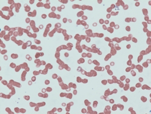
Rouleaux formation
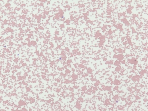
Red cell agglutination
- High Power:
-
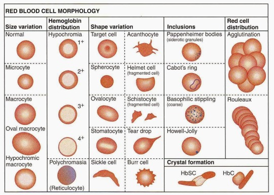
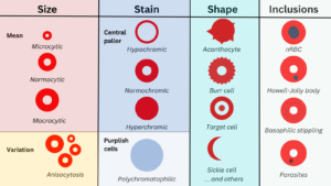
- Size:
- Mean cell size (relative to small lymphocyte):
- Microcytic
- Normocytic
- Macrocytic
- Variation in cell size (anisocytosis)
- Mean cell size (relative to small lymphocyte):
- Staining (color):
- Central pallor:
- Hypochromia
- Normochromia
- Hyperchromia
- Polychromatophilia
- Central pallor:
- Shape (poikilocytosis):
- Normal
- Spiculated cells:
- Acanthocyte (spur cell)
- Echinocyte (burr cell)
- Teardrop cell (dacrocyte)
- Sickle cell (drepanocyte)
- Schistocyte:
- Helmet cell
- Horn cell (keratocyte)
- Bite cell
- Blister cell
- Ovalocyte
- Spherocyte
- Inclusions:
- Nucleated red blood cells
- Howell-Jolly bodies
- Basophilic stippling
- Pappenheimer bodies
- Parasitic inclusions
- Malaria
- Babesia
- Size: