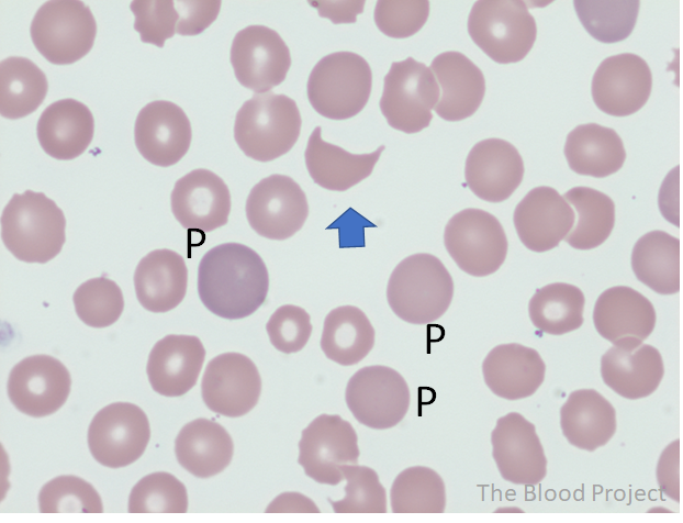
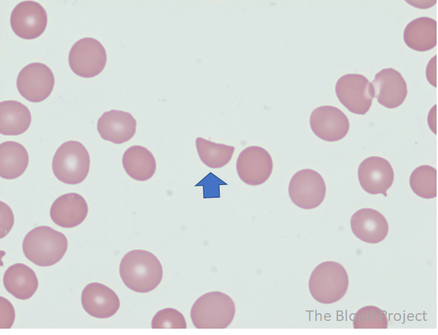
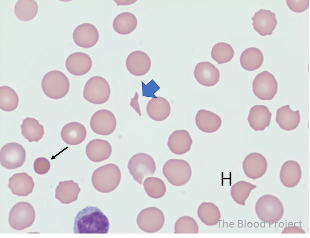
Aug

24

2021

Schistocytes

By William Aird

This peripheral smear is from a patient with thrombotic microangiopathy. The arrow points to a horn cell (also called a keratocye), which is characterized by short “flaps” at each end. These are formed by rupture of one or more peripheral pseudovacuoles and subsequent fusion of the cell membrane P, polychromatophilic cell (100x, oil).

Hemet cell (arrow) in a patient with valve hemolysis. Amputation of these cells results in a straight border which sharp angulated edges (vs. the “flaps” in the horn cell). The missing cell portion corresponds to the fragments that have been split off as a result of a break on a fibrin strand (100x, oil). 

Triangular cell (blue arrow) in a 46-year-old man with thrombotic thrombocytopenic purpura. Black arrow points to a microspherocyte (small-sized hyperdense red cell with deep staining and no central pallor). There is also a helmet cell in the lower right corner (H) (100x, oil).
ParameterProperties
Red blood cell, shape abnormalitySchistocytes
DefinitionSchistocytes, or schizocytes (from the Greek word schisto, broken or cleft) are circulating fragments of red blood cells or red blood cells from which cytoplasmic fragments have been lost. They lack central pallor and are often deeply staining.
Conditions associated with the shape abnormalityThrombotic microangiopathy (TMA), burns, valve hemolysis, march hemoglobinuria
Mechanism of formationSchistocyte formation results from mechanical damage to the red cell caused by fibrin strands on the endothelial surface, excess of turbulence of blood or thermal injury.
HistoryTerm was introduced in 1891 by Paul Ehrlich.1
Source/authorWilliam Aird
Reviewed and edited byParul Bhargava
ReferencesInt J Lab Hematol 2012;34:107