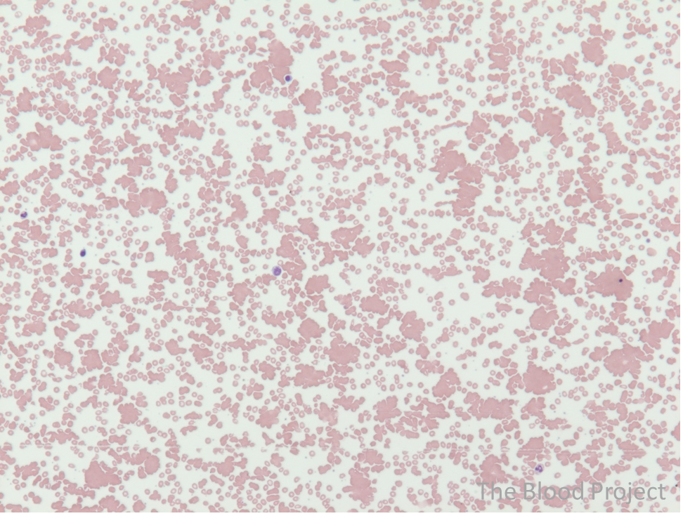
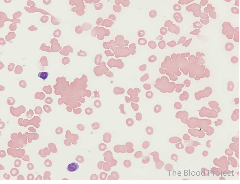
| Red cell agglutination | |
| Definition/description | Clumps of red blood cells, often of equal length and width, in contrast to linear array of red cells in rouleaux formation. |
| Also known as | Autoagglutination, clumping |
| Mechanism of formation | Usually caused be reactivity with anti-erythrocyte IgM antibodies called cold agglutinins. |
| History | In 1955, John Dacie described autohaemagglutination of red cells in the peripheral smear of a patient with “auto-immune hemolytic anaemia of the cold antibody type.” Am J Med. 1955;18:810-21. |
| Source/author | William Aird |
| References | Hematology Am Soc Hematol Educ Program. 2016;2016(1):226-231. |