| Parameter | Properties |
|---|---|
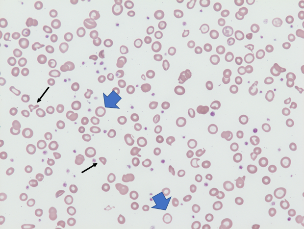
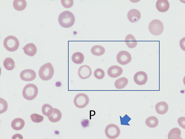
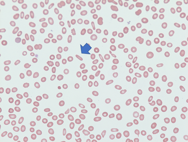
| Condition | Iron deficiency anemia |
| Findings | Hypochromia, pencil shaped red cells, microcytosis and hypochromasia. Pencil cells are hypochromic variants of elliptocytes having long axes at least triple the length of the cell’s short axis. Reactive thrombocytosis may also occur. |
| Differential diagnosis | Thalassemia minor and anemia of chronic disease |
| Mechanism | Decreased hemoglobin production leads to hypochromia. The blunted hemoglobin production results in a smaller-than-normal mature red cell. |
| Source/author | William Aird |
| Reviewed and edited by | Parul Bhargava |