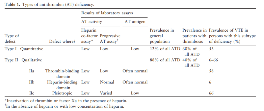
- Type I
- Quantitative deficiency – functional activity and antigenic levels AT are proportionately reduced.
- Most commonly caused by short deletions and insertions and less commonly by single base pair substitutions.
- The deletions vary from 1 to 30 base pairs in length and are scattered throughout the AT gene.
- Type II
- Qualitative deficiency – normal antigen levels are found in association with low AT activity due to a dysfunctional protein.
- Commonly arise secondary to single base pair substitutions.
- Much more common than type I.
- Three subtypes
- Type IIa – caused by mutations that affect AT’s reactive site (i.e. the region where AT binds to its target protease).
- Type IIb
- An abnormality of the heparin-binding domain of AT, interfering with AT activity only in the presence of heparin.
- Significantly lower risk for thrombosis than individuals with other types of AT deficiency.
- Type IIC – pleiotropic group of mutations near the reactive loop site, which may interfere with the mobility of the reactive loop site after heparin binding, thus influencing AT’s interaction with thrombin.
Jul
5
2022