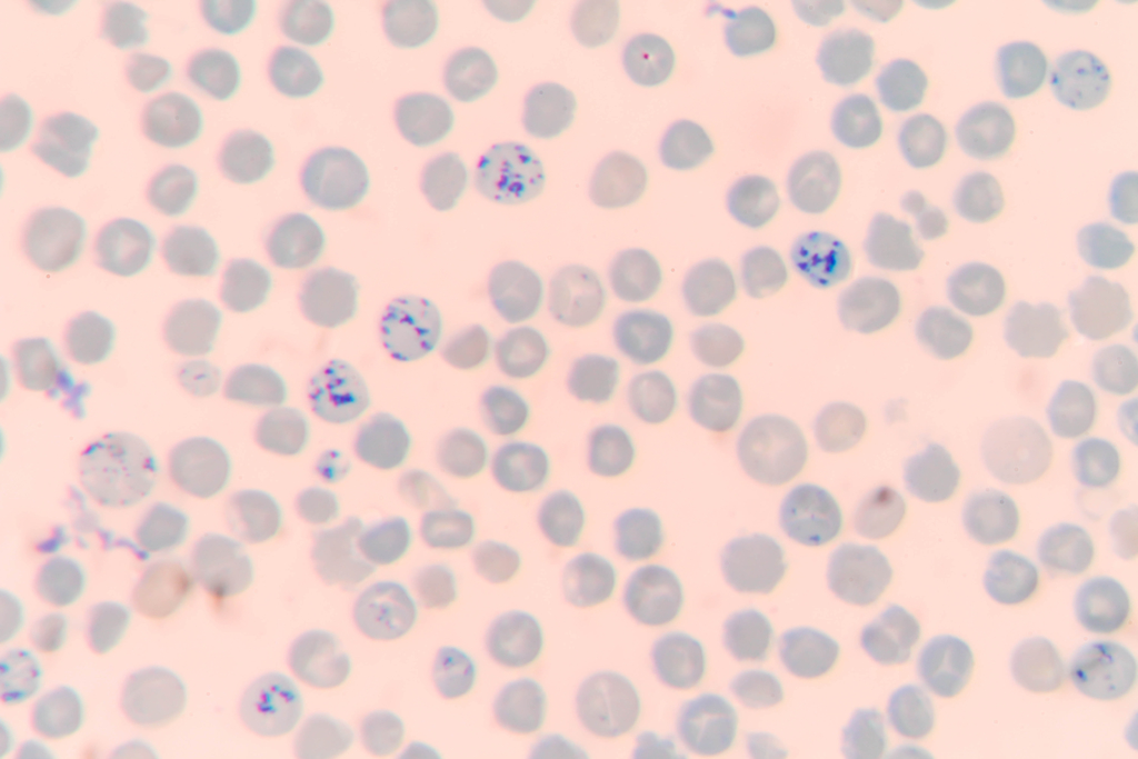
The traditional method of measuring the reticulocyte count is a manual method that uses supravital stains (such as methylene blue) to highlight the reticulum (RNA) network of this immature red cell fraction. A lab technologist uses a microscope to count the number of such cells relative to the number of mature red blood cells. The number of reticulocytes is reported as a percentage of total red blood cells.
Reticulocyte stain in which living red blood cells are incubated in methylene blue. reticulocytes are readily identified by the presence of a network of reticular material.
2) Automated analyzer:
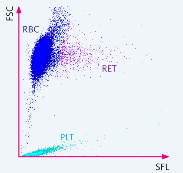
The number of reticulocytes can be measured directly by most automated analyzers by staining the remnant RNA with a fluorescent dye. The number of reticulocytes is reported as an absolute count.
Scattergram of the reticulocyte (RET) channel with a normal cell distribution. In the RET channel, the lysis reagent slightly perforates the cell membranes of red blood cells, white blood cells and platelets and so allows the fluorescence marker to penetrate the cell. The fluorescence marker labels the intracellular nucleic acids whereby the intensity of the resulting fluorescence signal is directly proportional to the nucleic acid content. Using the forward scattered light and the fluorescence signal, the reticulocytes can be separated from mature red blood cells. Source.