Fragmented RBCs can be detected by certain automated hematology analyzers based on the analysis of fraction of small red blood cells (RBCs) in the context of normal RBC volume indices (mean corpuscular volume and width). Their presence should prompt a peripheral blood smear review.
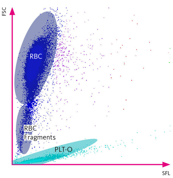
Figure: A specific area below the RBC area in the RET scattergram is used for identification of fragmented red blood cells. Due to the absence of nucleic acids in red blood cells the intensity of the measured side fluorescence signals (SFL) is extremely low. In addition, the high-angle forward scatter (FSC) is lower than that of intact red blood cells. Each cell is plotted in the RET scattergram based on its fluorescence intensity (SFL on x-axis) and its high-angle forward scatter (FSC on y-axis), which reflects characteristics of both cell size and cellular content. The triangle indicates the detection area for fragmented red blood cells (FRC). Source.