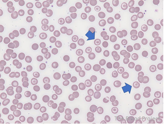
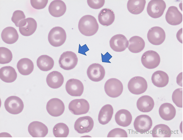
| Red cell shape abnormality | Target cell |
| Also known as | Codocyte (from the Greek term for “hat”) |
| Definition | Centrally located disk of hemoglobin surrounded by an area of pallor with an outer rim of hemoglobin adjacent to the cell membrane giving the cell the appearance of a bull’s eye or shooting. |
| Conditions associated with the shape abnormality | Seen in liver disease (macrocytic targets), iron deficiency anemia, thalassemia (microcytic target cells). Target cells may also be seen in hemoglobin C and E disease, and following splenectomy. |
| Mechanism of formation | Excess of cell membrane relative to cell volume |
| Source/Author | William Aird |
| Reviewed and edited by | Parul Bhargava |