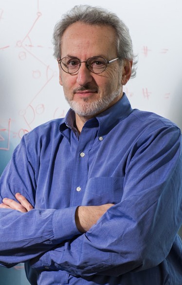
Donald E. Ingber, MD, PhD is the Founding Director of the Wyss Institute for Biologically Inspired Engineering at Harvard University and a chaired professor at Harvard’s schools of medicine and engineering and Boston Children’s Hospital. Don is a member of National Academy of Medicine, National Academy of Engineering, National Academy of Inventors, and the American Academy of Arts and Sciences. His transdisciplinary approach to science has led to major advances in biosensors, diagnostics, cell biology, cancer research, tissue engineering, systems biology, nanobiotechnology and translational medicine. In all his work, Don breaks down boundaries between science, art and design.
In this episode, Don Ingber talks with Helen Osborne about:
- How organ-on-chip and “human body on chips” technologies are built and how they realistically mimic human organ function by combining living cells, blood flow, and mechanical forces like breathing and stretch.
- The implications of these chips for hematology and clinical care, including modeling coagulation and thrombosis, predicting drug responses and toxicities, advancing personalized medicine, and reducing reliance on animal models.
- His transdisciplinary journey blending science, art, engineering, and design, and why breaking down disciplinary boundaries is essential for transformative discovery, innovation, and teaching.
Producer and audio editor: Adam Weiss, Relativistic Media
Transcript:
HELEN: Welcome to Talking About Blood. I’m Helen Osborne, host of this podcast series and a member of the Advisory Board for The Blood Project. I also produce and host my own podcast series, and it’s about health communication called Health Literacy Out Loud. Today, I’m talking with Dr. Donald Ingber, who is the founding director of the Wyss Institute for Biologically Inspired Engineering at Harvard University and a chaired professor at Harvard Schools of Medicine and Engineering and Boston Children’s Hospital. Don is a member of the National Academy of Medicine, National Academy of Engineering, National Academy of Inventors, and the American Academy of Arts and Sciences. His transdisciplinary approach to science has led to major advances in biosensors and diagnostics, cell biology, cancer research, tissue engineering, systems biology, nanobiotechnology, and translational medicine. In all his work, Don breaks down the boundaries between science, art, and design. Welcome to Talking About Blood.
DON: Thank you so much. Happy to be here.
HELEN: I am so intrigued about the many, many innovative ways you’re advancing science and medicine. One of them that really grabbed my attention was that organ chip technology. So please get us started. Introduce us all to you and to this organ on chips. And, you know, we’ll take it from there.
DON: Sure. So I’ve been working in science a long time in terms of the blood focus. I did my postdoc with Judah Folkman in Department of Vascular Biology at Harvard and Boston Children’s Hospital, Harvard Medical School. And I worked on angiogenesis. And in fact, my endowed professorship at Harvard Medical School is the Judah Folkman Professor of Vascular Biology. So there I’ve worked in for a very long time. I’ve worked in it from the endothelial cell angle, as well as from angiogenesis, as well as from blood coagulation. And it’s great to be here.
HELEN: We’re glad to have you here. Tell us more.
DON: Yeah. So, I guess I would say that my entire career, I’ve been interested in the idea that mechanical forces are as important as chemicals and genes for biology. And so that’s something I pursued for many years. That led me to develop ways that I could actually show experimentally that literally if you stretch a cell or round a cell, physically deform a cell, you could affect their function and growth and viability. And that led me to reach out to approaches outside of science from the computer microchip manufacturing world, where you have literally the same sort of photolithography-based approach that artists use to make photolithographs, artwork. That’s how people adapted to make microchips by designing on a computer patterns of, you know, all the different chip connectors and transistors, electrodes, all those little lines you see in pictures of microchips. They expose the material to light with a mask that creates the pattern, and the cool thing for me was that you control the feature size at the same nanometer to micrometer scale that living cells and tissues live at. So, starting 35 or more years ago, we started to explore the use of a novel form of that technique developed by my collaborator at Harvard, George Whitesides, that can essentially instead of having to be in a clean room to make every single chip with etching, you etch the substrate once and then you make a rubber stamp and that retains those same features. And so in around early 90s, we started to create culture environments where we can control cell shape by controlling their adhesions. The little dots we made that were micrometer size surrounded by non-adhesive regions. With that, we could actually show that stretching cells and rounding cells control whether they grow or they go into a suicide program or they differentiate. We did that with blood vessel endothelial cells, and we tried it with smooth muscle cells.
HELEN: Can you make this more vivid? For scientists might be able to keep up with you at all the levels you’re going at, or an engineer, but can you also make it so that those of us who aren’t in science and medicine can get more of a graphic picture of what it is that you’re creating?
DON: Sure. So about 35 years ago, we started to take microchip manufacturing techniques that have control over features in the nanometer to micrometer scale where living cells inhabit in tissues to create substrates where we can address the question, do stretching cells, a cell that stretches flat versus one that’s round, have different functionality. And so the idea was to create little adhesive islands because cells adhere to substrates by binding to extracellular matrix molecule. Matrix are the molecules that assemble the scaffolds that hold cells in tissues like eggs in an egg carton.
HELEN: Okay.
Don: Nowadays, we take those molecules, put them on a dish, and that lets the cell adhere to a dish. So, what we knew at that time was that cells attach and pull themselves outward by binding and deforming themselves. And the idea was if I could create round island on the size of a single cell that was big, the cells would stretch, flatten, and then if I could surround it by non-adhesive Teflon-like material, there’d be no resistance and it would stop and I’d have a pancake shape. So, if I made a smaller island, I’d have a cupcake shape. So, if I had a tiny island, I’d have a golf ball on the tee. And doing that, we could show that pancake cell proliferates. The cell that’s a golf ball and a tee goes into a suicide program, cell death called ‘apoptosis. And the one was in between actually differentiates, meaning specialized. So, for example, if I have a smooth muscle cell, you can control their contractility. If I have a capillary blood vessel cell and I make lines that would cause them to be moderate spreading, they form a capillary tube with a lumen down their center. Even though right next to it where there’s no patterning and the cells just are spreading normally, they’re proliferating. So that was how I got into bringing computer microchip manufacturing into biology. And this is sort of a nanotechnology approach because we control adhesion at the nanometer scale. Now, my collaborator started to use this technique, which actually had a rubber stamp that you mold on etched surfaces to create these patterns.
HELEN: Are you talking teeny, teeny, teeny, tiny ones?
DON: The patterns that we created are, you know, for the cell adhesion, were on the order of, you know, 10 to 100 micron size islands. But the We then started to create inside the rubber stamp, which is the size of a thumb drive. We’d make hollow channels that you could flow fluids. You can think of as little artificial blood vessel networks. Okay. So, these are less than a millimeter in diameter and we actually started to culture cells in them. Then years later, one of the people that I trained that became a professor at Michigan, Shuichi Takayama, was at a meeting and he presented what he called a ‘lung on a chip’.
HELEN: A lung on a chip, okay.
DON: But at that point, it didn’t have any cells in it. He was just flowing little droplets of fluid through the air space, through this air channel. And what was amazing to me, it kind of blew me away, was that it made a sound. This little device made a sound that was exactly the sound I was taught to listen for when I went to medical school when we learned how to use a stethoscope.
HELEN: Oh my goodness.
DON: Basically, it’s called a crackle. And if you hear that in the lungs… you know they have fluid on the lungs. And we used to, I remember we asked the professor, like, what causes that sound? And they go, I don’t know, fluid on the lungs. But this showed that it was like little droplets probably of mucus passing through air channels in the lung that were the size of about less than a millimeter in diameter. Anyway, the student who did that work applied to do a post-doc with me. And when he applied, this is Dan Huh, and I remember saying, “I love what you’ve done, but why don’t we make like a real living lung on a chip?” That led to what is now known as organs on chips. And what we do there is we have three hollow channels that are parallel to one another. The middle channel, we cut into a top and bottom half channel by putting a porous membrane. This is all made out of an optically clear, flexible rubber.
HELEN: And you said it’s about the size of a thumb drive?
DON: About the size of a thumb drive or an old school eraser from, if you remember.
HELEN: Oh, yeah. Okay. Good image. Thank you.
DON: And so, this middle membrane is porous. But we coat it with those extracellular matrix molecules I mentioned that mediate cell adhesion in our body. So, when we wanted to make the lung on a chip, what we do is we culture the cells that line the air sock of your lung on the top of this membrane, the cells that line the capillary blood vessel on the bottom of the membrane and what we just recreated what in science we call the alveolar capillary inner base. This is where you have gas exchange, where you have aerosol-based drug delivery, where you have influenza, where you have lung metastases. So, it’s like a major functional unit of the lung that we have, what you can think of like a living three-dimensional cross-section through it. But the cool thing is we could put air over the air sac cells. We could flow medium or even for short times, whole blood through the blood vessel channel. And then the final trick is because we have these side channels and it’s all flexible, we could apply cyclic suction and the tissues stretch and relax at the same rate and degree as when we breathe. And doing all this, we really can mimic human organ level responses to drugs, to diseases, toxins. It’s really quite amazing.
HELEN: Just the word I was thinking as you’re describing is mimicking it, that human organ response, including sound.
DON: That was an amazing one, yes. Wow. So, we now have built over 20 different organ chips of different organs, healthy versus diseased. We’ve linked them together by the blood vessel channels to create what we call a human body on chips. So, you can look at organ-organ interactions or predict how drugs will change over time in our bodies. It’s known as drug pharmacokinetics, you know, why a doctor prescribes a medication once a day, over three times a day. The doses go up and down in your blood and are very different in terms of safety and efficacy, depending on how they give it. We can mimic that in the chips because we have flow. Of course we could also enter a drug in the chip, measure its levels when you have multiple chips linked, and actually use computers to predict how the drug’s levels would vary in a human body, which would actually shortcut clinical trials design.
HELEN: Oh, my goodness. Wow. I’m awed by this. But I have some questions about it. Now, the audience for this podcast may be seasoned hematologists, long time in practice. They can understand what you’re talking about and are so appreciative, I assume, of this advanced technology. We also have listeners who might be newer in the science or medicine industry. I’m intrigued about that because you talked about bringing in someone in the earlier stages of his career who came up with something, you know, you didn’t know about and together you’ve had new findings. There also are listeners like me who just want to know more about this. So, for the seasoned professionals, would they already be knowing and using this organ on a chip or blood on a chip? Is this a tool that they have in everyday practice, or is this new information even for the seasoned professional?
DON: This is a new technology even for seasoned professionals. You know, most of funding in science is required you to do animal testing.
HELEN: Okay.
DON: Especially people who work on blood, right? It’s very hard to mimic the complexity of whole blood flowing through your body with the physical forces of shear forces and pulsatile distortions in your blood vessels and the interactions between platelets and immune cells and endothelial cells that line your blood vessels. Well, we could do that now with these devices. Now, there’s a company that I founded called Emulate that sells instruments and chips around the world to academics and companies. They probably have 600 of these instruments around the world. But it’s still very rare that it’s integrated into academic labs. More and more people are becoming aware of it, especially because recently the FDA announced that in the spring of this year, they announced that their goal is to replace animal testing in the next three to five years. They mentioned doing that using new approach methodologies. of which human organ chips were highlighted. And then NIH, a week later, published that they’re no longer going to accept applications, grant applications that rely exclusively on animal models, and they should include these sorts of new approach methodology. So, I think people will be becoming more aware of it.
HELEN: Are they accessible? I mean, so someone’s listening to this, and you talked about how it can be changing the science, the animal testing, how it can be changing the research on people that you might be doing, and you also talked about dosing. How would a practicing physician be able to use this in his or her practice?
DON: Well, I’ll give you a specific example. We just published a paper with a cancer surgeon at McGill in Canada. He has patients with esophageal cancer. They are given a complex drug regimen. They often respond, but then they become resistant and they have to choose new drugs and you don’t know what to choose. You kind of wing it. Right. So, he’s been collecting cells biopsies from these patients where he grows the stem cells from the cancer in structures called organoids, which are little balls of cells that grow in a gel. And then he grows the connective tissue cells that support the tumor underneath it. And we know, I’ve worked for years on this, but the idea that there are interactions between the stroma, the connective tissue cells, and the cancer that regulate each other’s growth. So, even though most people just study the cancer cell in the body, the two interact. What he did is he cultured the cancer cell. He took them out of the gel, these organoids that are stem cell-based, break them up, put them on the top of our membrane in our chip. He put the stromal cells on the bottom. He recreated that interface that’s in vivo. And now he could flow drugs in at the same timing that he gives it to patients, you know, whether it’s over six hours or two days, he could give it at the same dose as he gives in patients. And with that, you beautifully predict the patient responses, which drugs they’ll be sensitive to or resistant. And so, he’s beginning to use that to actually select drugs for individual patients. So, this is a new form of personalized medicine that actually is very, very precise.
HELEN: Wow. Well, I can’t come up with better words than ‘wow’ and ‘awesome’ and all of that.
DON: For the people that are hematologists who work on blood, let’s say blood thrombus or coagulation, clot formation. Another example there is we can flow whole blood for maybe 20 to 30 minutes through these chips without anticoagulants during that time because the healthy lining of your blood vessel, the endothelial cells are anticoagulant on their own. That’s why we don’t clot all the time in our body. So, we could flow them and we could do any imaging in these devices you can do in vitro or in vivo. And so, we can watch blood coagulation with fluorescent platelets and fluorescent fibrin. You could watch a fibrin clot forming or platelets aggregating. As an example, there was a drug that was a monoclonal antibody treatment designed for lupus, the clinical condition. It went through animal studies, no toxicity, got to a phase one clinical trial and there were major problems because people got clots in their lungs, blood clot. So not predicted in animals. When we take a human lung on a chip and we flow whole blood through it, we mimic that response. We also now are funded by the government to look at radiation injury, and we could see radiation having effects on blood coagulation in our lung chips. So, it’s really quite amazing what you can do.
HELEN: It indeed is. For people who want to learn more about this, you’re a very, very well-published author, right? So, there’s ways that they could be finding out more about that. And I’m just so impressed, aside from your science and your medicine and your innovation that you’re putting together, of how you’re so willing to bring in from other fields, from science, art, design you’re talking about, from engineering, you’re bringing it all together. It sounds to me as though you’re learning is somewhat reciprocal, too. Because it sounds as though you’re not only bringing together all these different parts and seeing what happens. Somebody else will tell you, “Oh, I heard this sound”, and then you’ll try to figure out about that. Or somebody else said, “I’m using this with a patient right now to figure ou. how to do the best dosing”. I’m very impressed with that process of learning and giving down. Indeed, that is reciprocal in there. We could keep going like this. People are going to have to read your papers to be learning more, but I just want to leave this podcast now with any recommendations or tips you’d want to offer to our listeners at these disparate levels, but we’re all interested in blood.
DON: So, I don’t think you make disruptive advances by just following linear paths in your own field. I think the breakthroughs in all parts of society are when you break boundaries between different disciplines or, you know, you see things in one discipline and you realize, my God, that could solve problems in the other, but nobody’s ever talked to each other. And that’s how I sort of lived my life since I’m very young. I went to Yale as an undergrad and I was a tough science major in molecular biophysics and biochemistry, but I decided to take an art class, sculpture class, because I saw kids walking around campus holding sculptures that to me looked like molecules or viruses. That led me one day to see a sculpture that moved like cells, which led to the rest of my career basically, because I said, “Oh, that’s how cells must be built” and turned out I was right. And that seeing a cell structure as a mechanical structure led me to realize that, well, then mechanical forces can affect biochemistry and that led to this whole idea that mechanical forces are important. And in our organ chips, we find again and again, that we don’t fully mimic clinical responses unless we have breathing motions in lung or peristalsis in the intestine or, you know, one beat per second in your kidney, which is each time your heart pulses, the glomerulus where you filter blood stretches a little bit. Only when we mimic these things on chip do we get the full clinical mimicry. So, my path has brought me between art and design, science. I’ve been very lucky in that my scientific innovations have been in museums. The Organ on a Chip won the International Design Award. It beat out a Google Car and a Frank Gehry building that year. And it’s in the Museum of Modern Art’s permanent design collection. My tensegrity about these structural models of how cells are built have been in the Design Museum in Berlin. So, I’ve been able to crisscross different fields and I’m the founding director of the Wyss Institute for Biologically Inspired Engineering at Harvard and the whole concept is really transdisciplinary, collaborative research where we bring people from totally different areas who’ve never worked on a problem before and come up with out-of-the-box solutions in a very short time. You know, it’s really amazing.
HELEN: It is amazing. I think that what Bill Aird, the founder of The Blood Project, is trying to do, maybe on a smaller scale than what you’re doing, but is really the transdisciplinary, the collaborative, as he’s doing things of different ways of learning and teaching about all elements of blood to a wide-ranging audience, bringing in the spoken word, as with these podcasts, bringing in poetry and art and the humanities as well as the science.
DON: Let me just say one last thing. I think that’s fantastic. So, turning point in my life was fourth, fifth, and sixth grade. I was in a special class for two years. We would do things like read Treasure Island. We would, every movie that was made of it, we would write a play. We’d make puppets. We’d build the stage. We’d put on the play for the entire school district, and it really was transformative in terms of learning how to see things from every possible perspective. Same thing from, different perspective. I think that’s a great way of teaching.
HELEN: Oh, I do too. And that’s why I do that in my own work in health literacy too. There’s no wrong answer. Let’s put all those pieces together. We’re better when we collaborate. Thank you. Thank you so much for being a guest on this podcast. I’ve just learned a lot and thoroughly enjoyed meeting you.
DON: Thank you so much. I really enjoyed it.
HELEN: As we just heard from Dr. Donald Ingber, it’s important to be putting all the many pieces of our lives and our interests together in a very collaborative way. To learn more about The Blood Project that in ways does just that, and to explore its many resources for professionals and trainees and patients, go to thebloodproject.com. I invite you to also listen to my podcast series about health communication at healthliteracyoutloud.com. Please help spread the word about this podcast series and The Blood Project. Thank you for listening. Until next time, I’m Helen Osborne.