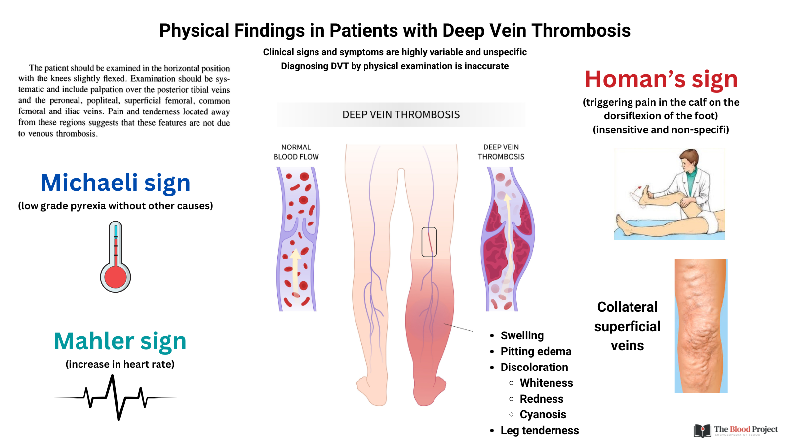
The most common physical examination findings associated with deep vein thrombosis (DVT) are unilateral leg swelling (due to venous congestion and edema), pain or tenderness (often in the calf or along the course of the deep veins), increased warmth, erythema (redness), and pitting edema. Additional findings may include a palpable cord (representing a thrombosed vein), dilated superficial collateral veins, and, less commonly, cyanosis of the affected limb. These findings are neither sensitive nor specific for DVT, as they can be seen in other conditions such as cellulitis, muscle injury, or chronic venous insufficiency. However, their presence should raise clinical suspicion, especially in the context of risk factors for venous thromboembolism.
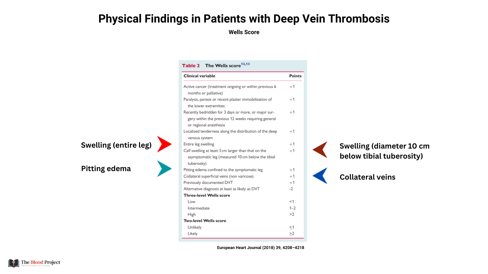
Other classic but less reliable signs include Homans sign (calf pain on dorsiflexion of the foot), which lacks sensitivity and specificity and is not recommended as a sole diagnostic maneuver. The combination of these findings, along with clinical risk assessment tools such as the Wells score, guides the need for further diagnostic testing.

For larger image, click here.
Examples of classical (largely historical) signs:
- Homan’s sign:1
- This classical sign is elicited by forcible dorsiflexion of the foot with the knee extended; calf pain upon this maneuver suggests DVT. However, it is neither sensitive nor specific, so is of limited diagnostic utility.
- How to perform:
- The patient lies supine with the knee fully extended.
- The examiner gently but briskly dorsiflexes the patient’s foot.
- If this causes deep calf pain or tenderness, the test is considered positive.
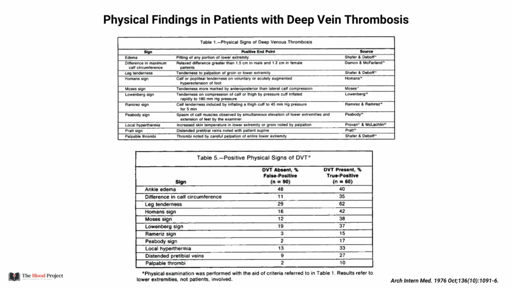
- Numerous studies have shown that Homan’s sign has low sensitivity and specificity:
- It occurs in about one-third of patients who actually have DVT.
- It can also be positive in patients without DVT (false positives seen in about 20% of cases).
- Positive Homan’s sign may also be seen in other conditions such as:
- Ruptured Baker’s cyst
- Gastrocnemius muscle spasm
- Cellulitis
- Neurogenic claudication
- Intervertebral disc herniation
- Lowenberg sign:2
- Pain in the affected calf at lower sphygmomanometer cuff pressure compared to the unaffected leg.
- How to perform:
- A blood pressure cuff is wrapped around the patient’s calf.
- The cuff is inflated progressively.
- The sign is positive if pain is elicited in the affected calf at a lower cuff pressure than in the unaffected leg, typically when the cuff is inflated to about 80 mmHg or lower in the affected leg.
- Normally, discomfort occurs only at pressures above 180 mmHg.
- Like other DVT signs such as Homan’s, the Lowenberg sign is neither highly sensitive nor specific and cannot confirm or exclude DVT on its own.
- Michaeli sign:
- Refers to a low-grade fever (subfebrile temperature) exceeding 37.5°C (99.5°F) without any other obvious cause.
- Michaelis described this as an early warning or premonitory sign before full thrombosis or embolic events may occur.
- Not specific and is rarely used alone to diagnose DVT.
For larger image, click here.

For larger image, click here.
Bottom Line:
The diagnosis of DVT today relies on clinical probability scoring (e.g., Wells score) and confirmation with compression ultrasonography. Eponymous signs like Homan’s, Moses, or Meyer are mostly of historical interest and not diagnostically reliable.
Complications of DVT:
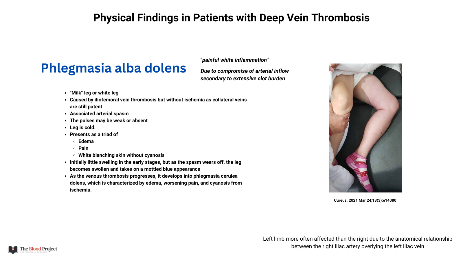
Phlegmasia alba dolens (“Painful white leg”)
- Definition:
- A rare, severe form of deep vein thrombosis (DVT) characterized by extensive occlusion of deep veins in the lower extremity, leading to massive swelling, pain, and pallor (whiteness) of the affected leg.
- Pathophysiology:
- Massive DVT → obstruction of major deep veins
- Superficial and collateral veins are still patent, so venous drainage is reduced but not completely blocked
- Increased interstitial pressure compresses arterioles, reducing arterial inflow → pallor
- Risk of progression to phlegmasia cerulea dolens (venous gangrene)
- Clinical features:
- Pain
- Swelling – Marked, tense edema
- Pale or white (alba) skin color
- Cool to the touch
- Pulses present but diminished
- Usually unilateral (left > right)
- Can evolve to phlegmasia cerulea dolens

For larger view, click here.
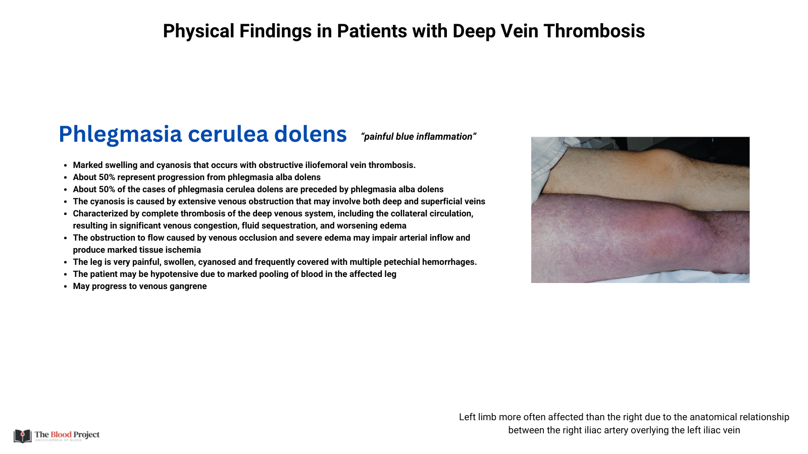
Phlegmasia cerulea dolens (“Painful blue leg”)
- Definition:
- A condition caused by massive thrombosis of both deep and collateral venous systems, leading to severe venous congestion, limb ischemia, and a characteristic bluish discoloration of the affected extremity.
- Pathophysiology:
- Complete obstruction of venous drainage (deep + superficial systems)
- Severe increase in interstitial pressure
- Leads to arterial compression, impaired inflow, and eventually tissue ischemia
- Can progress to venous gangrene and compartment syndrome
- Clinical features:
- Pain
- Swelling – Massive, tense edema
- Cyanotic (blue/purple) skin color due to venous stasis
- Cool to cold limb
- Pulses often absent or difficult to palpate
- Can evolve from phlegmasia alba dolens

For larger view, click here.