Aug

23

2021

Iron Deficiency Anemia

By William Aird

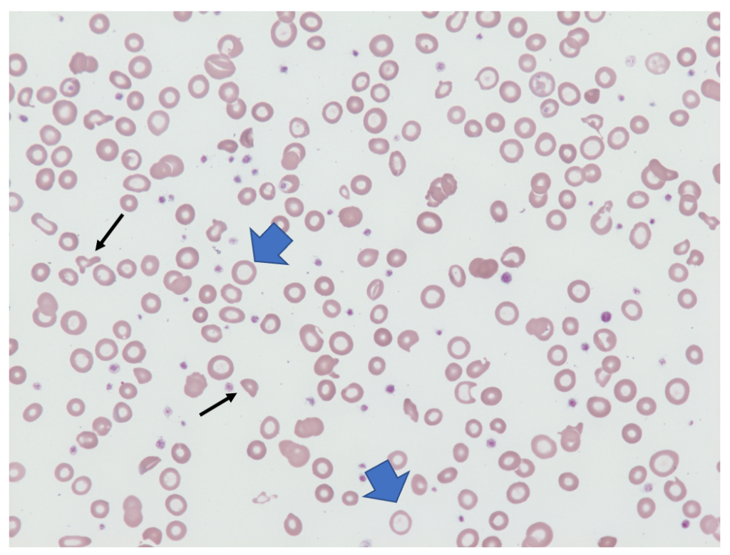
Hypochromic red blood cells (examples shown with blue arrows)  in a 24-year-old woman with severe iron deficiency anemia. Note the occasional fragmented red cell (black arrows). These cells, which are commonly seen in patients with severe iron deficiency anemia have retained their central pallor, which distinguishes them from the type of schistocytes seen in thrombotic microangiopathy (50x).

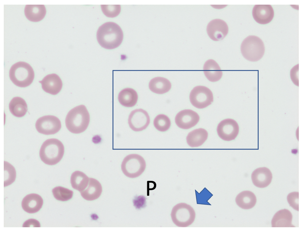
Hypochromic red blood cells (example shown with blue arrow)  in a 24-year-old woman with severe iron deficiency anemia. The boxed area illustrates significant variation in cell size (anisocytosis). P, platelet (100x, oil).

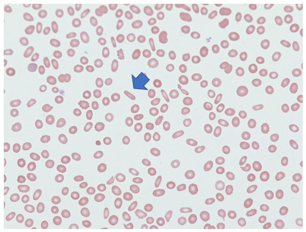
Pencil-shaped cells (one shown by blue arrow) in 44-year-old man with a history of iron deficiency anemia due to GI blood loss (50x).
ParameterProperties
ConditionIron deficiency anemia
FindingsHypochromia, pencil shaped red cells, microcytosis and hypochromasia. Pencil cells are hypochromic variants of elliptocytes having long axes at least triple the length of the cell’s short axis. Reactive thrombocytosis may also occur
Ddxbeta-thalassemia minor and anemia of chronic disease
MechanismDecreased hemoglobin production  leads to hypochromia. The blunted hemoglobin production results in a smaller-than-normal mature red cell
Source/authorWilliam Aird
Reviewed and edited byParul Bhargava