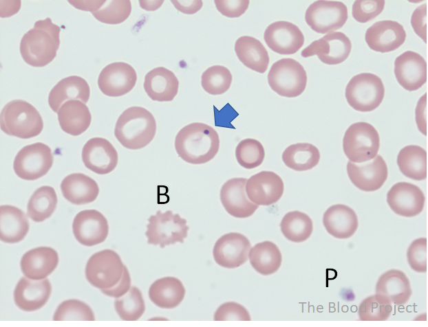
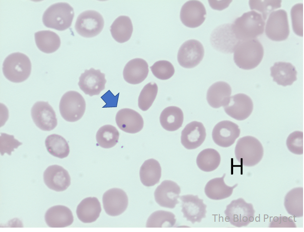
| Red blood cell, inclusion | Howell-Jolly body |
| Definition | Howell-Jolly bodies are small round purple inclusions in RBCs about 1 μm in diameter. Compared to Pappenheimer bodies, Howell-Jolly bodies are larger in size, have smooth outlines, typically one per RBC, and are comprised of DNA. |
| Ddx | Platelet overlying a red cell, Pappenheimer bodies, artifact. |
| Conditions associated with the inclusion | Asplenia (post-splenectomy, functional hyposplenia) |
| Mechanism of formation | They are formed in the process of red cell nuclear karyorrhexis or when an aberrant chromosome becomes separated from the mitotic spindle and remains behind when the rest of the nucleus is extruded. Normally, the spleen is very efficient in removing Howell-Jolly bodies from red cells, but if the spleen is missing or hypofunctioning, they may be readily found in the peripheral blood. |
| History | Howell 1890 showed extrusion of red cell nucleus; Jolly reported his findings of these inclusions in 1907 (learn more here). |
| Source/author | William Aird |
| Reviewed and edited by | Parul Bhargava |
| References | Am J Med Sci 2012;343:407 |