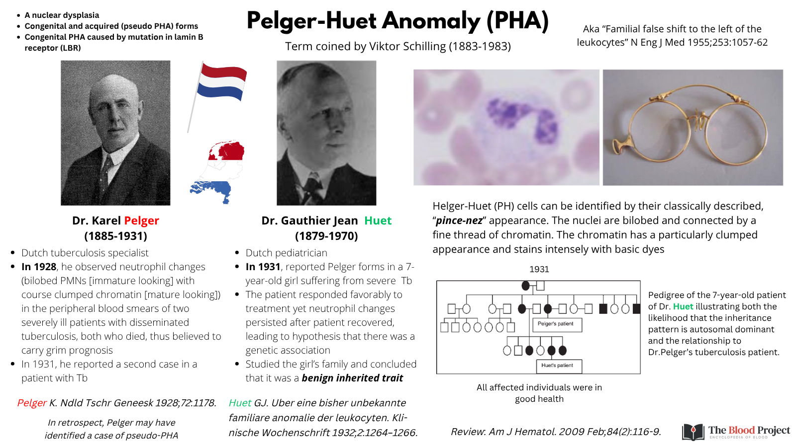
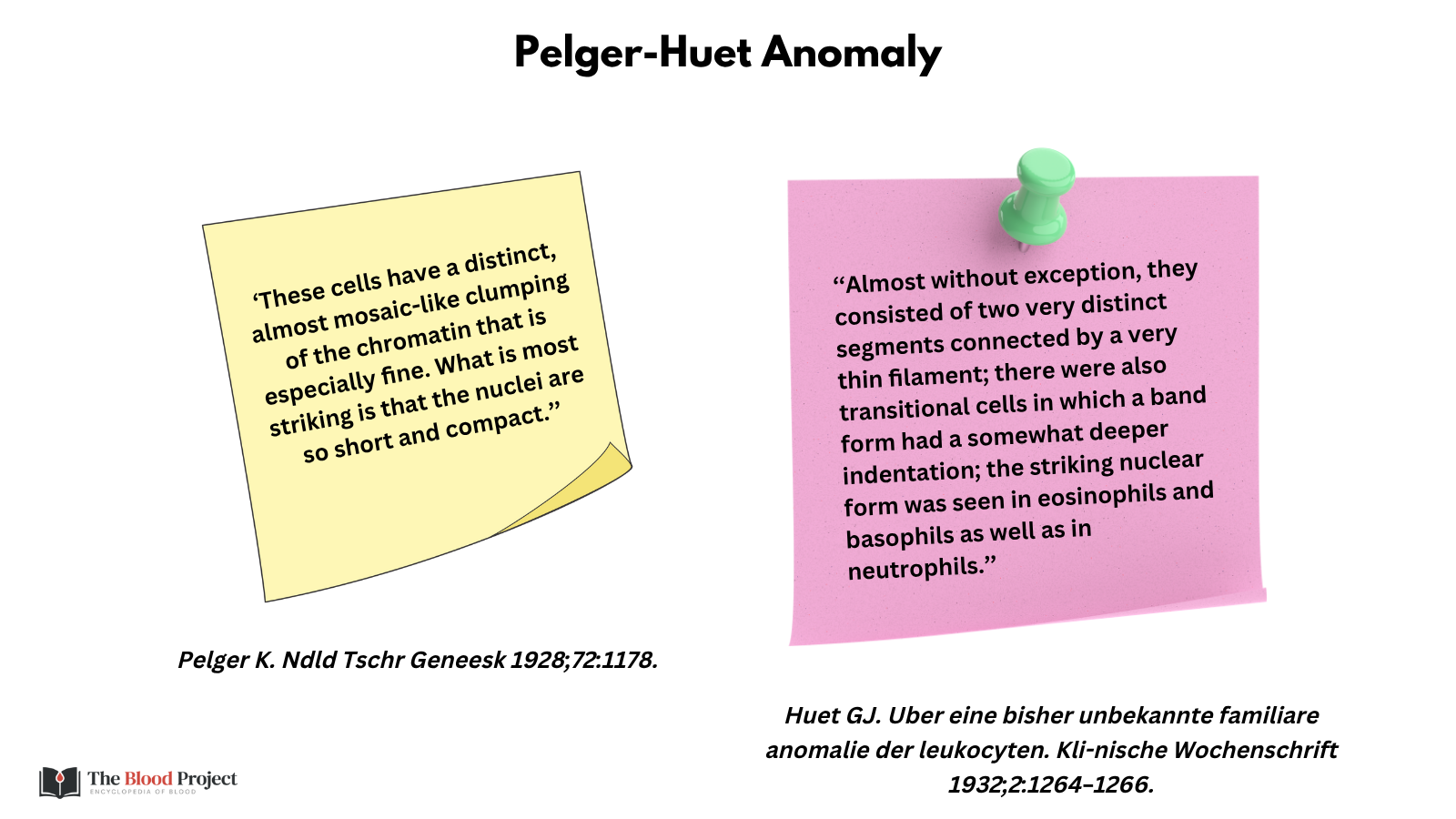
Named after Karel Pelger (1885-1931), a Dutch specialist in tuberculosis, and Gauthier Huet (1879-1970), a Dutch pediatrician. In 1928, Pelger published an account of a patient with Tb who had neutrophils with bilobed nuclei (a marker of immaturity) and coarse clumped chromatin (a marker of maturity). The patient died, and so the findings were believed to predict a poor prognosis. In 1931, Huet reported similar looking neutrophils in a patient with Tb. Only this time, the cell morphology persisted when the patient recovered. Huet studied her family and found similar forms in healthy relatives, indicating a benign inherited condition. Schilling termed the condition Pelger-Huet anomaly (PHA). Today, two types are recognized: 1) congenital PHA (caused by mutation in the LBR gene), and 2) acquired PHA (aka pseudo PHA), found most commonly in myelodysplastic syndromes (MDS). Neutrophil function is normal in both types.


Key Takeaways:
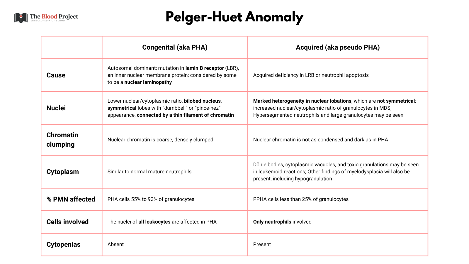
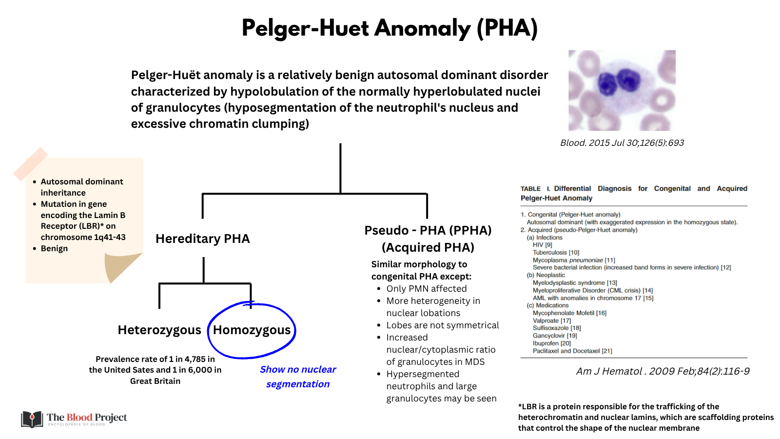
- Pelger-Huët anomaly (PHA) is a rare, benign hematological disorder involving the abnormal shape of neutrophil nuclei, most often appearing as bilobed, peanut-shaped, or dumbbell-shaped forms instead of the normal multi-lobed configuration. The defining feature is hyposegmentation—a reduced number of nuclear lobes—in granulocytes, particularly neutrophils.
- Inheritance: PHA has an autosomal dominant inheritance pattern, caused by mutations in the lamin B receptor (LBR) gene located at chromosome 1q41–q43.
- Hereditary (congenital) PHA is usually discovered incidentally during blood tests, as those who have it seldom exhibit any symptoms or increased susceptibility to infection.
- It is crucial to distinguish congenital PHA from pseudo–Pelger-Huët anomaly (PPHA), which is an acquired form often associated with conditions such as myelodysplasia, myeloid leukemia, infections, medication side effects, or radiation exposure.
- Named after Karel Pelger (Dutch hematologist), who first described the condition in 1928 and G. Huët (Dutch researcher), who further characterized it in 1932. Together, their names form the eponym Pelger-Huët anomaly, honoring their combined contributions to identifying and describing this distinctive morphological feature of neutrophils.