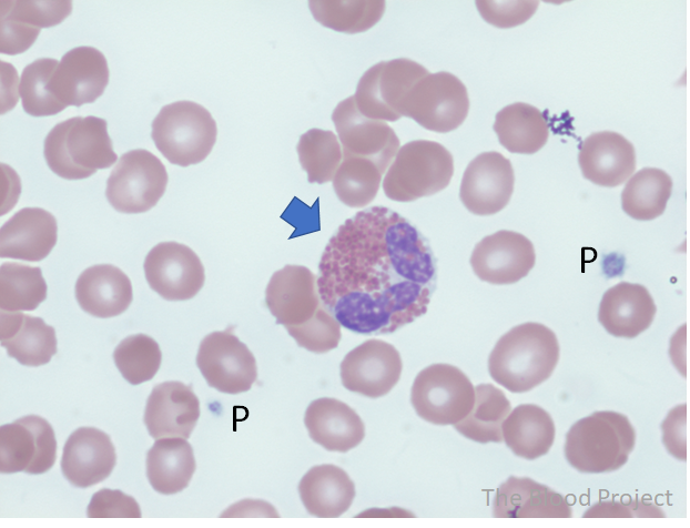
| White blood cell type | Eosinophil |
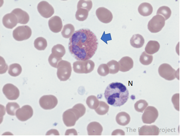
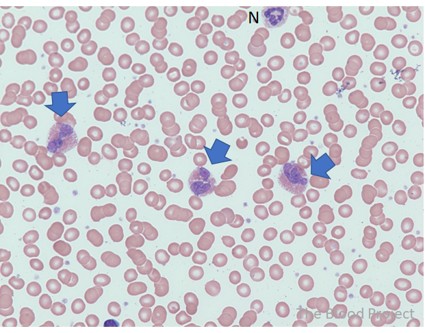
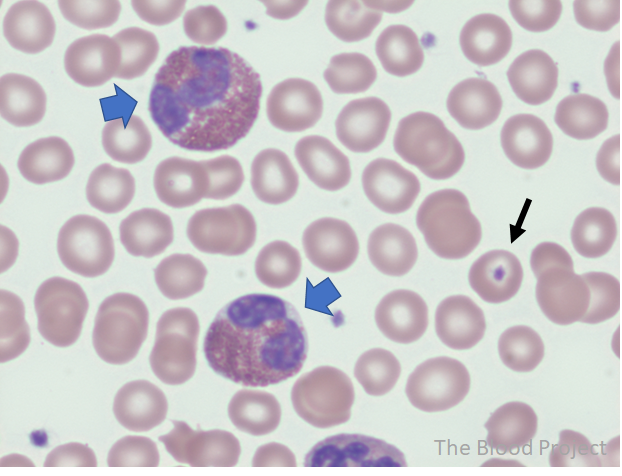
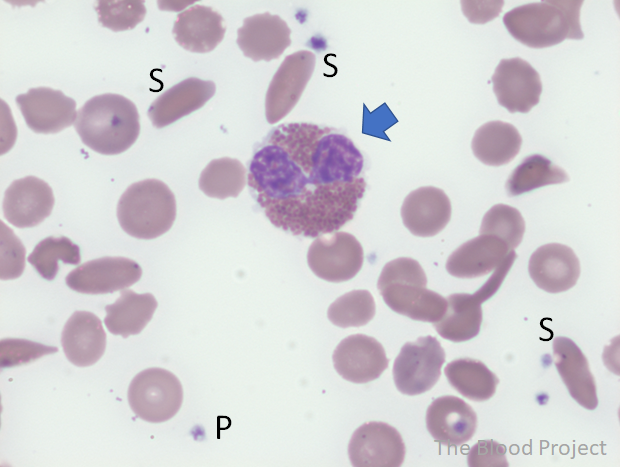
| Definition | Round to oval leukocytes that are present in the blood, bone marrow, and tissues of normal individuals. Recognized by their characteristic coarse orange-red cytoplasmic granulation. Their abundant cytoplasm is generally evenly filled by numerous large coarse, orange-red granules of uniform size. These granules rarely overlie the nucleus (which contains compact chromatin) and exhibit a refractile appearance with light microscopy due to their crystalline structure. In the most mature eosinophilic form, the nucleus is segmented into two or more lobes connected/separated by a barely visible thin filament. About 80% of segmented eosinophils have the classic two-lobed appearance. The rest have 3 lobes. Typically, these lobes are of equal size. |
| Conditions associated with the cell type | The most common causes of eosinophilia include allergy, parasitic infection, drugs. |
| Mechanism of formation | Eosinophils differentiate in the bone marrow, and then they migrate into blood, constituting about 1–6% of circulating leukocytes. |
| History | First identified by Paul Ehrlich in 1879 by virtue of their unique staining properties. |
| Source/author | William Aird |
| Reviewed by | Parul Bhargava |
| References | Clin Rev Allergy Immunol. 2016;50:125 |