| Parameter | Properties |
|---|---|
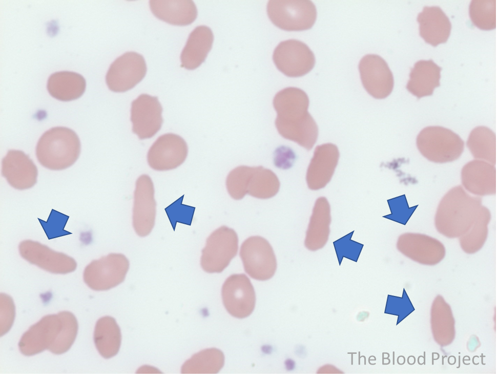
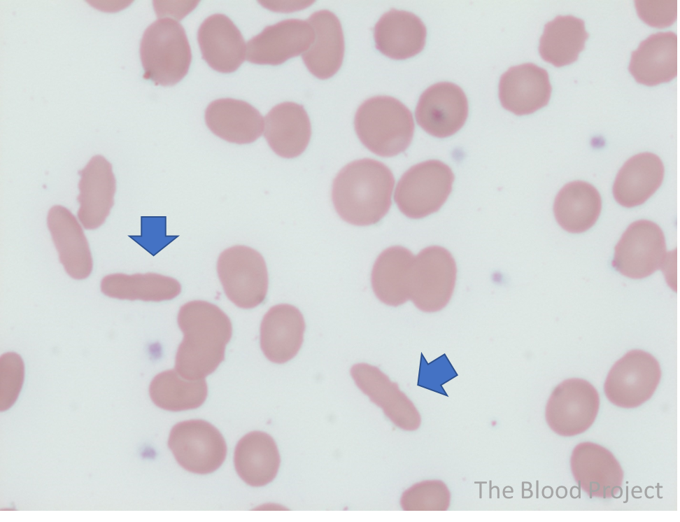
| Red cell shape abnormality | Elliptocyte |
| Also known as | Ovalocyte |
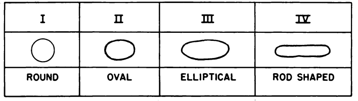
| Definition | Small or normal sized red cells that have an elongated appearance. Usually contain central pallor. The ends of the cells are blunt and not sharp like sickle cells. |
| Types | 1) Classical elliptocytes: sides are parallel, with long axis > 2x short axis. Large numbers may be seen in hereditary elliptocytosis. 2) Ovalocytes: egg-shaped red cells with long axis < 2x short axis. Large numbers may be seen in hereditary elliptocytosis. 3) Rod-shaped cells (pencil cells): elliptocytes with long axis more than 3 times the length of the short axis. Large numbers may be seen in hereditary elliptocytosis and severe iron deficiency. |
| Conditions associated with the shape abnormality | Hereditary elliptocytosis (% of elliptocytes 10-100%), thalassemia, iron deficiency. Rare elliptocytes (less than 1%) may be seen on a normal peripheral smear. |
| Mechanism of formation | Hereditary elliptocytosis is caused by hereditary membrane protein defects especially involving defective horizontal interactions in the membrane cytoskeleton. |
| History | According to Davidson and Strauss, writing in 1961: “The first record of elliptical human red corpuscles is usually credited to Dresbach (1904) but Lambrecht (1938) states that the anomaly was observed in 1860 by Goltz.” J Clin Pathol. 1961;14:615-2. |
| Source/Author | William Aird |