Labs

Let’s begin with the patient’s complete blood count (CBC):

WBCHbMCVPLT
22.810.69840

What’s what: WBC, white blood cell count; Hb, hemoglobin; MCV, mean cell volume; MCHC, mean cellular hemoglobin concentration; RDW-SD, red cell distribution width-standard deviation; platelets, PLT; Normal values: WBC 5-10 x 109/L, RBC 4-6 x 10¹²/L, Hb 12-16 g/dL, Hct 35-47%, MCV 80-100 fL, MCHC 32-36 g/dL, RDW-SD < 45%, platelets (PLT) 150-450 x 109/L

The following is the patient’s white cell differential:

For mature white cells, it is advisable to focus on absolute counts rather than percentages because higher-than-normal or lower-than- normal counts are defined by thresholds in absolute numbers. This applies to those cell types that are enumerated by automated counters, namely neutrophils (which include segmented and band forms), lymphocytes, monocytes, eosinophils and basophils. To define which of the above counts are altered in our patient, we can refer to our “cheat sheet”:

Expressed as cell number x 106/L

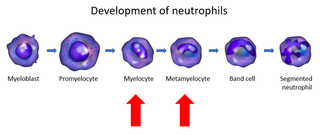
For less mature forms, namely myelocytes, metamyelocytes and promyelocytes in the granulocyte lineage, it is common practice to interpret the percentages relative to the total white cell count, because these are assessed by manual counting and because the presence of even a small number of these cells is abnormal.

How is a left shift defined?

a
Presence of increased % of neutrophils
b
Band neutrophils > 10%
c
Elevated number or percent of immature granulocytes, not including blasts
d
Increased nucleated red cells
e
Presence of Dohle bodies in neutrophils

Additional definitions of left shift:

DynaMed:

Left shift indicates an abnormally high proportion of immature neutrophils in the blood, defined as either:

  • Increased neutrophil bands in the peripheral blood (typically > 10% of white blood cells) by manual differential cell count
  • Elevated I/T-G ratio* by manual differential cell count
  • Elevated number or percent of immature granulocytes detected by automated cell analyzer, which typically represents promyelocytes, myelocytes, and metamyelocytes (but not blasts)

* Immature/total granulocyte (I/T-G) ratio defined as immature neutrophil count (myelocytes + metamyelocytes + bands +/- promyelocytes)/total neutrophil count; used as an indicator of a granulocyte left shift


Up-to-Date:

Left shift is an ill-defined term that refers to an increase in the percentage of band forms, generally accompanied by metamyelocytes and myelocytes.


Other definitions:

An increase in the ratio of nonsegmented/segmented neutrophils in the peripheral blood. J Clin Invest.1967;46:1943-1953.

A band/total neutrophil ratio or white blood cell count above 6% to 20% commonly used to indicate left shift. Clin Chim Acta. 2016 Jun 1;457:46-53.

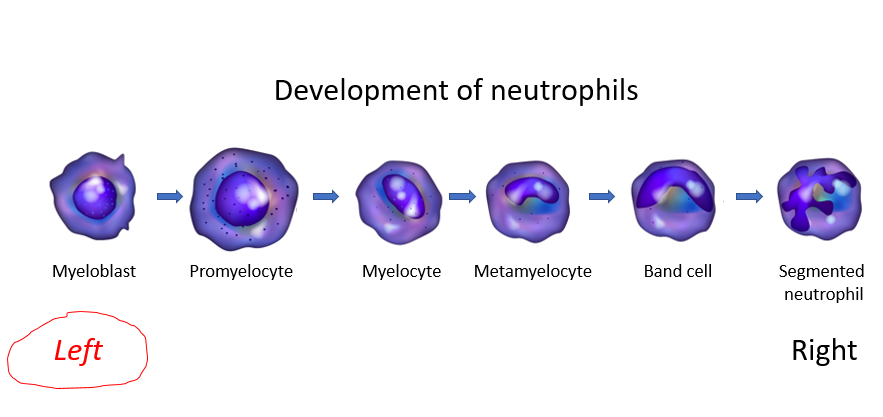
What is a band neutrophil?

According to the College of American Pathologists:

  • A band neutrophil is round to oval and 10 microns – 18 microns in diameter.
  • The nuclear:cytoplasmic ratio is 1:1:5 to 1:2 and the nuclear chromatin is condensed.
  • The nucleus is indented to more than half the distance to the farthest nuclear margin, but in no area is the chromatin condensed to a single filament.
  • The nucleus can assume many shapes, including:
    • Band-like
    • Sausage-like
    • S-shaped
    • C-shaped
    • U-shaped
  • The cytoplasm contains primarily specific (pink) granules.

In addition to bands, the differential also showed increased numbers of metamyelocytes and myelocytes. These are less mature forms of granulocytes, situated further to the left on the differentiation schematic:

Does the patient meet the definition of leukemoid reaction?

a
Yes
b
No

Leukemoid reaction typically refers to a white blood cell count > 50-100 × 109//L that occurs in response to reactive causes.

Does the patient meet the definition of hyperleukocytosis?

a
Yes
b
No

Hyperleukocytosis typically refers to a white blood cell count > 100 × 109//L, usually found in leukemias and myeloproliferative disorders.

The patient’s differential showed neutrophilia, lymphopenia, and eosinopenia. This combination of changes is called a stress leukogram. It occurs in patients with stress of any kind, including in the setting of severe infection.

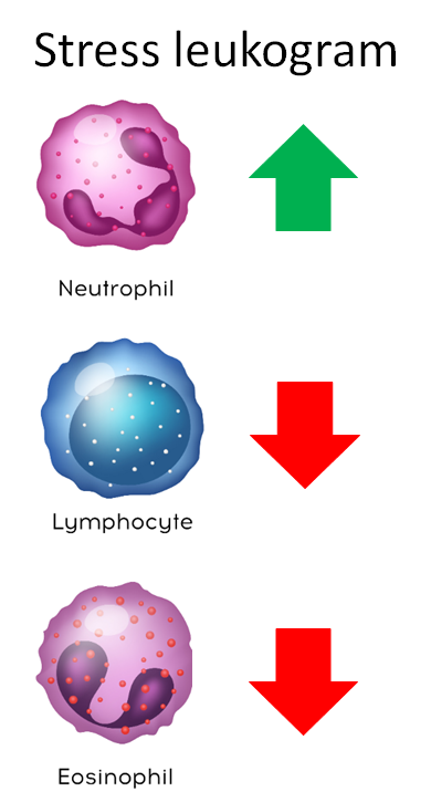
One way to remember this pattern is to consider that steroids (endogenous levels of which are increased in stress states) are used to treat lymphoma (by killing lymphocytes) and to treat hypereosinophilia (by killing eosinophils).

Monocyte counts tend to track with neutrophil counts in the stress response, but this is not always the case.

Based on what you know so far, your suspicion is highest for:

a
Trauma
Trauma may cause the blood picture shown so far, but there is no history of it in this case.
b
Anxiety
Anxiety can also cause a stress leukogram, but not with this degree of left shift.
c
Infection
The fact that the patient presents just 2 days after a procedure, has fever and hypotension, and a profound left shift makes infection the likeliest diagnosis.
d
Aortic aneurysm
The history is not typical for an aortic aneurysm.

The patient’s platelet counts are low. What are possible causes of her thrombocytopenia?

a
Thrombotic thrombocytopenia purpura (TTP)
The presentation is not typical of TTP.
b
Chronic immune thrombocytopenia (ITP)
The patient denies a history of thrombocytopenia, but this diagnosis is formally possible.
c
Infection without disseminated intravascular coagulation (DIC)
Correct. Infection may cause thrombocytopenia even in the absence of DIC. Proposed mechanisms include platelet sequestration along activated endothelium or in platelet-white cell aggregates, hemophagocytosis and direct effect of pathogens on platelet production, adhesion and aggregation.
d
DIC
Yes, DIC from any cause, including infection, is almost always associated with thrombocytopenia.
e
Cirrhosis
There is no history of cirrhosis, and no suggestive findings on physical exam, but the diagnosis is formally possible.

What changes might you see in the following labs – decreased or increased values?

Lab testResultComment
Serum creatinine
Serum anion gap
Serum albumin
Serum ferritin
PT
aPTT
Plasma D-dimers
Serum TIBC
Serum lactate
Plasma fibrinogen
PT, prothrombin time; aPTT, activated partial thromboplastin time; TIBC, total iron binding capacity

What changes might you see in the following labs – decreased or increased values?

Lab testResultComment
Serum creatinineIncreasedIf sepsis-associated acute kidney injury
Serum anion gapIncreasedIf lactic acidosis from hypoperfusion
Serum albuminDecreasedNegative acute phase reactant
Serum ferritinIncreased Positive acute phase reactant
PTIncreasedMay be elevated in DIC
aPTTIncreasedMay be elevated in DIC
Plasma D-dimersIncreasedMay be elevated in DIC
Serum TIBCDecreasedNegative acute phase reactant
Serum lactateIncreasedIf significant hypoperfusion from hypotension
Plasma fibrinogenDecreased or increased Low if DIC, high as positive acute phase reactant
PT, prothrombin time; aPTT, activated partial thromboplastin time; TIBC, total iron binding capacity; DIC, disseminated intravascular coagulation.

Here are the patient’s actual values (blue indicates increased, red indicates decreased):

Lab testPatient’s
results (normal range)
Comment
Serum creatinine4.3 mg/dL (0.4-1.1 mg/dL)Acute kidney injury
Serum anion gap26 mEq/L (10-18 mEq/L)Her lactate was 10.9 mmol/L (see below)
Serum albumin3.1 g/dL (3.5-5.2 g/dL) Negative acute phase reactant
Serum ferritin33,873 ng/ml (13-150 ng/ml)Positive acute phase reactant
PT18.4 sec (9.4-12.5 sec)Consistent with DIC
aPTT38.1 sec (25-36.5 sec)Consistent with DIC
Plasma D-dimers>21600 ng/ml (0-500 ng/ml)Consistent with DIC
Serum TIBC204 ug/dL (260-470 ug/dL) Negative acute phase reactant
Serum lactate10.9 mmol/L (0.5-2 mmol/L) Hypotension
Plasma fibrinogen 133 mg/dL (180-400 mg/dL)Consistent with DIC, especially in context of her acute phase response
PT, prothrombin time; aPTT, activated partial thromboplastin time; TIBC, total iron binding capacity; DIC, disseminated intravascular coagulation.

Let’s summarize the labs:

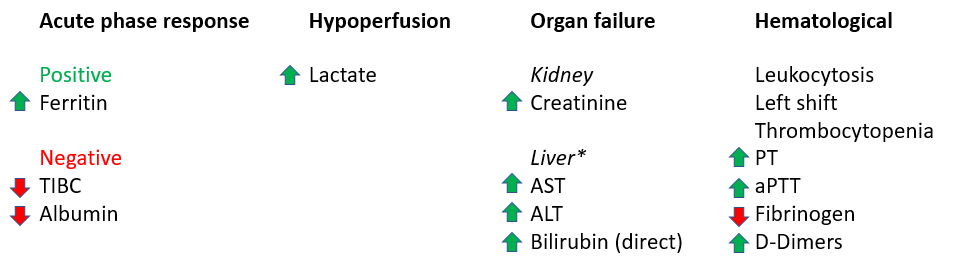
*To keep things simple, the patient’s liver function results are not shown, but they demonstrated evidence of shock liver with AST and ALT in the thousands IU, and a direct bilirubin that was was moderately elevated.

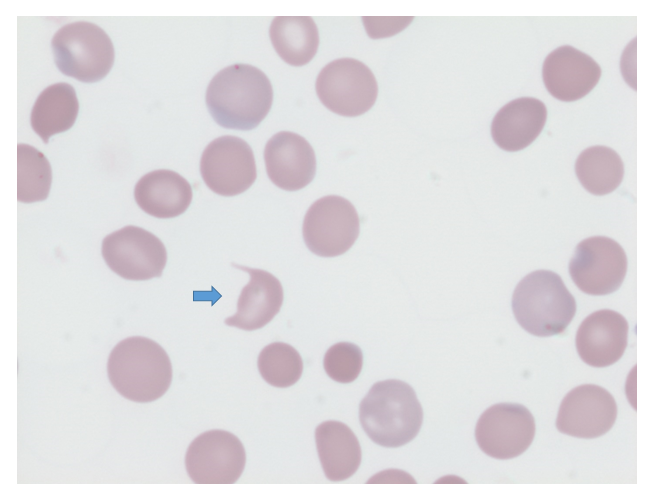
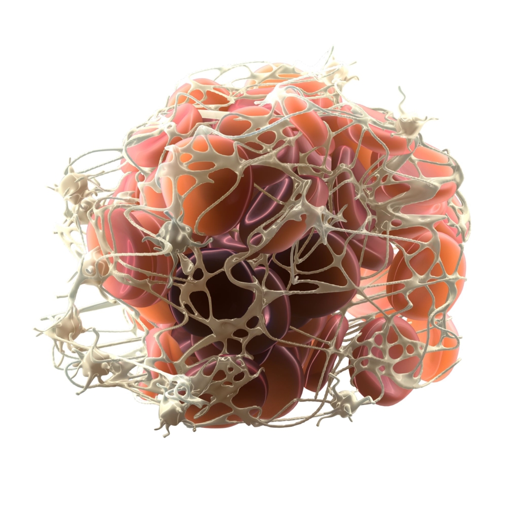
The patient’s peripheral smear shows presence of schistocytes, not unlike the following smear from a different patient.

Arrow indicates a schistocyte called a horn cell.

How would you put together the hematological data?

a
Patient is on coumadin and is not telling us
The coumadin might explain the elevated PT, but not the plethora of other hematological findings.
b
Patient has antiphospholipid syndrome (APS)
APS might explain concomitant thrombocytopenia, elevated aPTT and perhaps D-dimers (if she had a clot), but it would not explain the leukocytosis with left shift and the low fibrinogen.
c
Patient has disseminated intravascular coagulation (DIC)
Correct! The thrombocytopenia + decreased fibrinogen + elevated D-dimers + elevated PT in the context of probable urinary tract infection and sepsis are highly consistent with a diagnosis of DIC.

The ferritin is extremely high. Does that mean the patient has hemophagocytic lymphohistiocytosis (HLH)?

a
Most likely
b
Not necessarily
The higher the ferritin level, the more likely the diagnosis of HLH, but the risk of HLH remains relatively low even at ferritin levels >50,000. Most of the time, it represents an acute phase response from an underlying condition such as infection or cancer.

So, you are concerned about disseminated intravascular coagulation (DIC). Is there a single test that is, by itself, capable of diagnosing DIC?

a
Yes
b
No
There is no gold standard for the diagnosis of DIC and no single test that is, by itself, capable of accurately diagnosing DIC.

Should I use my gestalt or should I employ a clinical scoring system to make a diagnosis of disseminated intravascular coagulation (DIC) in a case such as this?

a
Gestalt (based on clinical experience)
b
Clinical scoring system
Clinical practice guidelines recommend using a DIC scoring tool to diagnose DIC.

The following are recommendations from two independent practice guidelines regarding the use of clinical scoring system:

Recommendation from the Harmonization guideline:


Recommendation from the Japanese Society of Thrombosis and Hemostasis :

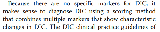
Bottom line: Use an established scoring system to make the diagnosis.

There are several clinical scoring systems for diagnosing disseminated intravascular coagulation (DIC). They use similar parameters, and each has been validated as a tool for diagnosing DIC and/or predicting poor outcome:

The ISTH subsequently published guidance based on harmonization of the recommendations from 3 of the guidelines.

ISTH, International Society of Thrombosis and Haemostasis; JMWH, Japanese Ministry of Health and Welfare; JAAM, e Japanese Association for Acute Medicine; JSTH, Japanese Society of Thrombosis and Hemostasis; SISET, Italian Society for Thrombosis and Hemostasis

The following table shows a comparison of the commonly used diagnostic scoring systems for disseminated intravascular coagulation (DIC):

From JSTH

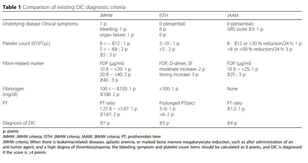
Note that for 2 of the 3 scoring systems (ISTH and JAAM), a diagnosis of DIC requires that patients have an underlying precipitating illness.

There are several common features and differences between these scoring systems:

Common features:

  • All but one (JAAM) use the same 4 parameters:
    • Platelet count
    • Fibrin degradation products
    • Fibrinogen
    • Prothrombin time (PT)

Differences:

  • Underlying disease is essential for ISTH and JAAM, but not for JMHW.
  • Fibrinogen is graded for JMHW, but not ISTH. It is not included in the JAAM score.
  • JAAM score takes into account the possibility that the platelet count is low in leukemia and other bone marrow disorders.

ISTH, International Society of Thrombosis and Haemostasis; JMWH, Japanese Ministry of Health and Welfare; JAAM, e Japanese Association for Acute Medicine; JSTH, Japanese Society of Thrombosis and Hemostasis; SISET, Italian Society for Thrombosis and Hemostasis

Let’s look at the 4 parameters in turn.

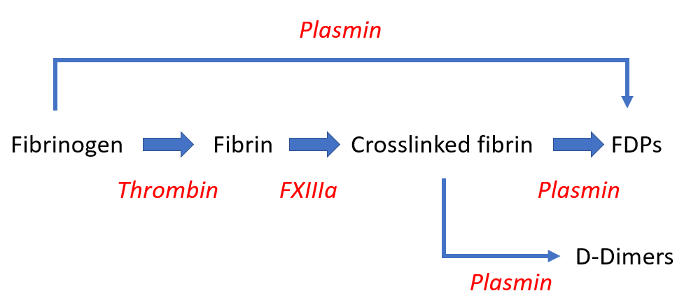
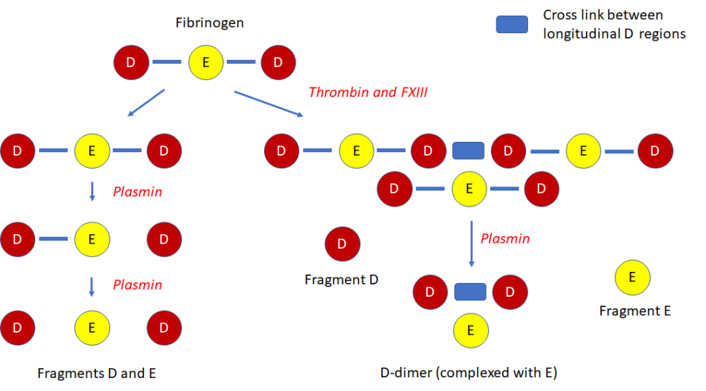
Fibrin-fibrinogen degradation products (FDPs), D-dimer

High sensitivity, low specificity (may be increased in cases of deep vein thrombosis, pulmonary thromboembolism, massive hydrothorax/ascites, and large subcutaneous hematomas).

D-dimer:

  • A degradation product of cross-linked fibrin.
  • The smallest circulating FDP that is specific for fibrinolysis.
  • Contains two cross-linked D fragments of the fibrin protein, hence its name.
  • Quantified by immunoassay.
  • Elevated in patients with disseminated intravascular coagulation (DIC), but high levels can also be found in patients with venous thromboembolism, recent surgery, or inflammatory conditions.

FDPs:

  • FDP assays measure the breakdown split products of either fibrinogen or fibrin.
  • Increased values indicate enhanced fibrinogenolysis or fibrinolysis.
  • Quantified by immunoassay.
  • Lacks specificity because in addition to DIC, high levels can be found with trauma, recent surgery, inflammation, venous thromboembolism, and many other conditions.
  • Because FDPs are metabolized in the liver and cleared by the kidneys, levels are influenced by liver and kidney function.
Fibrinogen is cleaved by thrombin to generate fibrin. Fibrin is then crosslinked by FXIII, resulting in an insoluble gel. Crosslinked fibrin is degraded by plasmin. This process, termed fibrinolysis yields soluble fibrin degradation products (FDPs). Fibrinogen may also be cleaved by plasmid to generate FDPs (fibrinogenolysis). Fibrin- and fibrinogen-derived FDPs are identical with the exception of D-dimer, which requires the action of plasmin on crosslinked fibrin.
A more granular look at the formation of FDPs and D-dimers from fibrinogen and fibrin. Fibrinogen is a dumbbell shaped molecule with 2 D domains flanking a single E domain. The sequential action of thrombin and FXIII results in the formation of crosslinked fibrin, with the crosslinks occurring between D domains on the longitudinal axis. Plasmin degrades fibrinogen and fibrin into fibrin degradation products (FDPs). Note that the D-dimer is unique to the breakdown of crosslinked fibrin. Other FDPs, namely D and E fragments are generated from plasmin-mediated degradation of both fibrinogen and fibrin. FDP assays detect all FDP fragments (from fibrinogen and fibrin breakdown), whereas the D-dimer assay measures D-dimers alone (from fibrin breakdown).

Thrombocytopenia:

  • High sensitivity, low specificity for diagnosis of disseminated intravascular coagulation (DIC).
  • Reported to occur in up to 98% of DIC cases.
  • Most patients with DIC have platelet counts < 100 × 109/L.
  • Platelet count < 50 × 109/L reported to occur in 10%-50% cases.
  • Low counts correlate with thrombin generation.

Fibrinogen:

  • Levels may decrease owing to consumption (increased conversion of fibrinogen to fibrin).
  • Levels may be further decreased when there is predominant hyperfibrinolysis (which can result in direct cleavage of fibrinogen).
  • Fibrinogen is an acute phase reactant, thus plasma levels may remain in normal range despite ongoing consumption.
  • Low fibrinogen level has high specificity, but low sensitivity for disseminated intravascular coagulation (DIC).
  • Overall sensitivity of low fibrinogen level reported to be only 28%, with hypofibrinogenemia occurring only in very severe cases of DIC.
  • Not a diagnostic criteria in the JAAM score.
Clotting is always initiated by tissue factor activation of factor VII (FVII) in the extrinsic pathway. The tissue factor is present on the subendothelial surface of the blood vessel wall and on the surface of activated circulating monocytes. Once activated, FVII activates FIX of the intrinsic pathway (not shown) and FX of the common pathway. FX, once activated, activates prothrombin, leading to generation of thrombin, and thrombin, in turn, cleaves fibrinogen to form fibrin. In disseminated intravascular coagulation (DIC), there is excessive activation of the extrinsic pathway. This leads to to increased fibrin formation and depletion of substrates, including (but not limited to) prothrombin and fibrinogen.

Schematic of fibrin mesh entrapping platelets (on the periphery) and red blood cells (in the center).

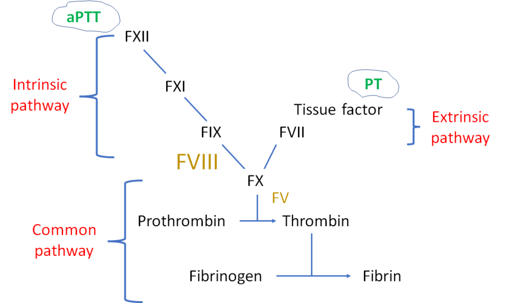
Prothrombin time (PT):

  • Increased in about one-half patients with disseminated intravascular coagulation (DIC) at some point during the course of the disease.
  • More sensitive than aPTT, which may be normal due to increased levels of factor VIII (FVIII), an acute phase reactant.
  • Not specific for DIC; elevated PT also occurs in patients with congenital factor VII deficiency, liver disease and vitamin K deficiency.
PT measures the integrity of the extrinsic pathway. It involves mixing patient’s platelet-poor plasma with thromboplastin (tissue factor + phospholipid), adding calcium, incubating at 37 degrees C and then measuring the time it takes for fibrin clot to form. The aPTT monitors the integrity of the intrinsic pathway. The PT is more sensitive than the aPTT for diagnosing DIC perhaps because factor VIII (FVIII) – which is part of the intrinsic pathway – is elevated as an acute phase reactant (shown in larger font).

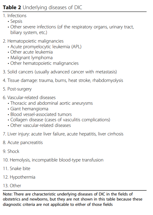
Recall that most guidelines require an underlying cause of disseminated intravascular coagulation (DIC). Here are some potential etiologies to look for:

From JSTH

The ISTH scoring system is simple! It consists of just 4 parameters based on widely available, routine coagulation tests.

ParameterScoreComment
Fibrinogen (g/L)>1 (0)
<1 (1)
Overall sensitivity of low fibrinogen level reported to be about 30%.
Prothrombin time (PT) (seconds)<3 (0)
3-6 (+1)
>6 (+2)
Elevated in 50%-60% of patients with DIC at some point during course of illness.
Platelet count (109/L)>100 (0)
50-100 (+1)
<50 (+2)
Thrombocytopenia reported to occur in up to 98% of DIC cases.
D-dimers or FDPsNo increase (0)
Moderate increase (+2)
Severe increase (+3)
Elevated FDPs and D-dimers are sensitive, but not specific for DIC.
DIC, disseminated intravascular coagulation; FDP, fibrin degradation product.

Data are presented as values with number of points in parentheses.

A total score of ≥5 is compatible with diagnosis of DIC.

Let’s apply the ISTH scoring system to our patient (right-most column):

ParameterScorePatient’s results (points)
Fibrinogen (g/L)>1 (0)
<1 (1)
1.3 (0)
Prothrombin time (PT) (seconds)<3 (0)
3-6 (+1)
>6 (+2)
7 (2)
Platelet count (109/L)>100 (0)
50-100 (+1)
<50 (+2)
40 (2)
D-dimers or FDPsNo increase (0)
Moderate increase (+2)
Severe increase (+3)
Severe increase (3)
Total score7

Data are presented as values with number of points in parentheses.

A total score of ≥5 is compatible with diagnosis of disseminated intravascular coagulation (DIC).

This patient’s score of 7 is diagnostic of DIC.

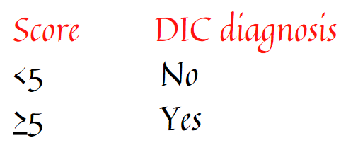
To reiterate, DIC is diagnosed when the ISTH DIC score is 5 or higher:

Learn more about the ISTH DIC score:

  • Original publication on the ISTH score can be found here.
  • Prospective validation study can be found here – sensitivity of the DIC score 95%, specificity 98%.
  • See ISTH calculator.

To summarize the testing section:

  1. The patient’s clinical presentation together with leukocytosis, stress leukogram, and left shift is consistent with infection.
  2. The ISTH disseminated intravascular coagulation (DIC) scoring system is consistent with a diagnosis of DIC.
  3. Likely diagnosis is DIC secondary to severe infection.
Prev
 1 / 0 
Next