Labs

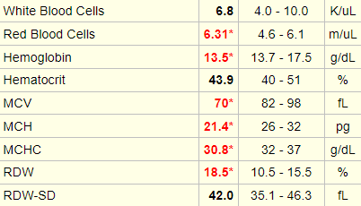
The following is the complete blood count (CBC) when the patient was first seen in the emergency room:

WBC (109/L)Hb (g/dL)Hct (%)MCV (fL)PLT (109/L)
33.920.869.381632

What’s what: WBC, white blood cell count; Hb, hemoglobin; MCV, mean cell volume; MCHC, mean cellular hemoglobin concentration; RDW-SD, red cell distribution width-standard deviation; platelets, PLT; Normal values: WBC 5-10 x 109/L, RBC 4-6 x 10¹²/L, Hb 12-16 g/dL, Hct 35-47%, MCV 80-100 fL, MCHC 32-36 g/dL, RDW-SD < 45 fL, platelets (PLT) 150-450 x 109/L

The following is a complete blood count from an individual with beta thalassemia. Note the increased red cell count, with a normal hematocrit (Hct). Hct = mean cell volume x red cell count, so if the red cells are small, as in this case, the bone marrow compensates by producing more of them to maintain a normal Hct. This condition used to be called microcytic erythrocytosis!

Our patient has elevation in all three cell lines: white cells, red cells and platelets. While we now use the term polycythemia to describe the red cells (and their effect on the Hct) alone, it was originally coined to describe a pan-increase in all three cell types (hence the qualifier poly).

Based on these lab values, what is the most likely diagnosis?

a
Polycythemia vera
b
Relative erythrocytosis
c
Hepatoma
e
Sleep apnea

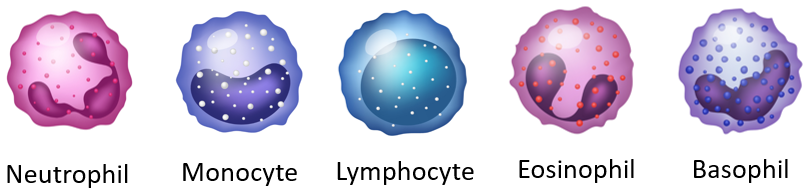
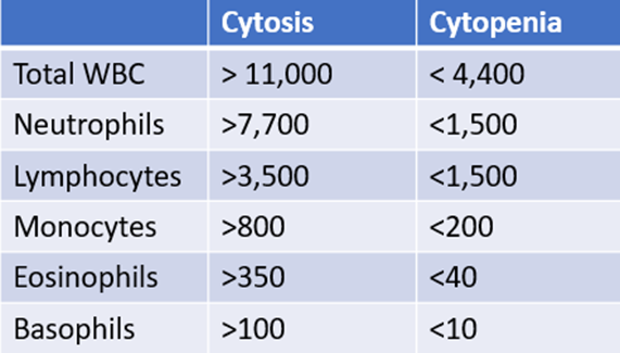
Let’s look at the white cell differential:

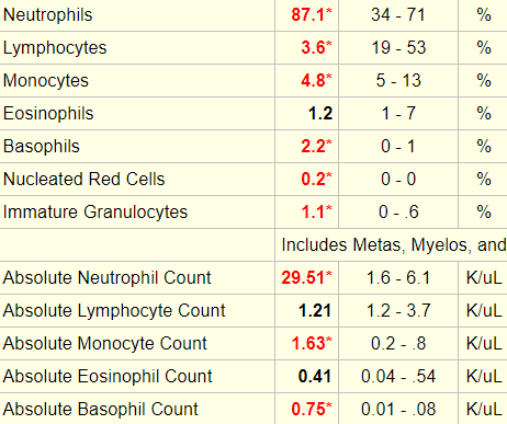
The following is “cheat sheet” of ranges that define lower- or higher-than-normal counts:
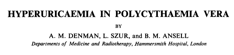
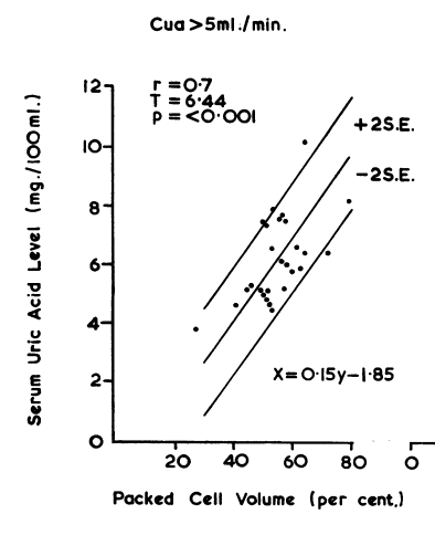
Expressed as cell number x 106/L.

Which additional lab tests would you predict might be abnormal (more than one answer may apply)?

a
Creatinine
b
Uric acid
c
Fibrinogen
d
Uncorrected PT and aPTT
e
LDH

The patient had a uric acid of 10.2 mg dL (normal 3.4-7.0 mg/dL).

High uric acid (complicated by gout and kidney stones) is associated with polycythemia vera due to the high turnover of red blood cells, which results in higher-than-normal uric acid production. 

Ann. rheum. Dis. (1966), 25, 340. Source.
Serum uric acid and packed cell volume were significantly and directly related in those patients in whom the urate clearance was normal.

As mentioned under the Physical Exam, the patient was seen by rheumatology who diagnosed her with crystal-proven gouty arthritis of the left elbow. She was treated initially with colchicine and NSAIDs and was subsequently started on allopurinol.

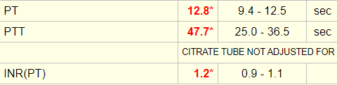
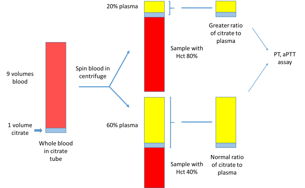
The patient’s initial coagulation screen showed the following:

The PT and aPTT are run on blood samples collected in sodium-citrate-containing blue top tubes. The sodium citrate is not a powder, but rather is in solution (there is a little “puddle” at the bottom of the tube). Sodium citrate serves as an anticoagulant by chelating calcium in blood. When the PT and aPTT assays are performed, a specified amount of calcium is added to the mixture to neutralize the effect of citrate and promote clotting. In patients with polycythemia, there is disproportionately less plasma in the blue top tube relative to the volume of citrate. As a result, the chelation effect is stronger, and the addition of calcium per the usual PT and aPTT protocol is not sufficient to completely inactivate it. This leads to an artifactually prolonged PT and aPTT. The artifact is corrected by reducing the amount of citrate in the collection tube.

The patient had a serum lactate dehydrogenase (LDH) of 315 IU/L (normal 94-250 IU/L).

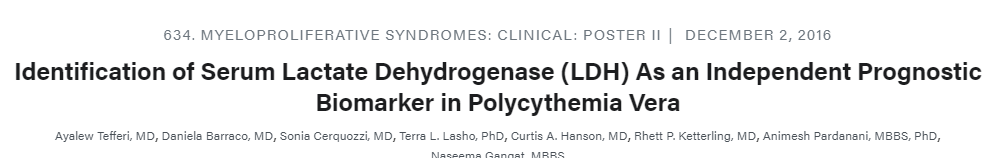
Serum lactate dehydrogenase (LDH) is a surrogate quantitative measure of cell turnover and tumor burden. The following is an abstract that reported the LDH level in 216 patients with polycythemia vera:

Increased serum LDH was recorded in 110 (51%) patients that included 26 (12%) patients with LDH ≥1.5 x UNL. A significant correlation was demonstrated between increased serum LDH and older age (p=0.007), female sex (p=0.04), leukocytosis (p=0.01), venous thrombosis history (p=0.005) and, interestingly, absence of active tobacco use (p=0.003).

For more information on Jak2 V617F, click here.

The Jak2 mutation was ordered when the patient was first seen, but this test takes several days to come back, so treatment decisions had to be made without knowledge of the result.

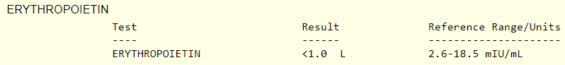
When the test result did return, it showed the following:

Is the Jak2 V617F mutation specific to polycythemia vera?

a
Yes
b
No
JAK2 V617F mutation is found with high frequency in Philadelphia chromosome-negative myeloproliferative neoplasms (>95% in polycythemia vera and approximately 50−60% in primary myelofibrosis and essential thrombocythemia). It can also occur in other myeloid neoplasms, such as refractory anemia with ring sideroblasts associated with marked thrombocytosis (RARS-T) and rarely in myelodysplastic syndrome, chronic myelomonocytic leukemia, atypical chronic myeloid leukemia and acute myeloid leukemia.

Another test that is commonly ordered in patients with polycythemia is a serum erythropoietin. What do you predict the serum erythropoietin will be in this case?

a
Low
b
Normal
c
High

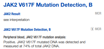
The erythropoietin level was undetectable!

Does the patient have polycythemia vera? Can this diagnosis be made prior to the Jak2 mutation result? To address this question, we need to consider the diagnostic criteria for polycythemia vera.

Diagnosis requires meeting either all 3 major criteria, or the first 2 major criteria and the minor criterion:

Major criteria

  • Hemoglobin > 16.5 g/dL (male), > 16 g/dL (female), or either of:
    • Hematocrit > 49% (males), > 48% (women).
    • Other evidence of increased red cell volume.
  • Bone marrow biopsy with hypercellularity for age with panmyelosis with prominent erythroid, granulocytic, and megakaryocytic proliferation.
  • JAK2 V617F or other mutation of similar function (for example, JAK2 exon 12 mutation).

Minor criteria

  • Serum erythropoietin level below normal reference range.

You will note that without the JAK2 V617F mutation status, and without a bone marrow biopsy, the patient does not formally meet the criteria for diagnosis of polycythemia vera. But in a case like this (extreme elevation in the Hct, elevated white cells and platelets, basophilia and an absence of any obvious secondary cause for the polycythemia), the pretest probability of polycythemia vera is so high that is should be treated as such, pending the results of the complete work-up.

Prev
 1 / 0 
Next