Preparing to see the patient

You have just been asked to consult on a patient in the hospital with possible hemolysis. You should be asking yourself several questions as you prepare to see the patient. For example:

  • What is the evidence for hemolysis?
  • How severe is the anemia?
  • What is the cause of the hemolysis?

On the following slides, we will address each of these questions in turn.

Question 1 – What is the evidence for hemolysis?

Which of the following hemolytic markers are released from red cells?

a
Haptoglobin
Haptoglobin is not released from lysed red cells. It is synthesized and released by hepatocytes. Serum haptoglobin levels decrease in hemolysis because haptoglobin binds hemoglobin (dimers) released by red blood cells and the resulting haptoglobin-hemoglobin complex is taken up by macrophages. The haptoglobin is degraded, not recycled.
b
Lactate dehydrogenase (LDH)
Red cells contain LDH, specifically isoforms LDH-1 and LDH-2.
c
AST
Red cells contain AST, but little ALT. Thus, an elevated AST:ALT ratio is consistent with hemolysis, though it can also be seen in alcoholic liver disease and rhabdomyolysis.
d
Bilirubin
Bilirubin is not released by red cells. Rather it is produced and released by macrophages upon degradation of hemoglobin.
e
Free Hb
This is a rarely measured marker of hemolysis that is indeed released by lysed red cells.

The next two slides show schematics of hemolytic markers that are directly released by red cells and those that are indirectly affected by red cell release of hemoglobin.

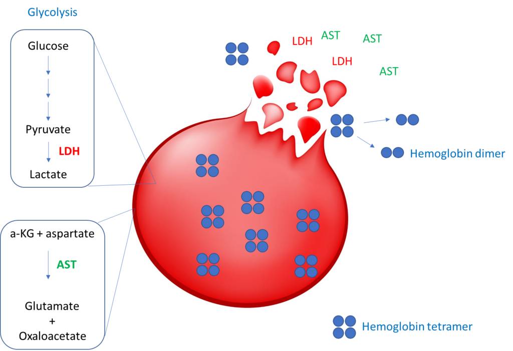
Hemolytic markers directly released from red blood cells:

Since red blood cells (RBCs) lack mitochondria they obtain all their energy needs by the equivalent of anaerobic glycolysis. This requires the conversion of pyruvate to lactate, a step that is catalyzed by lactic dehydrogenase (LDH). Aspartate aminotransferases (AST) is an enzyme involved in amino acid metabolism. It catalyzes the conversion of alpha-ketoglutarate (a-KG) and the amino acid, aspartate, to glutamate and oxaloacetate. Each mature RBC contains ~270×106 molecules of hemoglobin, accounting for approximately 90% of its dry weight. Hemolysis leads to the release of hemoglobin tetramers, which rapidly get degraded into dimers.

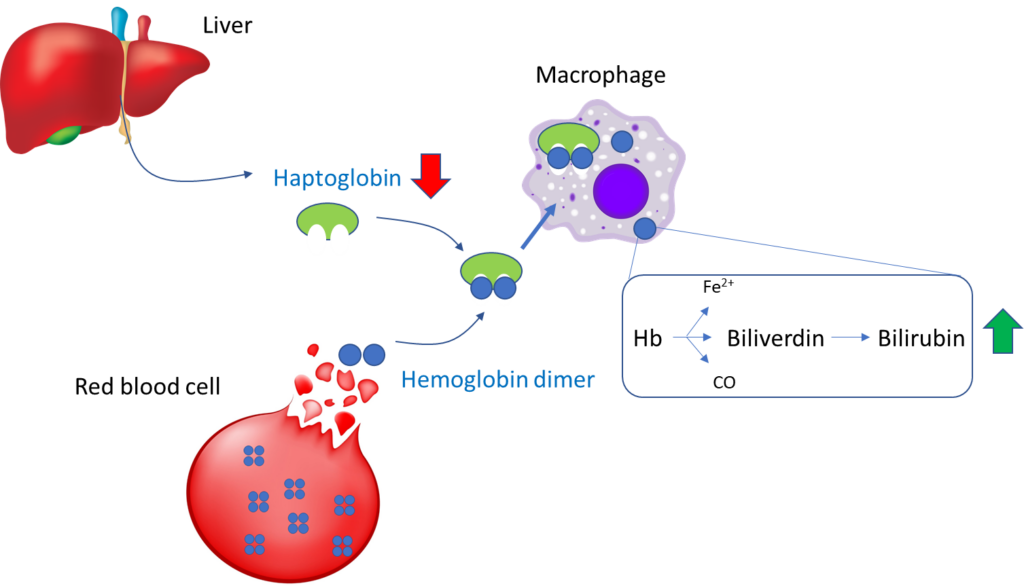
Indirect hemolytic markers (not released by red cells):

Lysis of red cells causes release of hemoglobin (Hb) tetramers, which are quickly broken down into dimers. Haptoglobin (Hp), which is made by liver hepatocytes (it is an acute phase reactant along with ferritin, fibrinogen, C-reactive protein and hepcidin), binds Hb dimers and the resulting Hb-Hp complex is endocytosed by tissue macrophages via a specific membrane receptor called CD163. Once inside the macrophage, haptoglobin is degraded (rather than recycled), which leads to reduced levels in the circulation. The hemoglobin is degraded to biliverdin (with release of carbon monoxide [CO], which can be assayed in exhaled breath) followed by indirect (unconjugated) bilirubin, which is released into the circulation, resulting in hyperbilirubinemia.

To reiterate, the following are considered serum markers of hemolysis:

  1. Serum LDH
  2. Serum bilirubin
  3. Serum AST
  4. Serum free Hb
  5. Serum haptoglobin

In fact, one study showed that mature erythrocytes contain 2,289 distinct gene products! Learn more here.

This is a schematic of the “top pathways” resulting from gene ontology term enrichment for biological function in red blood cells. The details are not important. The learning point here is that there is a whole research field dedicated to mapping the red cell proteome. Some of these markers, especially those that are specifically expressed in red cells and localized in the cytoplasm (as distinct from being attached to the membrane), may have the potential to serve as novel markers of hemolysis.

Note that with the exception of plasma hemoglobin, none of the hemolysis markers presently used is specific to red blood cells:

MarkerHemolysisLiver diseaseRhabdomyolysisComments
Increased LDH +++Also increased in megaloblastic anemia, HLH and LPD
Increased bilirubin++Also increased in megaloblastic anemia, HLH
Increased AST:ALT++ (especially alcoholic liver disease)+Also increased in megaloblastic anemia, HLH
Decreased haptoglobin++Also increased in megaloblastic anemia, HLH
Abbreviations: HLH, hemophagocytic lymphohistiocytosis; LPD, lymphoproliferative disorder.

No single pattern is absolutely diagnostic of hemolysis. The markers must be interpreted in the appropriate clinical context!

Question 2 – How severe is the anemia?

There are three possible ways to express anemia. Which the is the most relevant?

a
Red cell count
b
Hemoglobin
c
Hematocrit

You will probably agree that of the three parameters (red cell count, hemoglobin and hematocrit), the red cell count is the least useful measure of anemia. One of the reasons for this is that you could have all the red cells in the world, but if they are deficient in hemoglobin and/or smaller than normal in size, the oxygen carrying capacity of blood (which is a function of the hemoglobin concentration) may be impaired.

The same is true when thinking about hemoglobin vs. hematocrit (Hct). Hemoglobin carries oxygen. Hct is simply the fractional volume of blood that is comprised of red cells. It doesn’t tell us anything about hemoglobin levels and oxygen carrying capacity of the blood. In theory, red cells could be completely devoid of hemoglobin, yet maintain a normal Hct.

Consider three samples of blood that have been spun in a centrifuge to measure the hematocrit (Hct). Imagine the blood being composed of normal red blood cells (left), an equal number of cells that are much smaller than normal (center) and an equal number of poorly hemoglobinized cells (right). Note that a normal red blood cell number (center) or Hct (right) do not guarantee a normal Hb.

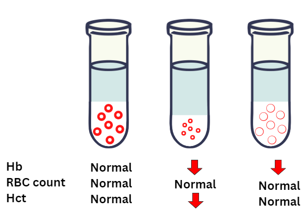
All of this is to say that we want to judge the severity of the patient’s anemia by examining her hemoglobin!

Question 3 – What is the cause of the hemolysis?

Now that you have thought about hemolytic indices, you turn your attention to etiology. Without knowing anything (beyond age and sex, and inpatient status) of the patient, and assuming she is indeed hemolyzing and the condition is acquired, which one of the following conditions are you most concerned about ruling out as soon as possible?

a
Drug effect
Drugs can cause severe hemolytic anemia, usually immune mediated, but it is rarely lethal.
b
Infection
Not an unreasonable answer, because infections can of course be fatal, but let’s hope that the primary team is on top of that!
c
Delayed transfusion reaction (DTR)
DTR is rarely fatal.
d
Thrombotic microangiopathy (TMA)
Undiagnosed thrombotic thrombocytopenia purpura (TTP), a form of TMA, has a mortality of up to 90%.
e
Paroxysmal nocturnal hemoglobinuria PNH)
PNH can present with severe anemia and/or thrombosis, but it does not carry the same initial high mortality rate as TTP.

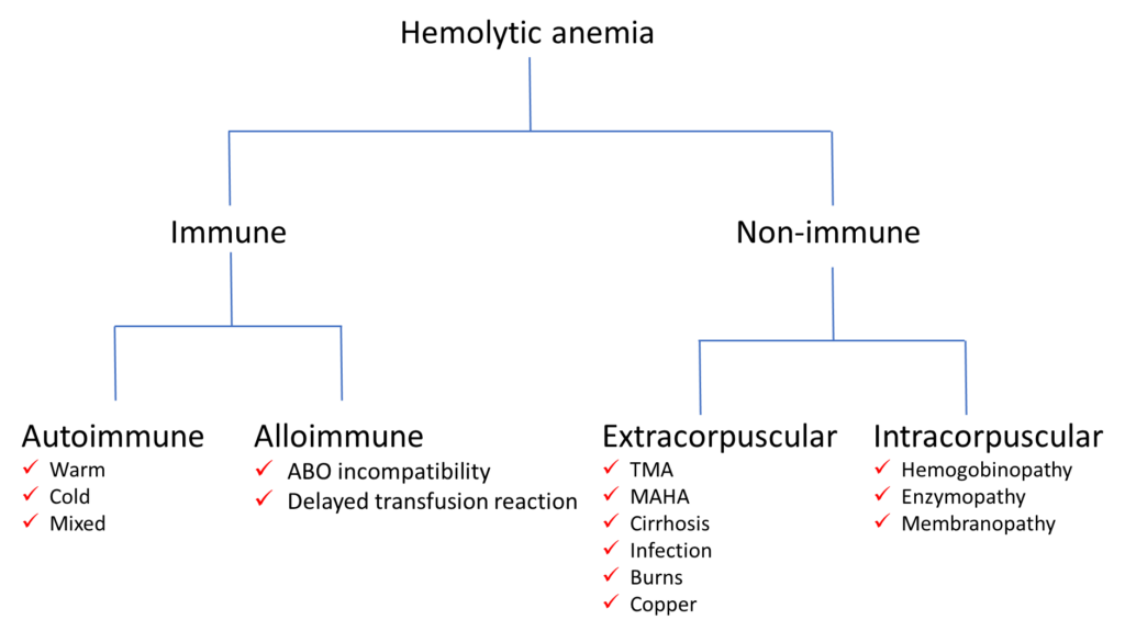
Differential diagnosis of hemolytic anemia

As you prepare to see the patient, it pays to have a broad outline of the differential diagnosis. Nothing too fancy or detailed. Something along the lines of the scheme shown below. We will discuss some of these conditions in a subsequent section when we consider the patient’s lab results.

One way to approach hemolytic anemia is by dividing it into immune and non-immune conditions. Immune causes are further divided into autoimmune and alloimmune causes, all of which are associated with a positive Coombs test (also called direct antiglobulin test). The non-immune category is divided into those causes in which the red cell is intrinsically normal, yet suffers collateral damage from a force outside the cell (extracorpuscular causes). By contrast, intracorpuscular causes of hemolytic anemia are referable to a defect in the erythrocyte, either in the hemoglobin molecule (thalassemia or sickle cell disease), key intracellular enzymes (especially G6PD and pyruvate kinase) or the cell membrane (for example, hereditary spherocytosis, hereditary elliptocytosis, and paroxysmal nocturnal hemoglobinuria [PNH]). Note that with the exception of PNH, the intracorpuscular causes are congenital/hereditary. TMA, thrombotic microangiopathy; MAHA, microangiopathic hemolytic anemia.

You are now primed to see the patient! In the next series of slides, we will consider the history and physical exam.

Prev
 1 / 0 
Next