Physical Exam

Which of the following physical findings might be seen in a patient with severe thrombocytopenia?

a
Petechiae
Correct. These are defined on the next slide.
b
Glossitis
Not associated with thrombocytopenia.
c
Splenomegaly
This might be found in hypersplenism (a cause of thrombocytopenia) or in cases of secondary ITP, for example in patients with ITP associated with chronic lymphocytic leukemia or lymphoma.
d
Ecchymoses
Correct. These are defined on the next slide.

Bleeding manifestations in skin and mucous membranes

Note how the following definitions vary significantly according to source!

International Working Group (IWG) definitions:

“The most frequent hemorrhagic manifestation in ITP is purpura. Purpura broadly encompasses any kind of mucocutaneous bleeding; it is commonly referred to as “dry” when bleeding is confined to the skin and “wet” when mucous membranes are also involved. The IWG recommends against the use of these terminologies because they lack precision.”

International Working Group on the standardization of bleeding assessment in immune thrombocytopenia; Blood. 2013;121(14):2596-60

The following are IWG definitions of bleeding in the skin:

LesionSite of bleedingDefinition
PetechiaeEpidermis and dermisRed (recent) or purplish (a few days old) discoloration in the skin with a diameter of 0.5-3 mm that does not blanche with pressure and is not palpable.
Ecchymosis (purpuric macule, bruises, or contusions)Epidermis and dermisFlat, rounded, or irregular red, blue, purplish, or yellowish green patch, larger than a petechia. Elevation indicated spreading of an underlying hematoma into the superficial layers of the skin.
HematomaSubcutaneous tissueBulging localized accumulation of blood, often with discoloration of overlying skin.
From the International Working Group on the standardization of bleeding assessment in immune thrombocytopenia; Blood. 2013;121(14):2596-60.

The following are IWG definitions of bleeding within visible mucous membranes such as the mouth:

Lesion
Petechiae, purpuric macules, and ecchymosisSame as for skin.
Bulla, vesicle, and blisterVisible raised, thin-walled, circumscribed lesion containing blood. Each bulla (>5.0 mm) is larger than a vesicle. Bullae, vesicles, and blisters should be counted together as bulla.
From the International Working Group on the standardization of bleeding assessment in immune thrombocytopenia; Blood. 2013;121(14):2596-60.

Up-To-Date definitions:

  • Petechiae:  
    • Small, flat (nonpalpable), red, discrete lesions (typically <2 mm in diameter) that do not blanch under pressure; these often occur in dependent areas of the body (lower legs in ambulatory patients; sacral area in recumbent patients).
    • Elsewhere in Up-To-Date, petechia are defined as small capillary hemorrhages that appear smaller than bruises and characteristically develop in crops in areas of increased venous pressure, such as the dependent parts of the body.
    • Petechiae are the smallest bleeding lesions (pinhead in size), and suggest problems with platelet number or function.
  • Purpura:
    • Refers to a lesion caused by coalescence of petechiae.
    • Purpura on the skin is sometimes referred to as “dry purpura.”
    • Purpura due to thrombocytopenia is nonpalpable and often occurs in dependent areas of the body. 
    • Hemorrhagic blisters in mucous membranes, such as the oral mucosa, are sometimes called “wet purpura”; this finding may be a predictor of more severe bleeding.
  • Bruise (ecchymosis) – a collection of blood beneath the skin resulting from extravasation of blood from surrounding vessels.

FIRST DERM definitions:

  • Petechiae are circumscribed deposits of blood (that is, small purpuric lesions) which are less than 0.5 cm in size (usually 1 to 2 mm).
  • Purpura:
    • Term used to describe a reddish discoloration of the skin or mucous membranes due to extravasation of red blood cells from blood vessels into the surrounding tissue.
    • Greater than 0.5 cm in size. 
  • Ecchymoses (bruises) are larger, deeper collections of blood under the skin (>1 cm in size).

Lotti T. Int J Dermatol. 1994;33:1-10

  • Purpura
    • Discoloration of the skin due to extravasation of red blood cells
    • Pressure by fingers or diascopy fails to blanch the lesion
    • Purpuric lesions may have different aspects and sizes:
      • Petechiae are small lesions up to 2 mm in diameter that often occur in crops.
      • Ecchymoses are larger, sometimes figurate lesions caused by extravasation of red blood cells

Referring to patients with thrombocytopenia, the 2019 American Society of Hematology Self-Assessment Program (7th Edition) states:

“Dependent areas and skin underneath tight clothing should be examined for petechiae and purpura, and oral mucous membranes should be examined for hemorrhagic bullae, which may be associated with an increased risk of severe bleeding at other sites.”

The following describes this patient’s physical exam when you see him:

Vital signs: Heart rate is 75/min, blood pressure 145/88

Head and neck: Purpuric lesions on roof of mouth and blood blisters on oral mucosa; no lymphadenopathy

Chest: Normal to inspection, palpation, percussion, and auscultation

Abdomen: Non-tender, no hepatosplenomegaly

CNS: Grossly normal

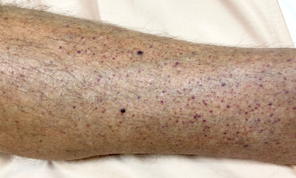
Skin: Petechiae on lower limbs

Petechiae on lower extremity. From Shutterstock, with permission.
Bruising (ecchymoses) on arm. From Shutterstock, with permission.
Prev
 1 / 0 
Next