Labs

Here is the patient’s complete blood count (CBC) when you see him in clinic:

WBCHbHctMCVMCHCRDW-SDPLT
5.211.24078 28425

What’s what: WBC, white blood cell count; Hb, hemoglobin; MCV, mean cell volume; MCHC, mean cellular hemoglobin concentration; RDW-SD, red cell distribution width-standard deviation; platelets, PLT; Normal values: WBC 5-10 x 109/L, RBC 4-6 x 10¹²/L, Hb 12-16 g/dL, Hct 35-47%, MCV 80-100 fL, MCHC 32-36 g/dL, RDW-SD < 45%, platelets (PLT) 150-450 x 109/L

Thrombocytopenia is commonly graded according to the platelet count:

  • Mild = 70-150 × 109/L
  • Moderate = 20-70 × 109/L
  • Severe = < 20 × 109/L

Thus, by definition, it is fair to say that the patient has microcytic, hypochromic anemia and severe thrombocytopenia.

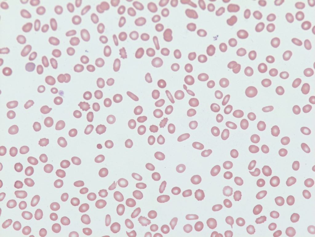
We established in the section on Differential Diagnosis that the three likeliest possibilities in this outpatient with severe thrombocytopenia and an unremarkable history include:

  • Immune thrombocytopenia
  • Pseudothrombocytopenia
  • Familial (congenital) thrombocytopenia

What would you expect to find for each condition on the peripheral smear? The answers are on the next slide.

ConditionFindings
Pseudothrombocytopenia
Familial thrombocytopenia (FT)
ITP
ITP, immune thrombocytopenia; LPD, lymphoproliferative disorder

What would you expect to find for each condition on the peripheral smear? The answers are on the next slide.

ConditionFindings
PseudothrombocytopeniaPlatelet clumping
Familial thrombocytopenia (FT)Giant platelets (in some types of FT)
ITP+/- Large platelets, but less than in FT
ITP, immune thrombocytopenia; LPD, lymphoproliferative disorder

The peripheral smear from our patient is shown here:

50x, oil

What is the mild microcytotic, hypochromic anemia, together with the presence of elliptocytes on the peripheral smear, most suggestive of?

a
Pernicious anemia
Mean cell volume (MCV) is typically high, and elliptocytes are not characteristically found.
b
Chronic liver disease
Mean cell volume (MCV) may be high. Peripheral smear more likely to show target cells and acanthocytes.
c
Iron deficiency anemia
Correct. Iron deficiency anemia is characterized by microcytosis (low mean cell volume [MCV]), hypochromia (low mean corpuscular hemoglobin concentration [MCHC]) and presence of elliptocytes (or pencil cells) on the peripheral smear. It may occur in patients with immune thrombocytopenia (ITP) owing to chronic blood loss.
d
Anemia of inflammation
Mean cell volume (MCV) and mean corpuscular hemoglobin concentration (MCHC) may be slightly low (in about 20% of patients). Peripheral smear not expected to show elliptocytes. However, rouleaux formation may be seen.

What is the most likely diagnosis?

a
Thrombotic thrombocytopenic purpura (TTP)
The clinical presentation is not consistent with TTP. While it is true that a patient with TTP may rarely present to the outpatient clinic, they will have evidence of microangiopathic hemolytic anemia, which this patient did not have.
b
Familial (congenital) thrombocytopenia
Absence of giant platelets argues against this diagnosis.
c
Immune thrombocytopenia (ITP)
Remains by far the likeliest diagnosis.
d
Alcohol abuse
The patient denies a history of excessive alcohol use.
e
Pseudothrombocytopenia
Ruled out by absence of platelet clumping on the peripheral smear.

What other labs would you like to order?

a
Antiplatelet antibodies
Antiplatelet autoantibodies are found In approximately 60% of all ITP patients, predominantly against platelet glycoprotein (GP) IIb/IIIa (~70%) and/or the GP Ib–IX–V complex (~25%). ELISA-based glycoprotein-specific direct platelet autoantibody testing has specificity > 80-90%, but low sensitivity. Not recommended for routine diagnosis owing to its poor sensitivity.
b
HIV serology
Recommended in initial work up of ITP.
c
Hepatitis C (HCV) serology
Recommended in initial work up of ITP.
d
H. pylori urea breath test or fecal antigen test
Recommended only if patient has abdominal symptoms or has been in an endemic region.
e
Bone marrow biopsy
Bone marrow examination is not diagnostic in patients with ITP and is performed only in those with other hematologic abnormalities and in those who do not have an adequate response to treatment.

There are two goals in working up a patient with suspected ITP:

  • Rule out other causes of thrombocytopenia
  • Test for causes of secondary ITP (underlying conditions that are associated with/cause ITP)

For details about ITP classification, click here.

Different guideline organizations and evidence-based resources recommend overlapping but distinct panels of tests for patients with suspected ITP. For details, click here. They can be broadly summarized as follows:

All patients should be tested with/for:

  • Complete blood count
  • Peripheral blood smear
  • HIV serology (a cause of secondary ITP whose treatment may normal platelet count)
  • HCV serology (a cause of secondary ITP whose treatment may normal platelet count)

Selected patients may be tested with/for:

  • H. pylori (a cause of secondary ITP whose treatment may normal platelet count)
  • Serum immunoglobulins (to rule out combined variable immunodeficiency)
  • Bone marrow biopsy
  • PT and aPTT

You have assessed the CBC and peripheral smear. You may have ordered liver function tests to evaluate for possible chronic liver disease as the cause of the patient’s thrombocytopenia. And perhaps you ordered a serum ferritin to confirm iron deficiency. Results for HIV and HCV serology will not be available until the next day.

What do you do next?

a
Explain to the patient you think he has immune thrombocytopenia (ITP), and send him home without treatment
b
Explain to the patient you think he has ITP, write a prescription for prednisone and send him home
c
Admit the patient for further treatment

Correct. Clinical practice guidelines recommend: 1) treating a patient with ITP if platelet count < 20-30 x 109/L, and 2) admitting such patients to the hospital for initial management.

The patient is admitted directly to the hospital

Prev
 1 / 0 
Next