Labs and Course in Hospital

The following is the complete blood count (CBC) at the time of admission:

WBC (109/L)Hb (g/dL)Hct (%)MCV (fL)RDW-SD (fL)PLT (109/L)
1.610.0298635.877

What’s what: WBC, white blood cell count; Hb, hemoglobin; MCV, mean cell volume; MCHC, mean cellular hemoglobin concentration; RDW-SD, red cell distribution width-standard deviation; platelets, PLT; Normal values: WBC 5-10 x 109/L, RBC 4-6 x 10¹²/L, Hb 12-16 g/dL, Hct 35-47%, MCV 80-100 fL, MCHC 32-36 g/dL, RDW-SD < 45 fL, platelets (PLT) 150-450 x 109/L

White blood cell differential:

Cell typePatient’s result
Neutrophil 1.39 x 10⁹/L
Lymphocyte 0.14 x 10⁹/L
Monocyte 0.06 x 10⁹/L
Eosinophil 0.00 x 10⁹/L
Basophil 0.00 x 10⁹/L

In case you are not sure how to characterize white blood cell (WBC) subtype counts, here is a helpful cheat sheet!

Notice how elevated (cytosis) or reduced (cytopenia) counts of the 5 subtypes of white blood cells are defined by ABSOLUTE COUNTS, not percentages!!

For those who have the habit of examining the percentages, consider the following examples:

LabelTotal WBC (x 109/L)Neutrophil %ANC (x 109/L)Lymphocyte %Lymphocyte count (x 109/L
Normal10.0606.0202.0
Patient 10.11000.100
Patient 220024.080160

Which the above case(s) is/are associated with neutropenia?

a
Patient 1
Correct! 100% of cells are neutrophils. Sounds impressive, but 100% of 0.1 is still only 0.1. This patient has severe neutropenia and is at increased risk for infections.
b
Patient 2
The percentage neutrophils is disconcertingly low, but 2% of 200 white blood cells in this patient with CLL is more than enough and does not come close to meeting the definition of neutropenia.

So, at this point we have a 44 year-old man with constitutional symptoms and pancytopenia.

The peripheral smear was unremarkable.

Other routine admission testing revealed:

  • Normal renal function
  • Normal electrolytes
  • Normal Alk
  • Normal bilirubin
  • Normal PT
  • Normal aPTT
  • AST 163 IU/L (normal 0-40 IU/L)
  • ALT 122 IU/L (normal 0-40 IU/L))

Now what? Let’s organize the differential diagnosis of pancytopenia in table format and consider likely diagnoses and next steps in lab testing:

CommentLab tests to consider
Underproduction
DrugsNo historyN/A
ToxinsNo historyN/A
InfectionsCannot rule outViral serologies
AlcoholNo historyN/A
RadiationNo historyN/A
AnorexiaNo historyN/A
PNHNo symptoms but cannot rule outPNH screen
BM infiltration
MalignancyCannot rule outTorso imaging, BM biopsy
MyelofibrosisPeripheral smear not typicalN/A
GranulomaCannot rule outBM biopsy
NutritionalCannot rule outVitamin B12/folate levels
Peripheral destructionNo history of autoimmune diseaseANA, hemolysis markers
SequestrationNo history of CLD but cannot rule outAbdominal imaging

Abbreviations: N/A, not applicable; BM, bone marrow; PNH, paroxysmal nocturnal hemoglobinuria; CLD, chronic liver disease

The following tests were performed:

TestPatient results (normal range)
Hemolysis (PNH, autoimmune pancytopenia)
Haptoglobin<10 (30-300 mg/dL)
LDH952 (40-130 IU/L
AST163 (0-40 IU/L)
ALT122 (0-40 IU/L)
Bilirubin0.6 (0-1.5 mg/dL)
Viral serologies
EBVIgG positive; IgM negative; viral load not detected
CMVIgG positive; IgM negative; viral load not detected
Nutritional status
Vitamin B12/folateNormal
Torso imagingNot done immediately

Abbreviations: LDH, lactate dehydrogenase

The haptoglobin is decreased and the LDH increased, and there is no convincing data of chronic liver disease. These data raise the possibility of hemolysis. What do you expect the reticulocyte count to be?

a
Low
b
High

The reticulocyte count was only 0.1 x 10⁹/L. This is an inappropriate response to anemia.

Other testing revealed an elevated serum ferritin 7706 ng/mL.

What are general mechanisms of an elevated ferritin (more than one answer may apply)?

a
Inflammation
Yes! Ferritin is an acute phase reactant.
b
Leakage of ferritin from cells
Correct. Especially in acute liver failure.
c
Iron overload
Either transfusional or non-transfusional

The results of this patient’s acute phase response:

MarkerPatient results (normal range)
Positive acute phase protein
CRP40.7 (0-5 mg/L)
Fibrinogen230 (180-400 mg/dL)
Haptoglobin<10 (30-200 mg/dL)
Negative acute phase protein
Transferrin (TIBC)128 (200-360 mg/dL)
Albumin3.3 g/dL (3.5-5.2 g/dL)

The elevated ferritin and CRP, and the low TIBC and albumin support the presence of inflammation. While haptoglobin is a positive acute phase reactant, it is highly sensitive to hemolysis and levels will be low when there is concomitant inflammation and hemolysis. The fibrinogen is inappropriately low relative to other acute phase proteins.

Note that an ESR was not obtained.

Based on the information so far, is there another diagnosis we should consider that was not included in our original differential diagnosis of pancytopenia?

But is hemophagocytic lymphohistiocytosis (HLH) associated with low haptoglobin and high LDH?

a
Yes
Phagocytosis of red cells leads to extravascular hemolysis!
b
No

True of false: the patient’s ferritin of 7706 ng/ml is diagnostic of hemophagocytic lymphohistiocytosis (HLH).

a
True
b
False
Among adult patients, elevated ferritin is neither sensitive nor specific for HLH. However, this diagnosis should certainly be considered in new onset pancytopenia in a patient with fevers and elevated markers of inflammation.

Which of the following findings is diagnostic for hemophagocytic lymphohistiocytosis (HLH)?

a
Soluble IL-2 receptor
This is a marker of T-cell activation but not specific to HLH.
b
Hyperferritinemia
Studies suggest that there is no level above which ferritin is specific for HLH, even at levels > 50,000 µg/L.
c
Hemophagocytosis
Hemophagocytosis While a pathologic hallmark of HLH, it is neither sensitive or specific. It is commonly found in critically ill patients in the absence of HLH.
d
Hypofibrinogenemia
While supportive in the diagnosis of HLH because fibrinogen is typically elevated in other inflammatory disorders, it alone is not diagnostic.
e
None of the above

At this point, the diagnosis of hemophagocytic lymphohistiocytosis (HLH) is on the table. Are you familiar with any diagnostic scoring system for HLH?

Let’s look at each scoring system in turn:

HLH-2004

dddddddddddddddddddddddddSource

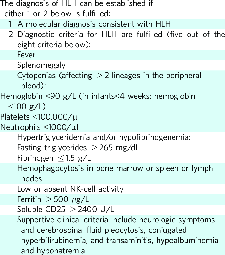
This patient met the following criteria:

  • Fever
  • Splenomegaly
  • Cytopenias
  • Increased triglycerides (results were 320 mg/dL)
  • Increased ferritin

Note: sIL2R and pathology take several days to come back. But in this case, we have 5/8 criteria, suggestive of HLH.

HScore

The calculator for the HScore can be found here.

This patient’s score was 254 suggesting > 99% probability of HLH.

Scoring breakdown:

  • Temperature > 102.9 (+49)
  • Splenomegaly alone (+23)
  • 3 lineagecytopenia (+34)
  • Ferritin > 6,000 ng/mL (+50)
  • Triglycerides 132.7-354 mg/dL (+44)
  • Fibrinogen > 250 mg/dL (+0)
  • AST > 30 U/L (+19)
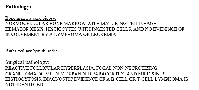
  • Hemophagocytosis features on bone marrow aspirate (+35)

We are admittedly concerned about the possibility of hemophagocytic lymphohistiocytosis (HLH). What additional studies would you want to pursue next given this concern?

a
Bone marrow biopsy
The finding of hemophagocytosis on histopathology is supportive of a diagnosis of HLH
b
Induced sputum for acid-fast bacilli staining
While tuberculosis has been reported as a trigger for HLH, this is an unlikely diagnosis in this patient without TB risk factors.
c
Genetic testing
Testing for genetic defects leading to HLH is controversial in adult patients because abnormalities are rarely detected. It may ultimately be considered in patients without a known trigger.
d
PET-CT
Lymphoma as a trigger of HLH is worth investigating in this young patient with axillary lymphadenopathy

Given leukopenia, cough, and fevers, the patient is started on broad-spectrum antibiotics. A CT torso is obtained to further evaluate possible pulmonary infection as well as axillary lymphadenopathy.

CT abdomen/pelvis showed:

  • No evidence of malignancy
  • Moderate splenomegaly (15.7 cm)

CT chest showed bilateral axillary lymphadenopathy.

Given concern for an underlying malignancy as the driver of his pathologic condition, the patient undergoes an excisional biopsy of an enlarged right axillary lymph node. He subsequently undergoes PET-CT to evaluate for other areas of FDG uptake.

FDG PET:CT – no abnormal FDG uptake identified

The bone marrow biopsy results showed hemophagocytosis.

The patient now meets 6/8 criteria:

  • Fever
  • Splenomegaly
  • Cytopenias
  • Increased triglycerides (320)
  • Increased ferritin
  • Hemophagocytosis

Recall that the remaining criteria include:

  • sIL2R (this would ultimately come back markedly elevated [3131 pg/mL])
  • Low NK cell function (rarely performed)

The soluble IL-2 receptor was 3131 pg/mL (normal range 175.3-858.2 pg/ml).

Prev
 1 / 0 
Next