Introduction
George Gulliver was a surgeon in the army and also associated with the Royal College of Surgeons. He was a specialist on the microscopic examination and measurement of blood cells. In the field of hematology, he is famous for having tabulated comparative measurements of red blood cells.
Biography
Who was George Gulliver?
- George Gulliver was born on 4 June 1804 in Banbury, Oxfordshire. He trained under local surgeons as an apprentice before entering St. Bartholomew’s Hospital in London, “where he soon became an especial favourite with teachers and pupils”,1 and where he held roles such as Prosector of Anatomy, Curator of the Museum, prosector to Abernethy and dresser to Sir William Lawrence.
Career & Honors
- 1826: Became a Member of the Royal College of Surgeons.
- 1827: Appointed hospital assistant to the forces; later served as surgeon to the Royal Horse Guards (“the Blues”).
- 1838: Elected Fellow of the Royal Society.
- 1843: Became a Fellow of the Royal College of Surgeons (“chosen for the Fellowship in recognition purely of scientific merits”).2
- 1852: Joined the council of the Royal College of Surgeons.
- 1861: Served as the Hunterian Professor of Comparative Anatomy and Physiology; “During the tenure of this office he gave courses of lectures on his favourite subjects, Blood, Lymph, and Chyle of Vertebrates, in which were related the results of his extensive researches on these subjects.”3
- 1863: Delivered the Hunterian Oration.
Scientific Contributions
Gulliver didn’t write a single comprehensive book, but he was prolific in research:
- Editorial Work:
- Edited the English translation of Gerber’s General and Minute Anatomy of Man and the Mammalia (1842), adding his own extensive notes and an appendix on blood, lymph, and related topics (see below).
- Edited The Works of William Hewson (1846) for the Sydenham Society, which included his annotations and a biography of Hewson.
- Contributed notes to Rudolph Wagner’s Physiology (translated by Dr. Willis, 1844).
- Lectures & Papers:
- His Hunterian lectures, titled Blood, Lymph, and Chyle of Vertebrates, were published in the Medical Times and Gazette between August 1862 and June 1863 (see below).
- Most of his published work appears in various periodicals, catalogued in the Royal Society’s Catalogue of Scientific Papers.
- Key Findings:
- First to publish comprehensive tables and observations on the shape and structure of red blood corpuscles across humans and other vertebrates.
- Made corrections to accepted views on blood coagulation, noted the fibrillar form of fibrin clots, clarified the nature of chyle and lymph nuclei, elucidated the thymus’s role in the lymphatic system, and offered insights on bone formation and repair.
- Contributed to pathology by highlighting the presence of cholesterine and fatty degeneration in organs and morbid tissues, and detailed features of fibrin clot softening and characteristics of tubercles.
- Also contributed to botany: studied raphides, pollen, and various plant tissues, demonstrating their taxonomic importance.
Later Years & Legacy
After retiring from military service, Gulliver devoted himself fully to scientific research. He became gradually enfeebled by gout, and many of his later papers were written when he was confined to his bed. He passed away in Canterbury on 17 November 1882, leaving behind a legacy of meticulous anatomical and physiological scholarship. He was survived by his son, also named George, who served as assistant physician at St. Thomas’ Hospital.
Summary
George Gulliver’s career was marked by intense scientific curiosity and dedication. His contributions to hematology, anatomy, pathology, and even botany established him as a respected figure in 19th-century British science. His editing and lecturing work expanded knowledge and honored predecessors like Hewson, and his empirical research advanced understanding across multiple biological disciplines.
Gulliver’s Iconic RBC Drawings
George Gulliver published some of his earliest measurements of mammalian blood corpuscles in 1840 in a paper entitled Mr. Gulliver’s Observations on the Blood Corpuscles or Red Discs of the Mammiferous Animals in the London and Edinburgh Philosophical Magazine and Journal of Science. In 1842, he published a more detailed analysis of both mammalian and non-mammalian red blood cells in an appendix to Elements of General and Minute Anatomy of Man and the Mammalia. He divided this appendix into two separate sections — one focused on mammals and the other on birds. While these chapters contain extensive tables and textual commentary that foreshadow some of his later findings, including notable species differences in red cell size, there is no illustration in this 1842 publication.
It wasn’t until two decades later, in 1862, that Gulliver published his iconic comparative figure showing red blood cells from a wide variety of vertebrate species. This image would become widely reproduced in textbooks and comparative physiology. The figure appeared in an 862 paper entitled: On the Red Corpuscles of the Blood of Vertebrata, and on the Zoological Import of the Nucleus, with Plans of their Structure, Form, and Size (on A Uniform Scale). In this paper, he described the relative form and size of the corpuscles in 171 species of the different classes and orders of the Vertebrate subkingdom. Gulliver was the first to publish comprehensive tables of the size, shape, and structure of red blood corpuscles in humans and animals. His microscopic drawings became iconic because they clearly depicted.

Gulliver’s Methods
- His specimen collection methods were actually quite traditional for a 19th-century comparative anatomist, but they’re not always spelled out in detail in the paper itself. Gulliver did not go on global specimen-collecting expeditions himself. Instead, he worked within London’s dense network of butchers, veterinary schools, zoological collections, and anatomical museums, supplementing with occasional self-collected samples from common species. His genius was in standardizing microscopic measurements across hundreds of animals, not in field collection per se. Here’s what’s known from his own writings and contemporary accounts:
- Source of Animals:
- Domestic animals (horses, cattle, sheep, dogs, etc.) he obtained from local farms, markets, butchers, and veterinary schools.
- Wild or exotic species he often accessed through zoological collections (such as the Zoological Society of London’s menagerie at Regent’s Park, opened in the 1820s), traveling naturalists, and specimens supplied by colleagues in comparative anatomy.
- Amphibians, reptiles, and fish he collected himself (frogs, newts, common fish) or received preserved material from natural history collections.
- Method of Collection:
- He usually drew fresh blood samples immediately post-mortem (from animals being slaughtered, dissected in veterinary/anatomical schools, or prepared as museum specimens).
- In smaller living animals (e.g. frogs), he sometimes did direct capillary puncture.
- Blood was diluted in saline or serum and examined with a simple light microscope; Gulliver had refined micrometry methods to measure cell diameters with remarkable accuracy for his time.
- Gulliver’s describes obtaining blood from:
- Right and/or right ventricles of the heart
- Portal and renal veins
- Forearms
- Vena cava
- Ear
- Tail
- Splenic vein
- Abdominal aorta
- Jugular vein
- Wound in the neck
- Renal artery
- Upper lip
- Microscopy & Measurement:
- He used one of the earliest micrometers (a calibrated glass scale fitted into the microscope) to measure red cells.
- “The instrument made use of, in these observations, as a compound microscope with an achromatic object glass of one eighth of an inch focal length, made by Ross, and furnished by him with a micrometer eyepiece divided into spaces corresponding to 1-4000th of an inch. The magnifying power afforded us exactly 800 diameters with a clear definition.”4
- “The corpuscles were thinly spread on glass and quickly dried, also floating in their own serum, and diluted when necessary with weak saline solutions, or sugar and water, or with urine.”5
- “In most cases the measurements were repeated by Mr. Siddall, an experienced microgapher, with another instrument by Ross, so as to avoid as much as possible accidental inaccuracies.”6
- His aim was comparative: establishing relative size and shape across taxa (mammals, birds, reptiles, amphibians, fish).
- His 1840s–1860s publications often list n values — meaning he made repeated measurements on blood smears from different individuals of the same species.
- Networks of Supply:
- Gulliver was closely associated with the Royal College of Surgeons and Hunterian Museum, which housed an enormous comparative anatomy collection. This gave him steady access to preserved and fresh animal material.
- He corresponded with zoologists, comparative anatomists, and physiologists who sent him samples. For rare species, he often analyzed museum-preserved blood (though he admitted fixation altered morphology slightly).
- Source of Animals:
“In the typical Victorian manner of a curious collector and collector of curiosities, Gulliver had measured countless red blood cells from any vertebrate he could get his hands on, with London Zoo his main hunting ground.” Tobias Warnecke
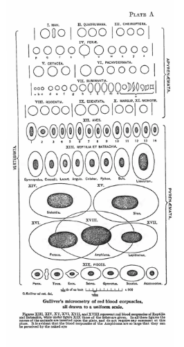
The Original Drawing by George Gulliver in 1862
- Each red blood cell is drawn to scale, showing the wide range of size and morphology across species.
- Labeled with numbers only — Gulliver included a key in the text, where the number corresponded to the species name.
- No superimposed “giant” red cell, which was added to modern versions. The bottom row simply contains the largest amphibian cells (e.g., Amphiuma, Proteus, Menobranchus).
- The drawing emphasized comparative measurement, not visual dramatization.
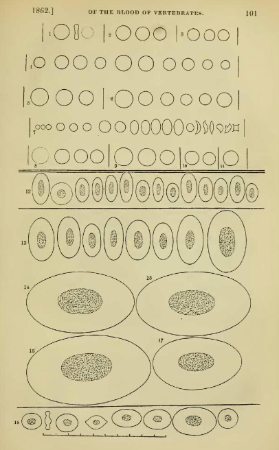
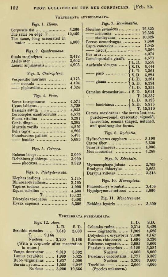
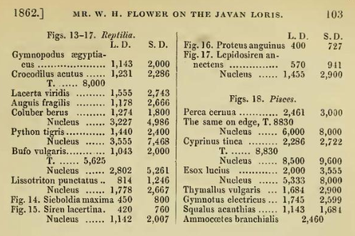
Description of the wood engraving (above): All the corpuscles are drawn to a scale of 1/4000th of an English inch, and are magnified about 920 times linear admeasurement. The scale is marked at the bottom of the engraving. Corpuscles only of average size are given; and but one corpuscle from each species of animal, with the few exceptions presently to be noticed. The corpuscles of Apyrenæmatous Vertebrates [mammals] occupy the upper part of the engraving, above the double line; and the different orders of these are separated by the short upright lines. The corpuscles of Pyrenzematous Vertebrates [non-mammalian vertebrates] occupy all the larger part of the engraving below the double line. At 12 is a row of birds’ corpuscles; 13-17, corpuscles of reptiles; and 18, a row of the corpuscles of fishes. The figures at 1 and 12, referring to structure, are fully explained at page 93. Of the Pyrenzmatous Vertebrates, the nuclei are shown much more plainly than they appear in the pure corpuscles; but the action of acetic acid exposes the nuclei as distinctly as they are here represented. The names of the animals are set down in the following table, according to the order in which the sketches of the corpuscles stand in the engraving. The following measurements of the corpuscles are all in vulgar fractions of an English inch ; but as the numerator is invariably 1, it is omitted throughout, and the denominators only are printed. T. denotes the thickness, L. D. the long diameter, and S. D. the short diameter of the corpuscles.
The Modernized Version (commonly reproduced in textbooks & online)
- Later adaptations added animal names directly under or beside the cells for easier interpretation.
- A very large Amphiuma erythrocyte (sometimes called the “giant red blood cell”) is drawn overlapping the bottom row of the largest four cells. This was not in Gulliver’s original — it’s an interpretive addition to highlight scale.
- This makes the modern version look a bit more dramatic, but it slightly distorts the layout compared to Gulliver’s careful comparative grid.
Noteworthy Passages from Gulliver’s 1862 Paper
- The object of this communication is to give a summary of the value and import of the red corpuscles of the blood as regards systematic zoology, deduced from my observations published, piecemeal, during the last twenty-three years, in the ‘Proceedings’ of this Society and elsewhere.
- The drawings now exhibited to the Society are selected from a much larger number in my possession, and are all on the same scale, exhibiting plainly to the eye the relative form and size of the corpuscles in 171 species of the different classes and orders of the Vertebrate subkingdom.
- Mammalian red cells:
- The [mammalian] corpuscle is slippery, soft, elastic, and viscid; it will assume a variety of forms, and quickly return to its regular shape; and the corpuscles will stick together, not only in the well-known piles, but also by their edges.
- The regular [mammalian] corpuscle has no nucleus—nothing at all like that so plain in the corpuscle of oviparous Vertebrata.
- The red corpuscle is a circular, flattened, biconcave disc, rounded at the margin. The biconcave form was inferred by Dr. Young and proved by Dr. Hodgkin and Mr. Lister. This concavity causes the central spot so long mistaken for a nucleus.
- There are certain exceptions, regular and irregular, to the circular and biconcave shape:
- The Camelide, as will be more particularly explained in the proper place, have oval corpuscles… it is in shape only that these red corpuscles resemble those of oviparous Vertebrata. The corpuscles generally of the Camelide have no nucleus, and so agree in structure, as they do also in size, with those of their mammalian allies.
- In certain Cervide, to be noticed presently, the angular, crescentic, and lanceolate corpuscles are in unusual abundance.
- And when we consider how pliant and elastic the Mammalian corpuscle is… we might expect rapid variations in its shape within certain limits; and such is the fact… accordingly, the corpuscles may be either swollen, puckered, or shrunk into a variety of figures, flat, tumid, like a shallow circular or oval cup, stellate, notched, granulated, mulberry-shaped, crescentic, angular, lanceolate, fusiform, comma-shaped, and other figures, defying definition… Dr. Richardson has well depicted a number of forms presented by the corpuscles in connexion with disease.
- It was the prevailing statement, after Hewson, that the size of the corpuscle is not at all connected with that of the animal… while confirming the accuracy of his statement as to animals of such different orders, I soon found that, in a really natural family, other things equal, the largest corpuscles will be generally found among the large species, and the smallest corpuscles among the small species, of that family… There are many exceptions to an exact relation between the sizes of the species and corpuscles.
- Few Mammalia have larger corpuscles than Man; among these may be noted the elephant, the whale, the great anteater.
- Non-mammalian red cells:
- The regular red corpuscle of oviparous Vertebrata is a cell or vesicle containing a nucleus, while the regular red corpuscle of Mammalia has no nucleus.
Why Iconic?
- They were among the earliest standardized illustrations of RBCs under the microscope.
- They highlighted the evolutionary and taxonomic value of red cell morphology.
- They influenced both hematology and comparative anatomy for decades.
Conclusion
Gulliver was the first to publish comprehensive tables of the size, shape, and structure of red blood corpuscles in humans and animals. His microscopic drawings became iconic because they clearly depicted:
- The biconcave disks of human RBCs (no nuclei).
- The elliptical, nucleated RBCs of birds, reptiles, and amphibians.
- Variation in cell diameter across species, from tiny mammalian cells (e.g., in musk deer) to very large nucleated amphibian cells (e.g., in salamanders).