For larger image, click here.
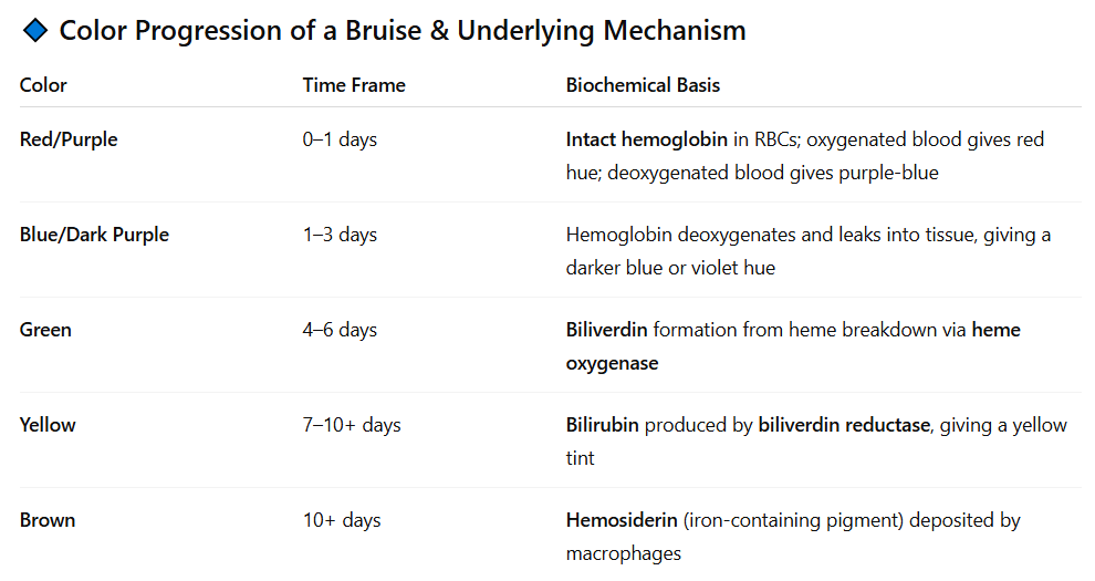
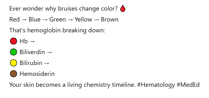
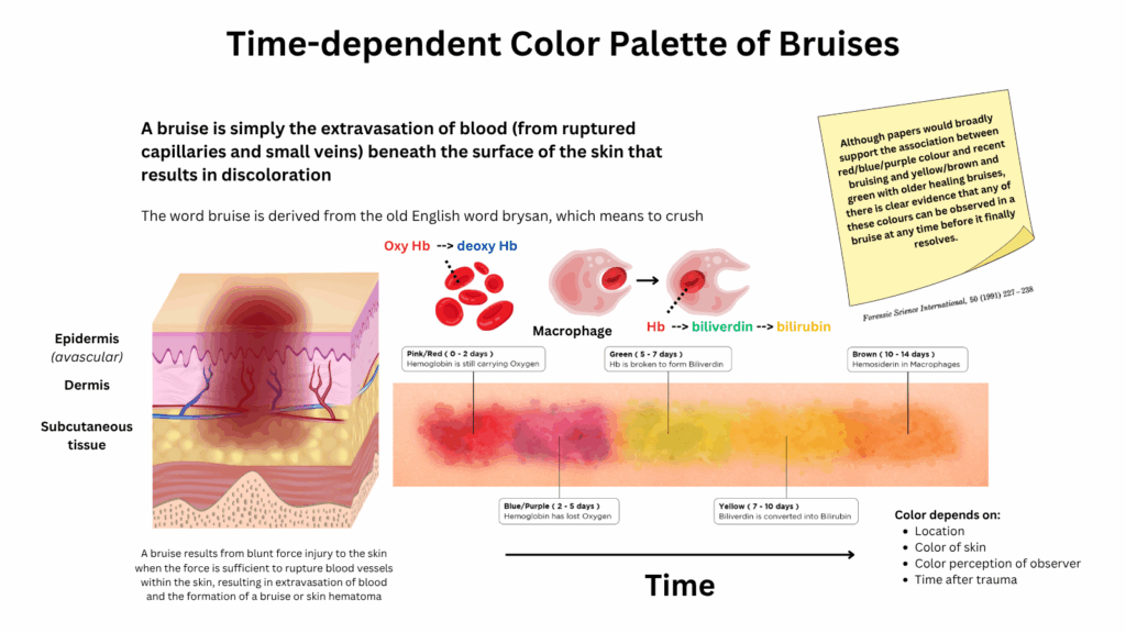
The colors of a bruise (ecchymosis) reflect the breakdown of hemoglobin in extravasated red blood cells and the sequential metabolism of heme pigments by tissue macrophages. Each pigment has a different absorption spectrum and reflects light differently, giving the bruise its characteristic color. These changes follow a predictable time course and are useful in estimating the age of a bruise.
Step-by-step breakdown of hemoglobin in a bruise:
- RBCs rupture in tissue → hemoglobin released
- Hemoglobin → heme + globin
- Heme → biliverdin (green) via heme oxygenase
- Biliverdin → bilirubin (yellow) via biliverdin reductase
- Iron from heme is stored as hemosiderin (brown)