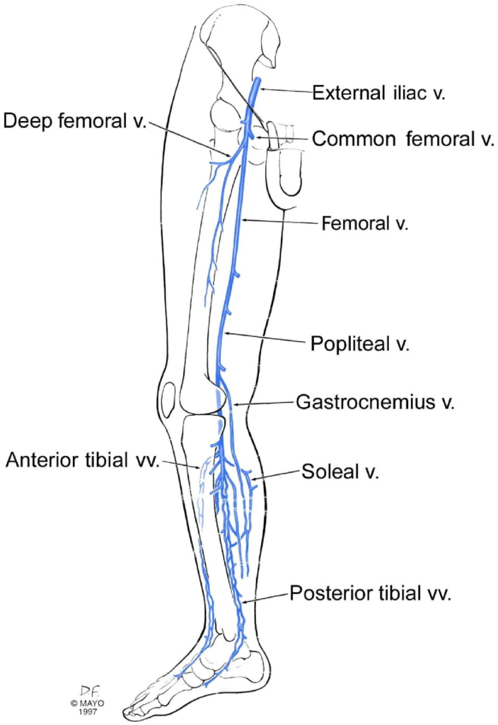
Deep veins of the leg. The lower limb consists of two main types of veins: superficial veins and deep veins. The superficial veins (not shown) are located within the subcutaneous tissue while the deep veins are found deep to the muscular fascia. The deep veins accompany the major arteries. Both types of veins contain venous valves, to prevent reflux of blood distally. In addition, small perforating veins (not shown) penetrate the muscular fascia and connect the superficial and deep veins. Communicating veins connect veins within the same system (i.e., deep to deep, superficial to superficial). The deep venous system of the calf includes the anterior and posterior tibialveins and the paired peronealveins. The soleal and gastrocnemial veins are intramuscular veins forming a plexus or sinus of veins that drain the gastrocnemius and soleal muscles and empty into the deeper named veins of the calf and popliteal fossa. Some consider these veins to be deep, others view them as superficial. The natural course and management of deep vein thrombosis involving the tibial and peroneal veins is much better understood compared with isolated gastrocnemius and soleal vein thrombi (IGSVT). The popliteal vein is formed by the confluence of the calf veins. The popliteal vein drains into the femoral vein (this was once called the superficial femoral vein, a term that has been abandoned because it is not a superficial vein). The femoral vein and deep femoral vein converge at the common femoral vein, which then drains into the external iliac vein. Source.
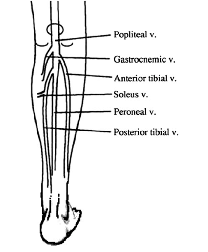
A graphic showing the gastrocnemius and soleal veins.Source.
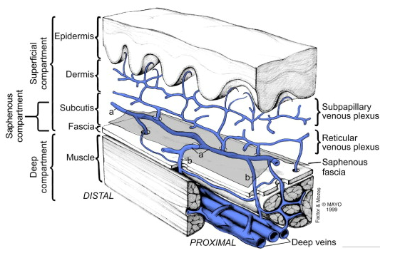
Venous systems of the leg. The skin and subcutaneous tissues are drained by the venous plexuses. Superficial veins (a) are connected to deep veins through perforators (b). The deep fascia covers the muscles, and the saphenous fascia invests the saphenous vein. Source.