| Spherocytes | |
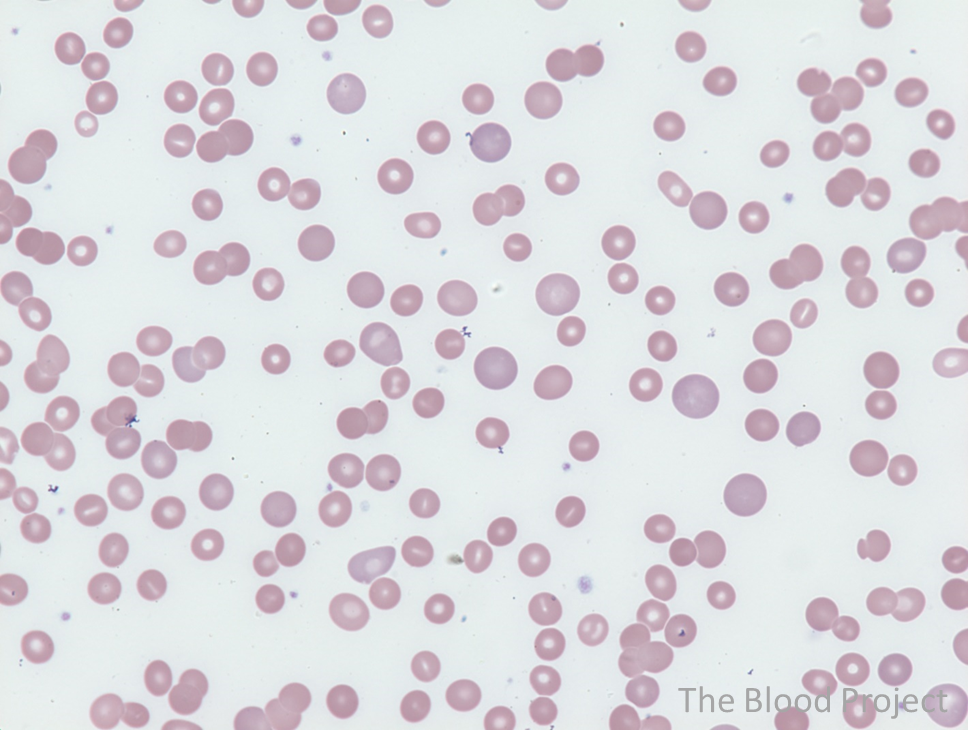
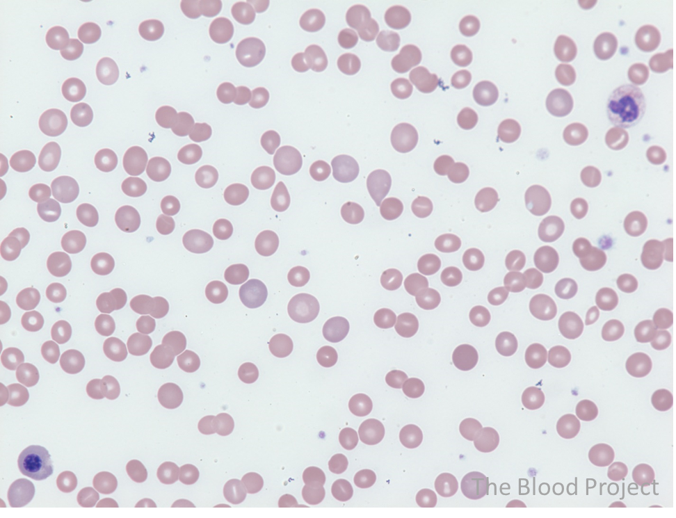
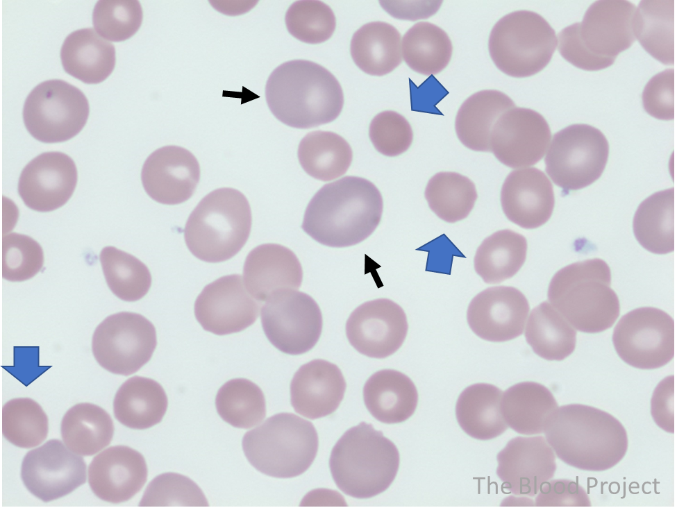
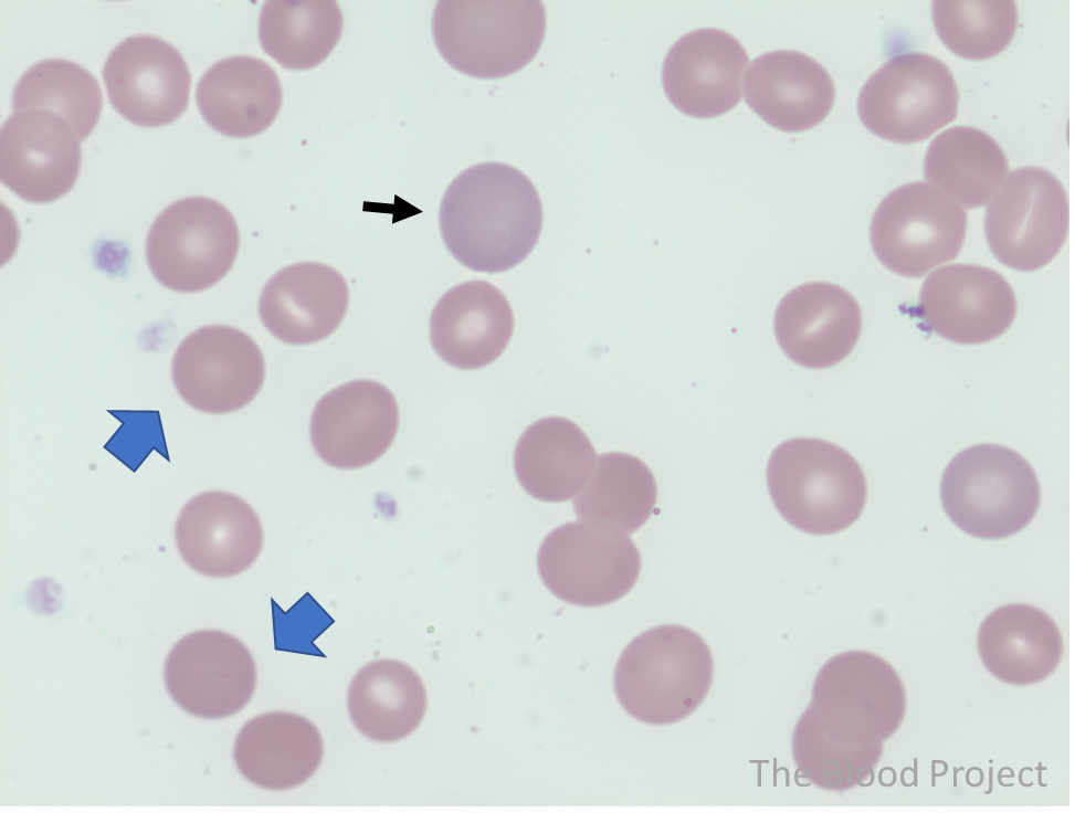
| Definition/description | Spherocytes are round, thicker and more densely staining than normal and lack central pallor. Typically appear slighter smaller than normal red cells. |
| Mechanism of formation | Occurs as a consequence of membrane loss, resulting in decreased surface area: volume ratio. |
| Other red cell phenotypes | |
| Polychromatophilia | Polychromatophilic cells appear slightly larger than normal red cells and their color is slightly more basophilic (blue-purple). They represent reticulocytes, reflecting the bone marrow response to anemia. |
| History | French investigators reported the presence of hemolysins in the sera of patients with hemolytic anemia as early as 1908. The direct Coombs (antiglobulin) test was introduced in 1945 and in 1946 was used to definitely diagnose idiopathic acquired hemolytic anaemia in 5 patients who demonstrated spherocytosis on their peripheral smear. |
| Source/author | William Aird |
| References | Lancet. 1946;1(6405):812-4. |