
Normal physiology reveals the disorder. Social normalization can hide it again.
Note: The video and audio linked above were generated with the assistance of AI. Clinical accuracy has been reviewed, but no AI-generated content can be guaranteed to be fully error-free.
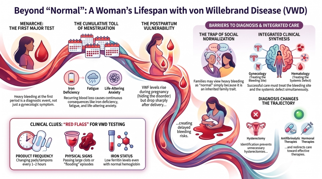
Why this spoke matters
Von Willebrand disease affects people of all sexes.
But it is often experienced, recognized, and managed differently in women.
The inherited defect is not inherently female. The hemostatic challenges often are.
Menarche, repeated endometrial shedding, ovulation, pregnancy, childbirth, and postpartum recovery place recurring demands on primary hemostasis. These are normal biological processes. In a person with reduced or dysfunctional von Willebrand factor, they may become the events that first reveal the disorder and then shape its burden for decades.
Yet physiology is only part of the story.
Heavy menstrual bleeding may be normalized within families, minimized by clinicians, concealed because of embarrassment, or treated as an isolated gynecologic problem without recognition of the underlying bleeding disorder. During that delay, iron deficiency, fatigue, school absence, impaired work, reproductive anxiety, and repeated procedures may accumulate.1
Understanding VWD in women therefore requires more than knowing how to treat a heavy period or cover a delivery.
It requires understanding the interaction of inherited biology, reproductive physiology, social interpretation, access to care, and the cumulative consequences of bleeding.
Normal physiology may reveal the disorder. Social normalization may hide it again.
Why women experience VWD differently
VWD is usually inherited in an autosomal pattern and affects males and females. Women, however, are more likely to encounter repeated hemostatic challenges through menstruation, pregnancy, and childbirth.2
A boy and girl with the same VWF defect may appear similarly affected during childhood.
Then menarche begins.
From that point onward, one patient may undergo a physiologic challenge every month, while the other may remain largely untested until trauma, dental extraction, or surgery.
This difference in exposure has consequences. Repeated menstrual blood loss may create cumulative iron deficiency and impaired quality of life. At the same time, menstruation may make the disorder more visible, but only when the bleeding is recognized as abnormal.
Women do not necessarily have a different inherited disorder. They encounter a different pattern of hemostatic exposure.
Normal physiology repeatedly tests primary hemostasis
The reproductive tract is not simply another bleeding site.
Endometrial shedding requires coordinated vasoconstriction, platelet adhesion, coagulation, tissue repair, and fibrinolysis. Local fibrinolytic activity is high, which helps explain why defects in primary hemostasis are often expressed strongly through menstrual bleeding and why antifibrinolytic therapy can be particularly effective at this site.
Menstruation is therefore not merely blood loss from a surface.
It is a recurring biological test of whether the patient can form and maintain a hemostatic plug in a highly fibrinolytic environment.
Menarche
Menarche may be the first major physiologic test of VWF-dependent hemostasis.
An adolescent who previously had only bruising or occasional epistaxis may suddenly experience prolonged bleeding, flooding, nighttime changes of menstrual protection, passage of large clots, iron deficiency, anemia, or a need for urgent medical care.
Heavy menstrual bleeding beginning at menarche should therefore be treated not only as a gynecologic symptom, but as a possible diagnostic event.3
It is not synonymous with VWD. Anovulatory bleeding, endocrine disorders, platelet dysfunction, other coagulation defects, pregnancy-related causes, and structural pathology may also contribute.
But when heavy bleeding begins with the first menstrual periods, especially when accompanied by iron deficiency, epistaxis, bruising, dental bleeding, or a family history, an inherited bleeding disorder deserves consideration.
Menstruation
Menstruation converts a hemostatic vulnerability into a recurrent exposure.
The clinical burden is rarely confined to the days of visible bleeding. Repeated blood loss may reduce iron stores, impair exercise tolerance and concentration, disrupt school or work, and produce constant anxiety about leakage, clothing, bathroom access, or travel.
A patient may appear well between periods while organizing much of her life around the anticipation and consequences of the next one.
The bleeding is episodic. Its consequences may be continuous.
Ovulation and gynecologic tissue injury
VWD in women is more than heavy menstrual bleeding.
Ovulation, ovarian cyst rupture, miscarriage management, endometrial biopsy, intrauterine procedures, and gynecologic surgery may all create additional hemostatic challenges. Hemorrhagic ovarian cysts, ovulation-associated bleeding, hemoperitoneum, and excessive bleeding after cervical or uterine procedures have been described.4
Structural and ovulatory gynecologic disorders such as fibroids, polyps, endometriosis, adenomyosis, and ovarian cysts may not be intrinsically more common because of VWD. Their bleeding manifestations may instead be more severe, more symptomatic, or more likely to prompt intervention.
The uterus is important.
It is not the whole story.
Why diagnosis is often delayed
Heavy menstrual bleeding is common.
That makes it an important clue, but not a diagnosis by itself.
The challenge is that abnormal bleeding may be difficult to recognize when it is experienced privately, begins early, and resembles the experiences of other women in the family.
A mother may tell her daughter:
“Our periods are just heavy.”
A clinician may say:
“That can be normal in adolescence.”
A patient may assume that changing protection every hour, bleeding through clothing, or missing school is simply part of menstruation.
In families with autosomal dominant VWD, abnormal bleeding may become the family standard against which normality is judged.
Inherited bleeding can be normalized precisely because it is inherited.
Stigma, limited menstrual-health education, fragmented care, and discomfort discussing menstrual blood loss may all contribute. Iron deficiency may be treated without addressing the source. Uterine bleeding may be managed without assessing systemic hemostasis. A normal PT or aPTT may be interpreted incorrectly as excluding a bleeding disorder.
It does not.
A normal PT is expected in VWD, and the aPTT may remain normal unless factor VIII activity (FVIII:C) is sufficiently reduced.5
Diagnostic delay is not neutral.
During the delay, the patient continues to bleed, lose iron, adapt her life, and sometimes undergo invasive treatment without a unifying diagnosis.
Heavy menstrual bleeding must be interpreted, not merely recorded
Asking, “Are your periods heavy?” is rarely sufficient.
Patients may answer no because they have never experienced anything else.
A more informative history asks about the behavior and consequences of the bleeding:
- How many days does bleeding last?
- Is there flooding or gushing?
- How often are pads, tampons, cups, or period underwear changed?
- Are nighttime changes or double protection required?
- Has bleeding soaked through clothing or bedding?
- Are large clots passed?
- Has school, work, exercise, travel, or sleep been disrupted?
- Has iron deficiency or anemia occurred?
- Have hormonal therapy, tranexamic acid, emergency care, or transfusion been required?
- Did heavy bleeding begin at menarche?
- Are epistaxis, bruising, dental bleeding, postoperative bleeding, postpartum bleeding, or a family history also present?
Flooding, changing protection at least hourly on heavy days, bleeding through clothing or bedding, large clots, nighttime changes, and iron deficiency are useful screening clues. None is diagnostic in isolation.6
A validated tool such as the ISTH Bleeding Assessment Tool can standardize the history and help determine which patients in lower-prevalence settings warrant laboratory evaluation. In patients with a strong phenotype or high pretest probability, however, a low BAT score should not be used to withhold testing.7
The score summarizes the history. It does not replace the history.
Testing must account for the conditions of measurement
VWF is an acute-phase reactant.
Acute bleeding, stress, anemia, inflammation, infection, pregnancy, and difficult phlebotomy may raise VWF and obscure a lower baseline. Testing during severe menstrual bleeding is not useless. A low value obtained during physiologic stress remains meaningful. But a normal or elevated result may need to be repeated when the patient has returned to baseline health.8
Initial VWD testing should include:
- VWF antigen
- a platelet-dependent VWF activity assay
- factor VIII activity (FVIII:C)
The results should be interpreted relationally and in the context of the bleeding phenotype. If the initial panel is nondiagnostic but suspicion remains, repeat testing and evaluation for platelet or other bleeding disorders may be required.
Recent laboratory guidance also emphasizes that assay methodology, sample handling, and activity-to-antigen ratios support interpretation but do not replace clinical judgment.
The laboratory result is part of the story. The conditions under which it was obtained are part of the result.
Iron deficiency is part of the bleeding phenotype
Iron deficiency is not merely a secondary laboratory abnormality.
It may provide objective evidence that recurrent blood loss has produced cumulative physiologic consequences, even when hemoglobin remains normal.
A patient may normalize her menstrual bleeding, underestimate its volume, or no longer describe it as burdensome.
Ferritin may tell a different story.
Iron deficiency without anemia may contribute to fatigue, reduced exercise tolerance, restless legs, cognitive complaints, and reduced quality of life, although these symptoms are nonspecific.9
Ferritin should be interpreted in clinical context, particularly when inflammation is present. A normal ferritin does not exclude clinically important heavy menstrual bleeding, and iron deficiency is not specific to menstrual loss.
But when recurrent menstrual bleeding and depleted iron stores occur together, the connection should not be ignored.
Assessment should include attention to iron stores, not only the complete blood count. Iron deficiency should be treated while ongoing blood loss is addressed.
Ferritin is part of the bleeding story.
A diagnosis can alter the trajectory of care
Without recognition of a bleeding disorder, a patient with heavy menstrual bleeding may undergo progressively intensive treatment directed only at the uterus.
Repeated hormonal changes, dilation and curettage, endometrial ablation, myomectomy, and hysterectomy may all be considered.
These interventions may be entirely appropriate when structural pathology, refractory symptoms, or patient preference justify them.
But they should not occur because an underlying hemostatic disorder was never considered.
Observational data indicate that women with VWD experience a higher burden of heavy menstrual bleeding and gynecologic intervention, including hysterectomy, than matched women without VWD.10
Diagnosis may redirect care toward antifibrinolytic treatment, hormonal or intrauterine therapy, iron replacement, coordinated hematology–gynecology care, and safer procedural planning.
Diagnosis may reduce avoidable escalation, improve selection of medical therapy, and make necessary procedures safer.
Care must treat both hemostasis and the bleeding site
VWD treatment alone cannot address every cause of uterine bleeding.
A patient with VWD may also have fibroids, adenomyosis, endometriosis, polyps, ovulatory dysfunction, pregnancy-related pathology, malignancy, or another gynecologic condition.
Likewise, gynecologic treatment alone may not adequately address an inherited defect in primary hemostasis.
The best care asks two questions:
Why is this site bleeding?
and
Why is this patient unable to stop it normally?
This dual approach is particularly important when bleeding becomes newly severe, changes pattern, develops after years of stability, or appears disproportionate to the known VWD phenotype.
The uterus may be the site of bleeding. VWD may be the reason the bleeding persists. Both require attention.
Reproductive choices are part of treatment
Women’s-health care in VWD should not reduce the patient to menstrual control.
Treatment choices intersect with contraception, fertility goals, desire for pregnancy, medication tolerance, sexual health, cultural or personal preferences, access, and cost.
Hormonal therapy and the levonorgestrel-releasing intrauterine system may reduce menstrual blood loss while also providing contraception. Tranexamic acid offers a nonhormonal approach and is especially useful because it counteracts the high fibrinolytic activity of the endometrium. Combination therapy may be needed when one strategy does not provide adequate control.11
The 2021 management guideline suggests hormonal therapy or tranexamic acid over desmopressin for women with VWD and heavy menstrual bleeding who do not wish to conceive. For those who wish to conceive, it suggests tranexamic acid over desmopressin.12
These are conditional recommendations, not mandates.
The choice of hormonal therapy depends on contraindications and patient-specific thrombotic risk. Insertion of an intrauterine device may itself require hemostatic planning in selected patients. Desmopressin is ineffective in type 3 VWD, generally avoided in type 2B VWD, and should not be relied upon unless response and suitability are established.
The aim is to align therapy with the patient’s bleeding burden, reproductive goals, preferences, and clinical context.
Fertility, inheritance, and reproductive planning
VWD is not generally considered a direct cause of infertility.
It may, however, complicate reproductive procedures and pregnancy planning.
Patients may have questions about inheritance, the likelihood and potential severity of disease in offspring, assisted reproduction, egg retrieval, miscarriage management, prenatal diagnosis in severe disease, delivery planning, and neonatal testing.
Inheritance counseling should reflect subtype.
Most type 1 and many type 2 forms are autosomal dominant, although penetrance and expressivity vary. Type 3 and type 2N VWD are typically autosomal recessive. Genetic findings, partner testing, and family history may alter counseling.
The goal is not to discourage pregnancy.
It is to replace uncertainty with anticipatory planning.
Pregnancy changes the laboratory phenotype
Pregnancy produces a physiologic rise in VWF and factor VIII.
This increase reflects hormone-responsive changes in endothelial synthesis and release, together with broader adaptations of pregnancy that shift hemostasis toward clot formation. In many women with type 1 VWD, VWF and FVIII rise substantially and may enter the normal range by the third trimester.
The response is subtype dependent.
In type 2 VWD, antigen may rise without reliable correction of the qualitative defect. The activity-to-antigen relationship, multimer abnormality, thrombocytopenia, or impaired factor VIII binding may remain clinically relevant. Women with type 3 VWD do not produce a meaningful endogenous VWF increase.13
Pregnancy therefore changes both hemostatic reserve and the interpretation of laboratory testing.
A normal third-trimester result may be essential for delivery planning. It does not necessarily represent the patient’s nonpregnant baseline or erase the diagnosis.
Pregnancy may normalize the number without curing the disorder.
Childbirth is a planned hemostatic challenge
Delivery should be approached as a coordinated clinical event rather than an unexpected test of hemostasis.
Planning may involve the patient, hematology, obstetrics, anesthesiology, transfusion medicine, and neonatology. The plan should integrate subtype, third-trimester platelet-dependent VWF activity and factor VIII activity (FVIII:C), prior bleeding and obstetric history, desmopressin response, availability of VWF concentrate, postpartum therapy, and fetal risk when severe VWD is possible.14
Neuraxial anesthesia requires separate consideration. The 2021 guideline suggests targeting VWF activity of 0.50 to 1.50 IU/mL when neuraxial anesthesia is otherwise considered appropriate, with maintenance above 0.50 IU/mL while the epidural is in place and for a period after removal. This is a conditional recommendation based on very-low-certainty evidence and should not be interpreted as a universal guarantee of safety.
Mode of delivery should generally be determined by obstetric indications. When the fetus may have severe VWD, traumatic fetal instrumentation should be avoided when clinically feasible.
The goal is not to convert every delivery into a hematologic procedure.
It is to ensure that the team knows what to do if normal obstetric hemostasis is insufficient.
VWD is only one contributor to obstetric bleeding. Uterine atony, retained placenta, genital tract trauma, operative delivery, and other obstetric causes still require prompt management.
Hemostatic treatment supports obstetric care. It does not replace it.
Postpartum physiology reverses the pregnancy gain
The postpartum period reveals why a reassuring delivery value is not the end of planning.
After childbirth, VWF and FVIII decline toward baseline. This may expose the underlying deficiency after the patient has left the hospital.
Women may experience immediate postpartum hemorrhage, excessive or prolonged lochia, secondary postpartum hemorrhage, or delayed bleeding days or weeks after delivery.
The 2021 management guideline suggests postpartum tranexamic acid for women with type 1 VWD or low VWF and notes that it may also be used in types 2 and 3 VWD when appropriate.15
Tranexamic acid does not replace VWF replacement or other hemostatic therapy when severe deficiency requires it. Nor does it replace evaluation for retained products, atony, trauma, infection, or other obstetric causes.
Discharge counseling should explain what bleeding is expected, what bleeding is excessive, whom to contact, how long therapy should continue, and when reassessment is required.
Delivery is a moment. Postpartum risk is a period.
Menopause changes the pattern, not necessarily the problem
Menopause removes one major recurrent source of blood loss.
For many women, heavy menstrual bleeding ends, iron balance improves, and a substantial part of the disease burden recedes.
But VWD does not become irrelevant.
Perimenopause may produce irregular or heavy bleeding. Postmenopausal bleeding requires anatomical and malignancy-focused evaluation and should not be attributed automatically to VWD.
Later-life care may also involve gastrointestinal angiodysplasia, surgery, malignancy, renal or cardiovascular disease, antiplatelet or anticoagulant therapy, and acquired modifiers of VWF function.
VWF often rises with age. Some women whose values were previously low or diagnostic for type 1 VWD may move into the reference range. Current guidance recommends reconsidering rather than automatically removing a previously confirmed diagnosis, assuming the original diagnosis was valid.16
A higher current level does not necessarily erase the historical bleeding phenotype or eliminate procedural risk.
Menopause may end menstrual bleeding. It does not end women’s health in VWD.
The patient may become parent, advocate, and caregiver
The life course of VWD may extend across generations.
A woman who struggled for years to receive her own diagnosis may later recognize symptoms in a child, pursue testing for relatives, advocate for safer procedures, explain inheritance to family members, and coordinate care across schools and healthcare systems.
Earlier recognition in the next generation may bring reassurance.
It may also revive memories of the patient’s own delayed diagnosis or create anxiety, guilt, financial burden, and caregiving responsibilities.
These are reported experiences, not inevitable consequences. But they remind clinicians that family evaluation is not merely a laboratory exercise.
Inheritance is biological.
Its meaning is personal.
Access to care shapes the experience of VWD
Biology alone does not determine outcome.
The burden of disease also depends on whether the patient can access clinicians who recognize abnormal menstrual bleeding, appropriate VWF testing, laboratory expertise, adolescent gynecology, hematology consultation, iron therapy, antifibrinolytic treatment, hormonal options, VWF concentrate, and coordinated obstetric care.
Fragmentation is particularly harmful.
The hematologist may understand the bleeding disorder but not the patient’s reproductive priorities. The gynecologist may understand uterine pathology but not the systemic hemostatic defect. The primary care clinician may repeatedly treat iron deficiency without addressing ongoing blood loss.
Women’s-health care in VWD is inherently multidisciplinary because the problem crosses biological and clinical boundaries.
Clinical synthesis
VWD in women is shaped by the interaction of biology, physiology, and interpretation.
The inherited defect reduces hemostatic reserve.
Reproductive physiology repeatedly tests that reserve.
Social normalization may delay recognition.
Repeated blood loss may produce iron deficiency and cumulative functional impairment.
Reproductive goals influence treatment.
Pregnancy temporarily changes the laboratory phenotype.
Delivery tests planning.
The postpartum fall restores vulnerability.
Menopause changes the pattern of bleeding without eliminating the need for VWD-informed care.
The central clinical question is not only:
How do we stop this episode of bleeding?
It is also:
Why was the bleeding not recognized earlier?
What has repeated bleeding already cost this patient?
How can treatment be aligned with her reproductive goals and stage of life?
What can be prevented before the next physiologic challenge occurs?
For many women, VWD is not revealed by abnormal physiology. It is revealed by normal physiology and obscured when abnormal bleeding is mistaken for a normal female experience.
Evidence anchor
| Evidence stream | What it shows | Why it matters | Main limitation |
|---|---|---|---|
| Women’s lived-experience literature | Menstrual bleeding may be normalized within families, minimized by clinicians, and associated with diagnostic delay, stigma, impaired quality of life, and complex reproductive decision-making.17 | Biology alone does not determine whether bleeding becomes diagnosis, treatment, or silence. | Much of the evidence is qualitative or review based and may reflect selected populations with access to specialty care. |
| Heavy menstrual bleeding and gynecologic cohort studies | Women with VWD or low VWF experience substantial heavy menstrual bleeding, iron deficiency, postpartum bleeding, and gynecologic intervention.18 | Laboratory descriptions such as “mild” may underestimate cumulative women’s-health burden. | Retrospective design, referral bias, variable definitions of heavy menstrual bleeding, and incomplete adjustment for gynecologic pathology. |
| Iron and quality-of-life literature | Heavy menstrual bleeding, iron deficiency, pain, and recurrent bleeding are associated with impaired health-related quality of life.19 | Outcomes should include fatigue, participation, function, and patient-defined burden, not only major hemorrhage. | Heterogeneous populations, instruments, and outcome definitions; causality cannot always be established. |
| Pregnancy and postpartum studies | VWF and FVIII often rise during pregnancy, particularly in type 1 VWD, but postpartum bleeding risk persists as levels fall and obstetric causes interact with the hemostatic defect.20 | Pregnancy and delivery require subtype-specific, prospective planning rather than reliance on the historical diagnosis alone. | Most evidence is observational; definitions of postpartum hemorrhage, treatment strategies, and target levels vary. |
| International management guideline | The 2021 guideline provides recommendations for heavy menstrual bleeding treatment, neuraxial anesthesia, postpartum tranexamic acid, and shared decision-making, with related good-practice statements supporting gynecologic assessment and multidisciplinary care.21 | Provides a current framework for aligning hemostatic treatment with reproductive goals and obstetric care. | Many recommendations are conditional and based on low- or very-low-certainty evidence. |
Clinical guidance: women’s health requires integrated care
Based primarily on the ASH/ISTH/NHF/WFH 2021 diagnosis and management guidelines, with laboratory interpretation informed by the 2024 British Society for Haematology guideline and broader contemporary reviews.
Recognize heavy menstrual bleeding as a possible hematologic presentation
- Ask about flooding, rapid product changes, nighttime bleeding, clots, leakage, duration, iron deficiency, and interruption of daily life.
- Consider VWD and other bleeding disorders when heavy menstrual bleeding begins at menarche, causes iron deficiency, requires treatment, or coexists with other mucocutaneous or procedure-related bleeding.
- Do not rely on the patient’s use of the word heavy.
- A low BAT score should not prevent testing when the bleeding history or clinical context creates a high pretest probability.
Test in context
- A normal PT or aPTT does not exclude VWD.
- Initial VWD testing should include VWF antigen (VWF:Ag), platelet-dependent VWF activity, and factor VIII activity (FVIII:C).
- A normal or elevated VWF result obtained during acute bleeding, anemia, inflammation, stress, or pregnancy may require repeat testing during baseline health.
- If VWD testing is unrevealing but suspicion remains, consider platelet and other inherited bleeding disorders.
Evaluate both the bleeding disorder and the reproductive tract
- VWD does not exclude fibroids, adenomyosis, endometriosis, polyps, malignancy, or ovulatory dysfunction.
- Structural pathology does not exclude VWD.
- New, changing, refractory, or postmenopausal bleeding requires site-specific evaluation.
Assess and treat iron deficiency
- Obtain ferritin when chronic menstrual or other mucosal blood loss is suspected.
- Treat documented iron deficiency even when hemoglobin is normal.
- Interpret ferritin in clinical context, particularly when inflammation is present.
- Reassess bleeding control when iron deficiency recurs.
Match treatment to reproductive goals
- Discuss hormonal and nonhormonal options.
- Consider efficacy, contraception, desire for pregnancy, contraindications, side effects, treatment burden, and patient preference.
- Involve gynecology and hematology when bleeding is refractory or invasive treatment is being considered.
Plan pregnancy before delivery
- Clarify subtype and previous treatment response.
- Reassess platelet-dependent VWF activity and factor VIII activity (FVIII:C) during the third trimester.
- Develop a written plan for delivery, anesthesia, hemostatic treatment, and postpartum care.
- Address fetal and neonatal considerations when severe disease is possible.
Anticipate postpartum decline
- Do not assume that adequate third-trimester levels eliminate postpartum risk.
- Provide instructions for delayed bleeding after discharge.
- Use postpartum tranexamic acid when appropriate.
- Evaluate obstetric causes of hemorrhage in parallel with hemostatic treatment.
Continue VWD-informed care after menopause
- Reassess the diagnosis and current VWF levels when clinically relevant, but do not erase a well-established historical phenotype solely because levels rise with age.
- Evaluate postmenopausal bleeding anatomically.
- Anticipate new questions involving surgery, gastrointestinal bleeding, cardiovascular disease, and antithrombotic therapy.
Practical takeaway: Women’s-health care in VWD should follow the interaction between the bleeding disorder, the reproductive tract, the patient’s goals, and her stage of life. Control the bleeding, investigate the site, identify and treat iron deficiency, and plan before the next challenge.
Reflect & Apply Case
A 16-year-old has experienced heavy menstrual bleeding since menarche.
She changes protection every one to two hours on the heaviest days, wakes at night to change it, passes large clots, and has stopped playing soccer because of fatigue.
Her hemoglobin is normal.
Her ferritin is 8 µg/L.
Her mother reports having had “the same kind of periods” and underwent hysterectomy at age 38.
What is the most important abnormality in this case?
Is it the menstrual bleeding?
The iron deficiency?
The family history?
The interruption of daily life?
Or is it the fact that each generation has interpreted the preceding generation’s abnormal bleeding as normal?
No single finding is sufficient.
The mother’s history does not prove inherited VWD. The ferritin does not establish the source of blood loss. Heavy menstrual bleeding is not specific for a bleeding disorder.
But together they describe a phenotype that cannot be dismissed.
The clinician must recognize the bleeding pattern, document its functional consequences, evaluate iron loss, assess gynecologic and endocrine contributors, and test appropriately for an inherited bleeding disorder. If initial VWF testing is normal during acute bleeding or physiologic stress, testing may need to be repeated during baseline health.
The task is not simply to prescribe iron.
Nor is it simply to order a VWF panel.
It is to understand why normal female physiology has produced a life organized around blood loss.
And it is to change what happens next.
Test your thinking
An interactive quiz for this topic is being developed and will be added soon.