
An evolutionary interpretation of primary hemostasis
Note: The video and audio linked above were generated with the assistance of AI. Clinical accuracy has been reviewed, but no AI-generated content can be guaranteed to be fully error-free.
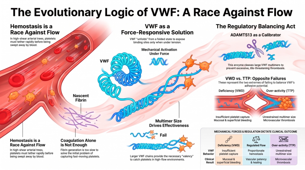
Why this spoke matters
Most discussions of VWF begin in the present tense.
Platelet adhesion.
Factor VIII stabilization.
Multimers.
ADAMTS13.
Bleeding.
Thrombosis.
But a deeper question sits underneath all of these topics:
Why did vertebrate biology require such a molecule in the first place?
VWF is biologically unusual.
It is:
- enormous
- multimeric
- mechanically responsive
- regulated by force
- exquisitely dependent on vascular context
Most coagulation proteins function primarily through enzymatic amplification.
VWF behaves differently.
Its biology is intimately tied to flowing blood.
That architecture invites a broader evolutionary question.
What selective pressures might favor the emergence of a force-responsive adhesive system?
This essay approaches that question cautiously.
Evolutionary biology rarely allows direct historical reconstruction. We infer selective pressures from comparative biology, molecular structure, and present-day function rather than observing evolutionary transitions directly.
Still, the architecture of VWF strongly suggests that vertebrate hemostasis eventually faced a problem that coagulation alone could not fully solve:
how to tether platelets rapidly in fast-moving blood.
Before fast blood
The mechanical demands on hemostasis are different in slow-moving fluid systems.
When flow is sluggish, soluble proteins can diffuse locally, adhesive interactions have time to stabilize, and coagulation reactions can organize before mechanical forces disrupt them.
But vertebrate circulation gradually altered that environment.
Closed circulatory systems allowed blood to move under greater pressure and with greater directional control. Branching arterial trees distributed blood rapidly through increasingly specialized tissues. Small arteries and arterioles exposed blood components to rising shear forces.
In this setting, vascular injury created a new physical challenge.
Platelets could no longer rely solely on passive contact with injured surfaces.
They had to tether rapidly before flowing blood swept them away.
Hemostasis became, in part, a race against flow.
That shift may help explain why vertebrate hemostasis evolved increasingly sophisticated mechanisms for platelet capture under force.1
Why coagulation alone may not have been enough
Coagulation stabilizes hemostasis.
But fibrin generation is not ideally suited to solving the earliest problem created by rapid flow:
capturing platelets quickly enough to establish a stable surface reaction.
In fast-flowing blood, the first challenge is mechanical.
A platelet must slow down.
Tether.
Resist detachment.
Only then can firmer adhesive interactions and downstream coagulation proceed efficiently.
This challenge is especially relevant in:
- small arteries and arterioles
- branching vascular beds
- stenotic regions
- mucosal microvasculature
- disturbed-flow environments
Under such conditions, weak adhesive interactions may fail before stable clot formation can begin.
VWF appears well suited to this problem.
Not because it simply “makes platelets sticky,” but because its multimeric structure allows platelet tethering under conditions in which ordinary adhesion would be mechanically fragile.2
From this perspective, VWF can be understood as part of a broader evolutionary transition in which hemostasis increasingly became a problem of adhesion under force.
The emergence of mechanical responsiveness
One of the most distinctive features of VWF is that it is not constitutively maximally active.
Its behavior changes under force.
That property is central to the molecule’s biologic logic.
Circulating VWF is generally autoinhibited. But when VWF becomes tethered or immobilized under flow, tensile forces can promote conformational changes that expose platelet-binding and proteolytic domains.34
This creates an elegant biologic compromise.
A permanently hyperadhesive molecule would create intolerable thrombosis.
An entirely inert adhesive molecule would fail under rapid flow.
VWF occupies the middle ground.
Its adhesive function increases preferentially in the very environments where rapid platelet capture becomes most important.
That does not mean VWF literally “senses” the circulation in an intentional way.
But it does mean that its biologic behavior depends on force, flow, tethering state, and vascular geometry.
The molecule is mechanically responsive in a highly useful physiologic sense.5
Why multimerization matters
VWF is not a small adhesive ligand.
It is assembled into multimers of striking size heterogeneity.
That organization appears highly relevant to the problem of platelet tethering under flow.
Large and high-molecular-weight multimers provide greater adhesive valency and are generally the most effective forms of VWF for platelet-dependent hemostasis under shear conditions.67
This helps explain why:
- loss of high-molecular-weight multimers impairs hemostasis
- ultra-large multimers can become excessively thrombogenic
- ADAMTS13 regulation becomes necessary
- endothelial control of release matters biologically
Multimerization therefore is not simply decorative molecular complexity.
It is directly related to the challenge of maintaining platelet adhesion in flowing blood.
Comparative biology and evolutionary interpretation
Comparative biology supports the idea that VWF acquired increasing structural complexity during vertebrate evolution.
Hagfish VWF appears structurally simpler than mammalian VWF and lacks the A3 collagen-binding domain important for platelet adhesion under high-shear conditions.8
Importantly, the claim here is not that evolution had a single explicit goal.
Evolution does not plan ahead.
Nor can current evidence reconstruct a precise historical sequence explaining exactly why each structural feature emerged.
But comparative biology does support a more modest and defensible interpretation:
as vertebrate circulatory systems became more specialized and mechanically demanding, increasing VWF complexity became increasingly compatible with the hemostatic challenges imposed by rapid flow.
That framing preserves the evolutionary insight without forcing the biology into an overly teleologic story.
The price of such a system
Every adaptation creates new vulnerabilities.
The same properties that make VWF effective under force also create thrombotic risk.
Large multimers can become excessively adhesive.
Force-dependent conformational change can amplify platelet tethering.
Newly released ultra-large multimers are particularly thrombogenic if not properly regulated.9
This helps explain why vertebrate hemostasis also required regulatory counterbalances.
ADAMTS13 limits excessive VWF multimer size and restrains excessive shear-dependent VWF activity by cleaving unfolded VWF within the A2 domain.1011
Endothelial cells regulate VWF release through storage within Weibel–Palade bodies.12
The result is not merely a single adhesive protein.
It is an integrated regulatory system balancing:
- platelet recruitment
- bleeding prevention
- vascular patency
- thrombosis avoidance
- mechanical responsiveness
What VWD reveals
Von Willebrand disease reveals something fundamental about vertebrate hemostasis.
Without VWF, the vulnerabilities of rapid-flow circulation become visible.
Bleeding occurs disproportionately at surfaces where platelet tethering must happen quickly and repeatedly:
- mucosal surfaces
- superficial vascular beds
- sites of recurrent minor injury
- procedural surfaces
This pattern is not arbitrary.
It reflects the importance of VWF in primary hemostasis under mechanically demanding conditions.13
In this sense, VWD does more than identify deficiency of a plasma protein.
It exposes a normally hidden feature of vertebrate circulation itself:
coagulation alone is not enough.
Fast-moving blood required force-tolerant platelet capture.
Clinical synthesis
An evolutionary framework does not replace mechanistic hematology.
It organizes it.
From this perspective:
- high-shear circulation created new mechanical demands for hemostasis
- platelet capture became a problem of adhesion under force
- VWF emerged as a multimeric system specialized for these conditions
- mechanical responsiveness reduced constitutive thrombosis
- multimerization increased adhesive effectiveness under flow
- ADAMTS13 became necessary to regulate excessive shear-dependent VWF activity
- VWD exposes the dependence of vertebrate hemostasis on rapid platelet tethering
At the bedside, this framework encourages clinicians to think not only about coagulation proteins, but also about the physical environment in which hemostasis occurs.
Where is blood moving rapidly?
Where are local flow conditions mechanically stressful?
Where must platelets tether before coagulation can stabilize injury?
Those questions help explain why VWD produces the phenotype that it does.
And they help reveal why vertebrate circulation may ultimately have required a molecule like VWF at all.
Evidence anchor: why VWF is specialized for hemostasis under flow
Summary derived from mechanobiology studies, structural reviews, comparative biology, and clinical stress-test observations. The evidence supports the central concept that VWF is not passive glue, but a force-sensitive adhesive system whose activity depends on multimer size, tethering, shear, and regulation by ADAMTS13.
| Evidence stream | What it shows | Why it matters | Main limitation |
|---|---|---|---|
| VWF under force | VWF function changes under mechanical force. Tethering, collagen binding, and multimer extension expose adhesive functions that support platelet capture under flow.14 | Flow is not just a threat to hemostasis. It is part of the signal that activates VWF at the right place. | Free-flow unfolding should not be overstated; robust activation generally depends on immobilization, tethering, or force transmitted through the multimer. |
| Multimer size | Larger VWF multimers are more effective for platelet adhesion and aggregation under shear because they provide multivalent binding capacity and greater force responsiveness.15 | Size is not incidental. It is part of VWF function. | Larger multimers are useful only when appropriately regulated. |
| ADAMTS13 regulation | ADAMTS13 cleaves VWF when force exposes the A2 domain, limiting multimer size and adhesive potential.16 | The same force-responsive system that permits platelet capture also exposes VWF to restraint. | In vivo ADAMTS13 regulation is more complex than simple cleavage-on-demand. |
| Comparative biology | Hagfish VWF suggests that VWF arose early in vertebrate evolution and was structurally simpler than mammalian VWF, notably lacking the A3 collagen-binding domain.17 | Modern VWF can be understood as a layered vertebrate system, not a molecule that appeared fully formed. | Comparative data support evolutionary interpretation, but do not prove every ancestral function. |
| Clinical stress tests | Severe aortic stenosis can produce acquired loss of high-molecular-weight VWF multimers and bleeding, demonstrating the sensitivity of VWF biology to pathologic shear.18 | Disease reveals the design: VWF works because it is mechanically responsive, but that same responsiveness can fail under abnormal flow. | Aortic stenosis is a clinical model, not an evolutionary experiment. |
Interpretive note: These evidence streams point in the same direction: VWF is best understood as a conditional adhesive system. Its function depends on where it is, how large it is, whether it is tethered, what force it experiences, and whether ADAMTS13 restrains it. The evolutionary lesson is not that VWF is simply “sticky,” but that it is sticky in context.
Reflect & Apply Case
A resident asks why von Willebrand disease predominantly causes:
- epistaxis
- heavy menstrual bleeding
- bleeding from superficial wounds
- mucosal bleeding
rather than the deep tissue bleeding pattern typical of severe hemophilia.
A useful starting point is not simply factor nomenclature.
It is vascular mechanics.
Where in the circulation must platelets tether rapidly under flow?
Where are vascular injuries repetitive, superficial, and mechanically exposed?
Where does early platelet capture matter before fibrin formation can fully stabilize the developing clot?
Viewed through this lens, VWD reveals more than a bleeding disorder.
It reveals the evolutionary logic of primary hemostasis itself.
Test your thinking
An interactive quiz for this topic is being developed and will be added soon.