
When quantity is low but biology is variable
Note: The video and audio linked above were generated with the assistance of AI. Clinical accuracy has been reviewed, but no AI-generated content can be guaranteed to be fully error-free.
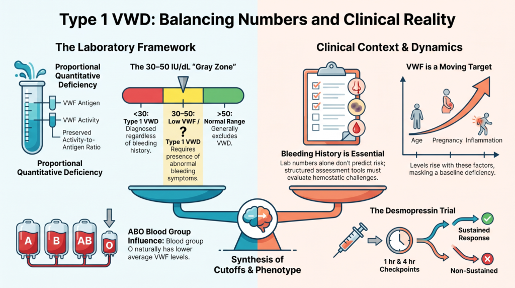
Why this spoke matters
Type 1 VWD sounds simple: there is less von Willebrand factor, the VWF that is present is mostly functional, VWF antigen and platelet-dependent activity are reduced in parallel, and multimers are usually present. That sounds straightforward, but type 1 VWD is where some of the hardest diagnostic questions in VWD live.
How low is low enough? When is low VWF a disease, and when is it a bleeding risk factor? Why do some patients with similar VWF levels bleed differently? Why do levels change with age? Why can a person have normal VWF today and still have a meaningful bleeding history?
Type 1 VWD is the commonest form of VWD, accounting for most diagnosed cases, but it is also the subtype that most clearly teaches that VWD is not diagnosed by a number alone.1
The basic definition
Type 1 VWD is a partial quantitative deficiency of VWF. “Partial” means VWF is reduced, not absent. “Quantitative” means the main problem is amount, not intrinsic function.
In typical type 1 VWD:
- VWF antigen is low.
- Platelet-dependent VWF activity is low.
- The activity-to-antigen ratio is usually preserved.
- FVIII may be normal or mildly reduced because VWF stabilizes FVIII.
- The multimer pattern is usually normal in distribution, although reduced in intensity.
This distinguishes type 1 from type 2 VWD, where VWF function is disproportionately abnormal, and from type 3 VWD, where VWF is absent or nearly absent.2 That definition is necessary, but it is not sufficient.
Type 1 is a quantitative category, not a single biology
Type 1 VWD is often spoken of as if it were one disorder. It is better understood as a category: a shared laboratory pattern that can arise from more than one biological mechanism.
Some patients make less VWF. Some secrete VWF less efficiently. Some clear VWF faster. Some have pathogenic VWF variants, while others have no identifiable VWF coding variant. Some have levels shaped strongly by ABO blood group and other modifier loci. Some have low VWF plus another bleeding modifier, such as a platelet, gynecologic, vascular, or fibrinolytic contributor.
This heterogeneity explains why type 1 VWD is clinically variable.3 The common laboratory pattern is quantitative deficiency; the underlying biology is not always the same.
The threshold problem
VWF levels are continuously distributed across the population. There is no natural biological cliff at 30 IU/dL or 50 IU/dL. Thresholds are necessary for diagnosis, communication, research, and access to care, but thresholds are tools. They are not the disease itself.
Historically, many frameworks distinguished:
- VWF below 30 IU/dL: more clearly type 1 VWD.
- VWF 30 to 50 IU/dL: often described as “low VWF,” especially when bleeding symptoms were present.
This distinction remains biologically useful. Patients with VWF levels below 30 IU/dL are more likely to have VWF-linked disease biology, identifiable VWF variants, familial segregation, and a clearer inverse relationship between residual VWF level and bleeding.4
Patients with VWF levels between 30 and 50 IU/dL are more heterogeneous. Some bleed significantly, some do not, and some have bleeding that is partly VWF-dependent but also influenced by other hemostatic, gynecologic, vascular, or local factors.
The 2021 ASH/ISTH/NHF/WFH diagnostic guideline changed the clinical framing by recommending type 1 VWD for patients with VWF levels below 30 IU/dL regardless of bleeding, and for patients with abnormal bleeding and VWF levels below 50 IU/dL.5 That guideline choice can improve access to care, but it does not eliminate the biologic heterogeneity of the 30 to 50 IU/dL group.
Low VWF and type 1: why the debate persists
The debate over “low VWF” is not semantic. It reflects real uncertainty. One concern is underdiagnosis: patients, especially women with heavy menstrual bleeding or postpartum bleeding, may experience long delays before receiving a diagnosis and a usable treatment plan. Another concern is overdiagnosis: mild mucocutaneous bleeding is common in the general population, and by definition a fraction of healthy people will have VWF levels below the lower limit of the reference range.6
For patients with bleeding and VWF in the 30 to 50 IU/dL range, the practical question is not simply whether this is “really” disease. The better question is whether this person has a reproducible hemostatic vulnerability that should change care. The label should serve that question.
Bleeding history is not optional
Type 1 VWD should not be reduced to laboratory values. Bleeding history is central, and typical symptoms include:
- epistaxis
- easy bruising
- oral cavity bleeding
- bleeding from minor wounds
- heavy menstrual bleeding
- post-dental bleeding
- postoperative bleeding
- postpartum bleeding
Bleeding assessment tools help structure the history and reduce subjectivity. They are especially useful in low-pretest-probability settings to decide who needs specific VWD testing, but they do not determine VWF causality.7
A low BAT score can be misleading in patients with limited hemostatic challenges, especially children. Adults may have accumulated higher scores because they have had more opportunities to bleed, and prior treatment can influence the score. Menstrual bleeding may be normalized within families. A score is a structured history, not a substitute for interpretation.
The key exposure question is: what hemostatic challenges has the patient actually faced?
Family history helps, but imperfectly
Family history can support type 1 VWD, but it can also mislead. Type 1 VWD often shows autosomal dominant inheritance with variable penetrance. A parent may carry a VWF variant but have few symptoms, and family members may share low VWF levels without sharing identical bleeding risk.
A family history of heavy menstrual bleeding may be highly relevant, but it may also be underreported because symptoms are normalized. Conversely, a relative’s bleeding may reflect anticoagulation, antiplatelet therapy, fibroids, childbirth complications, or another disorder.
The most useful family history asks:
- who bled?
- how did they bleed?
- what hemostatic challenges occurred?
- were VWF levels measured?
- was the diagnosis confirmed?
- did treatment work?
Family history is evidence. It is not a shortcut.
Why similar levels do not mean similar bleeding
Two patients can have a VWF activity of 38 IU/dL and very different lives. One may have little bleeding. Another may have severe heavy menstrual bleeding, iron deficiency, delayed post-dental bleeding, and postpartum hemorrhage.
This mismatch occurs because bleeding is not determined by VWF alone. Bleeding reflects:
- VWF level
- FVIII level
- VWF clearance
- blood group
- platelet function
- vascular integrity
- fibrinolysis
- hormonal and gynecologic factors
- local anatomy
- medications
- procedural exposure
- pregnancy and postpartum physiology
- inflammation
- comorbid disease
This is why type 1 VWD is not a simple linear scale. VWF level matters, but it does not carry the whole phenotype.8
ABO, modifiers, and moving targets
ABO blood group is one of the best-known modifiers of VWF levels. People with blood group O have lower VWF levels on average than non-O individuals, which can move some patients into the low or borderline range.9 But ABO does not “explain away” bleeding. It helps interpret the level; it does not replace the diagnosis. ABO-specific reference ranges are not a substitute for clinical interpretation.10
VWF is also an acute-phase reactant. Levels may increase with stress, inflammation, exercise, pregnancy, aging, estrogen exposure, malignancy, and other physiologic states. A patient may have low VWF at baseline but appear normal during inflammation, pregnancy, acute bleeding, or later life. Conversely, a single borderline low value in a person without bleeding symptoms may not prove disease.
This is why repeat testing is not a nuisance; it is part of the biology. Diagnosis should generally be based on repeat testing on two separate occasions, ideally when the patient is at baseline health and not during acute inflammation, exercise, pregnancy, or the postoperative period.11
Laboratory pattern in type 1
The laboratory pattern of type 1 VWD is relational. The core pattern is proportional reduction:
- VWF antigen is reduced.
- Platelet-dependent VWF activity is reduced.
- The activity-to-antigen ratio is usually preserved.
- FVIII may be normal or mildly reduced in typical type 1.
- Multimers are usually normal in distribution.
The key point is proportionality. Disproportionate activity reduction relative to antigen should prompt evaluation for type 2 VWD, using assay- and laboratory-specific cutoffs. Disproportionately low FVIII should prompt consideration of type 2N VWD or hemophilia A. Nearly absent VWF raises the distinction between severe type 1 and type 3 VWD. Thrombocytopenia or enhanced ristocetin-induced platelet agglutination raises concern for type 2B VWD or platelet-type VWD.12
Type 1 VWD is not simply “low VWF.” It is low VWF in the right pattern and the right clinical context.
Why screening tests can mislead
Routine hemostasis tests may be normal in type 1 VWD. A normal platelet count does not exclude VWD. A normal PT is expected. A normal aPTT does not exclude VWD because FVIII may be normal or only mildly reduced. PFA-100/200 closure time may be abnormal, but it may also be normal in mild type 1 VWD or low VWF. The bleeding time should not be used.13
Specific testing is required when the clinical question is VWD:
- VWF antigen
- platelet-dependent VWF activity
- FVIII activity
Additional tests, such as VWF collagen binding, multimer analysis, RIPA, FVIII-binding assay, or genetic testing, are used when the initial pattern suggests a qualitative subtype or another disorder.
Assay evolution and preanalytical risk
Historically, VWF platelet-binding activity was measured by the ristocetin cofactor assay. That assay is variable, depends on ristocetin, and can be affected by VWF sequence variants that impair ristocetin-mediated binding without causing an equivalent defect in VWF function in vivo.
Newer assays, including VWF:GPIbR and VWF:GPIbM, improve precision and avoid some limitations of the classic ristocetin cofactor assay. The 2021 diagnostic guideline suggests newer platelet-binding activity assays over VWF:RCo when available.14 Even better assays, however, do not eliminate the need for interpretation; no static assay fully reproduces VWF function under flow at a site of vascular injury.
Preanalytical error can also create or hide type 1 VWD. Samples collected, transported, stored, frozen, thawed, or processed incorrectly can distort results. Cold transport of whole blood can reduce VWF and FVIII activity and may lead to misclassification. The 2024 BSH laboratory guideline emphasizes baseline-state testing, ambient-temperature transport, prompt processing, and interpretation by laboratories with appropriate expertise.15
A surprising result should not be treated as a verdict. It should be checked against the clinical setting, the sample pathway, and prior results.
Type 1C: when the problem is clearance
Some patients with type 1 VWD make and release VWF but clear it unusually fast. This increased-clearance phenotype is often called type 1C VWD. The classic example is Vicenza-type VWD, but increased clearance is not limited to one variant.
These patients may have a brisk initial rise after desmopressin followed by a rapid fall. That matters clinically because a patient who “responds” at 1 hour may not be protected several hours later. The 2021 diagnostic guideline suggests using a desmopressin trial with 1-hour and 4-hour post-infusion testing to confirm increased clearance, rather than relying only on the VWF propeptide-to-antigen ratio.16
Type 1C teaches a central principle: the peak level is not the whole response. Duration matters.
Genetic testing: helpful, but not usually the first answer
Genetic testing is not routinely required for most type 1 VWD diagnoses. It is more useful for selected problems:
- confirming type 2 subtypes
- distinguishing type 2N VWD from mild hemophilia A
- distinguishing type 2B VWD from platelet-type VWD
- characterizing type 3 VWD for family counseling
- occasionally helping distinguish congenital VWD from acquired von Willebrand syndrome
For type 1 VWD and low VWF, genetic testing is more difficult. Candidate VWF variants are found more often when VWF levels are below 30 IU/dL and less often in the 30 to 50 IU/dL range. Even when variants are found, pathogenicity may be uncertain. Low VWF biology is often polygenic and modified by ABO and other loci.17
A negative genetic test does not exclude type 1 VWD, and a variant of uncertain significance does not prove it.
Pregnancy, aging, and reassessment
Type 1 VWD often improves numerically during pregnancy. VWF and FVIII may rise substantially, especially later in gestation, which may reduce delivery bleeding risk in some patients and allow neuraxial anesthesia when levels are adequate. Pregnancy normalization does not cure type 1 VWD, however. After delivery, VWF and FVIII fall toward baseline, and postpartum hemorrhage risk may persist, especially when the bleeding history is significant.
The 2021 management guideline suggests checking VWF and FVIII in the third trimester, planning delivery and anesthesia prospectively, and using tranexamic acid postpartum in women with type 1 VWD or low VWF levels.18 The patient may look “normal” late in pregnancy and vulnerable postpartum. The timeline matters.
VWF levels also often rise with age. Some people diagnosed with type 1 VWD earlier in life later have VWF levels in the normal range. If the original diagnosis was weak, based on borderline labs during childhood and minimal bleeding, reassessment may be appropriate. If the patient had a strong bleeding phenotype, reproducibly low historical levels, and treatment-responsive bleeding, normalization with age should not automatically erase risk.
The 2021 diagnostic guideline suggests reconsidering rather than simply removing the diagnosis in patients with previously confirmed type 1 VWD whose VWF levels normalize with age.19 Recent discussions of “undiagnosing” VWD emphasize that reassessment should be thoughtful, centered on bleeding history, repeated laboratory testing, and the clinical consequences of removing the label.20
A diagnosis should not be eternal by inertia, but it should not be removed just because the number improved.
Treatment logic in type 1 VWD
Treatment is not based on the subtype name alone. It is based on:
- bleeding phenotype
- baseline VWF and FVIII levels
- prior response to treatment
- procedure or bleeding site
- urgency
- age and comorbidities
- pregnancy or postpartum status
- contraindications to desmopressin
- availability of monitoring
For many patients with type 1 VWD, the main treatment options are:
- local measures
- tranexamic acid
- hormonal therapy for heavy menstrual bleeding
- desmopressin
- VWF concentrate when needed
Desmopressin is particularly useful in many patients with type 1 VWD because it releases endogenous VWF and FVIII from endothelial stores. But it is not automatically appropriate. It should be avoided or used cautiously in patients at risk for hyponatremia, very young children, older patients with vascular disease, patients with seizure disorders, and patients with cardiovascular disease. It is not appropriate as sole therapy for major surgery at critical sites, and it may be ineffective or short-lived in increased-clearance phenotypes.21
A desmopressin trial tells you whether the patient can release enough VWF and FVIII, and whether the response lasts.
Procedures: match the plan to the challenge
Minor mucosal procedures are not the same as major surgery. A dental extraction is not the same as neurosurgery, and a skin biopsy is not the same as tonsillectomy.
For selected patients with type 1 VWD, baseline VWF activity above 30 IU/dL, and a mild bleeding phenotype, tranexamic acid alone may be reasonable for minor mucosal procedures. For higher-risk procedures, therapy may need to raise VWF activity to hemostatic levels with desmopressin or VWF concentrate, often with tranexamic acid as an adjunct. For major surgery, guidelines generally emphasize maintaining both VWF activity and FVIII at hemostatic levels for an appropriate postoperative period.22
The question is not simply whether the patient has type 1 VWD. The question is what hemostatic support this specific challenge requires.
Clinical synthesis
Type 1 VWD is a partial quantitative deficiency of VWF, but the word “partial” carries the lesson. Partial deficiency. Partial penetrance. Partial correlation between level and bleeding. Partial stability over time. Partial answers from laboratory testing.
The diagnosis is strongest when the laboratory pattern, bleeding history, family history, hemostatic exposure, and treatment response point in the same direction. It is weakest when a single borderline VWF level is asked to carry the whole explanation.
Type 1 VWD is therefore best understood as a relational diagnosis. The key question is not simply how low the VWF level is, but also:
- is the reduction reproducible?
- are antigen and activity reduced proportionally?
- has the patient had meaningful bleeding challenges?
- does the bleeding phenotype fit?
- are physiologic modifiers influencing the level?
- would the diagnosis change care?
The best diagnosis of type 1 VWD is not the one that worships a threshold. It is the one that helps the patient bleed less.
Evidence anchor: why type 1 VWD lives at the border of numbers and bleeding
| Evidence stream | What it shows | Why it matters | Main limitation |
|---|---|---|---|
| Population distribution of VWF | VWF levels vary continuously across the population, without a natural biological cliff at 30 or 50 IU/dL.23 | Diagnostic thresholds are useful tools, not biological boundaries. | Borderline levels may reflect disease biology, physiologic variation, or bleeding risk modification. |
| Type 1 cohort studies | Patients with VWF below 30 IU/dL are more likely to have identifiable VWF variants, familial segregation, and clearer VWF-linked disease biology; patients with VWF 30–50 IU/dL are more heterogeneous.24 | The same laboratory range may contain different biological and clinical states. | Referral cohorts may enrich for symptomatic patients. |
| Bleeding assessment studies | Bleeding assessment tools help standardize history but are influenced by age, sex, hemostatic exposure, treatment history, and symptom type.25 | Bleeding history is essential, but it must be interpreted rather than simply scored. | A bleeding score cannot prove VWF causality or future procedural risk by itself. |
| Laboratory and preanalytical evidence | VWF levels are affected by acute-phase responses, pregnancy, exercise, aging, inflammation, stress, sample handling, and assay variability.26 | Repeat testing at baseline health and proper sample handling are part of diagnosis, not bureaucratic repetition. | Even repeated results require clinical interpretation. |
| Desmopressin and treatment response | Many patients with type 1 VWD respond to desmopressin, but magnitude and duration vary, especially in increased-clearance phenotypes.27 | A treatment trial can clarify whether endogenous VWF release is adequate and sustained for a planned challenge. | A good peak response does not always mean durable protection. |
Interpretive note: Type 1 VWD is not diagnosed by a number alone. The evidence supports a threshold-informed but context-dependent approach that integrates VWF level, bleeding history, assay pattern, physiologic state, and treatment implications.
Guideline perspective: how to use the type 1 label
Guidelines help structure the diagnosis of type 1 VWD, but they do not remove the need for judgment. The 2021 ASH/ISTH/NHF/WFH diagnostic guideline recommends diagnosing type 1 VWD in patients with VWF below 30 IU/dL regardless of bleeding, and in patients with abnormal bleeding and VWF below 50 IU/dL.28
That recommendation is clinically important because a diagnosis can improve access to education, treatment, procedural planning, reproductive care, and emergency planning. But it should not be interpreted as saying that patients with VWF levels of 30–50 IU/dL do not all have the same biology, bleeding risk, or treatment need.
For type 1 VWD, practical guidance is to:
- interpret VWF antigen, platelet-dependent activity, and FVIII together
- look for proportional reduction of antigen and activity
- repeat testing when results are borderline or obtained during non-baseline physiologic states
- interpret bleeding scores in light of age, sex, treatment history, and hemostatic exposure
- consider modifiers such as blood group, inflammation, pregnancy, aging, medications, and comorbid disease
- use desmopressin trials when response and duration will affect management
- match treatment to the hemostatic challenge, not simply to the diagnostic label
- avoid removing a well-supported diagnosis solely because VWF normalizes with age
The useful type 1 diagnosis is transparent about uncertainty. A practical formulation might be:
“This patient has type 1 VWD by current criteria, with VWF levels in the 30–50 IU/dL range and clinically significant mucocutaneous bleeding. The biology may be heterogeneous; management should be based on bleeding phenotype, repeated levels, prior treatment response, and procedural risk.”
The practical lesson is simple: thresholds help name the problem, but care is guided by pattern, phenotype, and consequence.
Reflect & Apply Case
An older adult carries a childhood diagnosis of type 1 VWD. Current VWF antigen and activity are normal, and the patient asks whether the diagnosis still applies.
A useful reassessment would ask:
- Was the original diagnosis based on reproducibly low VWF levels?
- Was there a convincing mucocutaneous bleeding phenotype?
- Were the original samples collected at baseline health?
- Has the patient had major hemostatic challenges, and what happened?
- Did desmopressin or VWF-directed therapy prevent or treat bleeding?
- Would removing the diagnosis improve care, or could it remove needed procedural protection?
The answer may be “the diagnosis should be reconsidered,” but not automatically “the diagnosis is gone.” In type 1 VWD, the current level is important, but the patient’s longitudinal bleeding phenotype is also evidence.
Test your thinking
A short quiz on type 1 VWD.