
Preparing for predictable bleeding
Note: The video and audio linked above were generated with the assistance of AI. Clinical accuracy has been reviewed, but no AI-generated content can be guaranteed to be fully error-free.
Why this spoke matters
Surgery is where uncertainty becomes practical.
And consequential.
A patient may live for years with mild symptoms. Then a dentist, surgeon, anesthesiologist, obstetrician, or proceduralist asks a simple question:
Can we do this safely?
In VWD, that question cannot be answered by diagnosis alone.
“Type 1 VWD” is not a perioperative plan.
“Low VWF” is not a perioperative plan.
“Responds to desmopressin” is not always a perioperative plan.
The procedure matters. The site matters. The duration of bleeding risk matters. The patient’s prior bleeding matters. The VWD mechanism matters. The available monitoring matters.
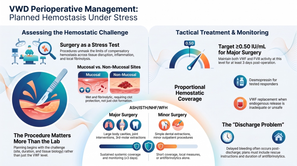
Surgery and procedures should therefore be managed as hemostatic stress tests that can be anticipated, modeled, and covered.
The goal is not to react to bleeding.
The goal is planned hemostasis under stress.
Surgery unmasks hemostatic reserve
Surgery does not create VWD.
It exposes the limits of compensatory hemostasis.
A patient may live for years with mild epistaxis, bruising, heavy menstrual bleeding, prolonged bleeding after minor cuts, or a family history that normalized bleeding symptoms without ever encountering a hemostatic challenge large enough to reveal the defect clearly.
Then surgery changes the equation.
Now the body must maintain hemostasis across exposed microvasculature, sustained tissue disruption, inflammation, local fibrinolysis, repetitive mechanical stress, and postoperative wound instability.
Daily life may be forgiving.
Surgery is not.
This is why peri-procedural bleeding can become the moment the disorder declares itself clinically, even if the biology had been present all along.1
The first question: what kind of challenge is this?
The first peri-procedural question is not:
What is the VWF level?
It is:
What hemostatic challenge are we trying to cover?
Procedures differ enormously. A superficial skin biopsy is not a tonsillectomy. A colonoscopy without intervention is not polypectomy. A procedure in a compressible site is not the same as a procedure in a critical space.
Perioperative planning begins with the challenge, not the laboratory value.
The ASH/ISTH/NHF/WFH management guideline emphasizes that individualized therapy plans should consider the bleeding risk of the specific procedure, and explicitly notes the risk of overtreating some low-risk procedures if every patient is pushed to the same laboratory target plus tranexamic acid.2
That is the central principle.
Procedural planning begins with the procedure.
Major versus minor is useful, but not enough
Guidelines often distinguish major from minor surgery.
That distinction helps. Major surgery generally requires sustained systemic hemostatic coverage and monitoring. Minor procedures may be managed with shorter coverage, adjunctive therapy, local measures, or sometimes antifibrinolytics alone in selected patients.
But “major” and “minor” are often misleading categories if used alone.
The ASH/ISTH/NHF/WFH guideline considered major surgery to include procedures requiring opening of large body cavities, procedures where severe hemorrhage is possible, interventions involving joints, third-molar extractions, and interventions where the patient’s life is at risk. Minor surgery included simple dental extractions and other outpatient procedures not otherwise specified as major.3
Simple dental extraction may be minor. Impacted third-molar extraction can carry higher bleeding risk and was treated as major surgery in the ASH evidence framework.4
That classification is useful.
But expert planning still asks:
- is the site mucosal?
- is bleeding compressible?
- is delayed bleeding common?
- is the wound surface large?
- will fibrinolysis be prominent?
- can local measures be applied?
- will the patient need postoperative anticoagulation or VTE prophylaxis?
- can VWF and FVIII be monitored quickly?
The label “minor” does not automatically mean “low risk.”
The label “major” does not automatically dictate one protocol.
Different procedures create different hemostatic demands
Not all procedures stress the hemostatic system in the same way.
Bleeding risk is not determined solely by operative size or technical complexity.
It is shaped by tissue biology.
Mucosal procedures such as tonsillectomy, dental extraction, sinus surgery, gynecologic procedures, childbirth, and endoscopy with intervention create a distinctive problem. These surfaces are wet, mobile, exposed, and fibrinolytic. A clot may form, then dissolve or dislodge before healing is complete.
Orthopedic surgery creates a different challenge. It may involve extensive tissue dissection and substantial blood loss, but the local fibrinolytic environment differs from the oral cavity, uterus, or tonsillar bed. In severe VWD, rehabilitation and early postoperative movement may extend the period of hemostatic vulnerability.
Cardiac surgery and mechanical circulatory support create yet another problem. Severe aortic stenosis, ventricular assist devices, extracorporeal circuits, and cardiopulmonary bypass may alter VWF biology itself through high shear, causing loss of high-molecular-weight multimers and an acquired VWD-like state.5
The point is not to memorize a procedure list.
The point is to ask what kind of tissue environment the clot must survive.
Treat the patient, not just the operation
Procedure risk must be integrated with patient risk.
The patient-level assessment can be grouped into three domains.
Hemostatic biology
- VWD type or best working diagnosis
- baseline platelet-dependent VWF activity, VWF antigen (VWF:Ag), and factor VIII activity (FVIII:C)
- quantitative versus qualitative defect
- documented desmopressin response
- durability of desmopressin response
- desmopressin contraindications
- likely need for VWF replacement
- history of inhibitor or alloantibody
Bleeding history
- prior procedure-related bleeding
- prior similar procedures without bleeding
- delayed bleeding after discharge
- transfusion, packing, cautery, reoperation, or factor treatment
- response to prior antifibrinolytics or local measures
Modifiers and competing risks
- antiplatelet or anticoagulant therapy
- NSAID use
- SSRI or SNRI use
- supplements that may affect bleeding
- perioperative VTE prophylaxis needs
- thrombotic risk factors
- pregnancy or postpartum state
- renal disease, liver disease, anemia, thrombocytopenia, or another hemostatic modifier
The procedure creates the challenge.
The patient determines the reserve.
The plan must account for both.
Prior bleeding history matters
A previous surgical history can be highly informative.
But it must be interpreted carefully.
No bleeding after a minor procedure does not guarantee safety for major surgery. Bleeding after a poorly controlled dental extraction does not necessarily predict uncontrolled bleeding with a better plan.
A prior transfusion, reoperation, delayed hemorrhage, or prolonged oozing carries more weight than vague “easy bleeding.”
The most useful procedural history asks:
- what procedure was done
- what hemostatic support was used
- whether antifibrinolytics were given
- whether local measures were used
- when bleeding occurred
- how long it lasted
- whether transfusion, reoperation, packing, cautery, or factor treatment was needed
Bleeding after an unplanned procedure is less informative than bleeding despite a carefully designed hemostatic plan.
The question is not simply:
Did the patient bleed?
It is:
What does the prior event teach us about the next challenge?
The role of desmopressin
Desmopressin may be useful for selected procedures in responsive patients.
It is best suited when the patient has a known adequate response, the response is durable enough for the planned challenge, the procedure is short or limited, the site is not critical, there is no contraindication, fluid restriction and sodium-risk mitigation are feasible, and adjunctive therapy can be added when useful.
But desmopressin has limits.
Patients with type 3 VWD should not receive it as effective therapy because there is little or no VWF reserve to release. Patients with type 2B VWD generally should not receive it because of increased platelet binding and risk of thrombocytopenia. Many patients with type 2 VWD require VWF concentrate rather than desmopressin.
Major surgery, especially at critical sites or when sustained coverage is required, generally should not rely on desmopressin as sole therapy.6
Desmopressin is not a procedural shortcut.
It is a tool only when response, duration, safety, and procedure risk align.
The role of VWF replacement
VWF replacement is needed when endogenous release is absent, inadequate, unsafe, or too short-lived.
It is often required for type 3 VWD, many type 2 variants, major surgery, critical-site procedures, severe baseline deficiency, inadequate or short-lived desmopressin response, desmopressin contraindication, and high-risk procedures requiring sustained coverage.
Replacement therapy restores VWF function and often supports FVIII as well.
The choice of product and dosing depends on the patient, baseline FVIII, urgency, product availability, monitoring capacity, and thrombotic risk. For emergency treatment with VWF-only products, FVIII may be needed if baseline FVIII is low or unknown, because VWF-only therapy stabilizes endogenous FVIII over time rather than supplying immediate FVIII directly.7
This is why replacement is not merely “give factor.”
It is matching VWF and FVIII kinetics to the operative timeline.
Early intraoperative, early postoperative, delayed postoperative, and healing phases may require different levels or durations of support.
Major surgery: sustained coverage
For major surgery, ASH/ISTH/NHF/WFH suggests maintaining both VWF activity and FVIII activity at ≥0.50 IU/mL for at least 3 days, with longer coverage individualized by procedure, bleeding history, and postoperative risk. The guideline also suggests against targeting FVIII alone.8
The reason is mechanistic.
FVIII alone does not correct the primary VWF defect. VWF participates in platelet adhesion, vessel-wall interaction, and FVIII stabilization. Patients with type 2 or type 3 VWD may not achieve adequate hemostasis if only FVIII is maintained.
“At least 3 days” is a minimum duration, not a predefined endpoint.
Some procedures require longer coverage. Some require site-specific monitoring. Some require attention to delayed bleeding after discharge.
Minor procedures: avoid both under- and overtreatment
Minor procedures are where VWD management can become either too aggressive or too casual.
For minor surgery or minor invasive procedures, the 2021 guideline suggests increasing VWF activity to at least 0.50 IU/mL with desmopressin or factor concentrate plus tranexamic acid, rather than increasing VWF levels without tranexamic acid.9
But the guideline also suggests tranexamic acid alone over increasing VWF levels in selected patients with type 1 VWD, baseline VWF activity above 0.30 IU/mL, a mild bleeding phenotype, and minor mucosal procedures.10
This pair of recommendations captures the tension.
Some minor procedures need systemic support.
Some do not.
Tranexamic acid alone is a proportional option only in selected patients: type 1 VWD, baseline VWF activity above 0.30 IU/mL, mild bleeding phenotype, and a minor mucosal procedure.
A superficial skin biopsy in mild type 1 VWD may not require the same plan as a dental extraction in a patient with prior prolonged oozing.
The goal is proportional hemostatic coverage.
Mucosal procedures need clot protection
Many common procedures in VWD involve mucosal surfaces:
- dental extraction
- oral surgery
- tonsillectomy
- nasal procedures
- gynecologic procedures
- endoscopy with intervention
Mucosal sites are fibrinolytic and mechanically exposed.
A clot may form and then fail.
That is why antifibrinolytics are often central, not optional. In mucosal procedures, clot protection is as important as clot formation.
The guideline places high value on the combined effect of raising VWF and using tranexamic acid for minor surgery, given their different mechanisms of action and the generally favorable adverse-effect profile of tranexamic acid.11
Local measures also matter.
Dental proceduralists may use gelatin sponges, fibrin glue, tranexamic acid rinse, suturing, and other local strategies as part of an individualized plan.12
There is one important site-specific caution. Antifibrinolytics should be used cautiously, and often avoided, in gross hematuria or suspected renal or ureteral bleeding because clot retention or obstruction may occur.13
The procedural lesson is simple:
if the bleeding risk is local and mucosal, the plan must protect the clot locally.
Emergency procedures
Elective procedures allow time for classification, desmopressin trials, product selection, monitoring arrangements, and multidisciplinary planning.
Emergency procedures may not.
In urgent surgery, the priority is not perfect diagnostic precision. The priority is safe hemostatic coverage for the immediate challenge.
If VWD is known or strongly suspected and the procedure is high risk, clinicians may need to provide VWF-directed therapy before all details are clarified. Product choice should account for urgency, baseline or unknown FVIII level, and whether immediate FVIII correction is needed.
Do not wait for perfect classification if urgent hemostatic coverage is required.
But do document what was given, what levels were obtained, and what happened. Emergency procedures often become future evidence.
Monitoring: what are we trying to know?
Monitoring should answer a clinical question.
Monitoring without a decision linked to the result has limited value.
Before surgery, monitoring may ask whether baseline VWF and FVIII levels are adequate, whether desmopressin produced the expected response, and whether the planned replacement dose is sufficient.
During and after surgery, monitoring may ask:
- are VWF activity levels adequate?
- are FVIII levels adequate?
- is FVIII accumulating?
- is the patient bleeding?
- is the wound stable?
- is the treatment duration adequate?
- is thrombosis risk increasing?
Monitoring is especially important for major procedures, repeated replacement dosing, severe VWD, type 2 or type 3 disease, and high thrombotic-risk patients.
The guideline notes that local availability of rapid FVIII and VWF testing is critical for postoperative management, and that elective high-risk procedures may require alternate arrangements if real-time monitoring is not available.14
Monitoring literature similarly emphasizes that treatment monitoring should integrate VWF activity, FVIII activity, dosing timing, treatment goal, bleeding status, and clinical context rather than treating a laboratory value as the whole answer.15
This is a systems issue, not merely a lab issue.
A safe plan requires the ability to know whether the plan is working.
When monitoring is limited
Not every hospital can rapidly measure VWF activity.
Not every procedure can be delayed.
Not every patient can be moved.
In these settings, the plan must be adapted.
Options may include:
- performing a preoperative pharmacokinetic study when feasible
- using a center with VWD expertise for elective high-risk procedures
- choosing a product and dosing plan with predictable prior response
- arranging post-dose FVIII and VWF testing when possible
- using adjunctive tranexamic acid and local measures
- extending observation when delayed bleeding risk is meaningful
- creating clear discharge and rescue instructions
A plan that cannot be monitored should be more conservative when stakes are high.
Uncertainty should modify the plan.
It should not be ignored.
Thrombosis and excessive correction
VWD patients bleed.
But perioperative treatment can create thrombotic concern in selected settings.
Risk may increase with older age, immobility, cancer surgery, orthopedic surgery, cardiovascular disease, high FVIII levels, repeated VWF/FVIII concentrate dosing, extended high VWF and FVIII levels, and concomitant antifibrinolytic use in high-risk contexts.
The guideline notes that, in patients at higher risk of thrombosis, it may be desirable to avoid the combination of extended elevated VWF and FVIII levels above 1.50 IU/mL with extended tranexamic acid use.16
This does not mean withholding needed hemostasis.
It means calibrating hemostasis to avoid both bleeding and overcorrection.
The discharge problem
Procedural bleeding does not always happen in the operating room.
It may occur after discharge.
This is especially true for mucosal procedures, dental extraction, tonsillectomy, and postpartum-like tissue remodeling contexts.
Delayed bleeding is part of the procedural risk, not a complication outside the plan.
A procedural plan should include:
- duration of antifibrinolytic therapy
- what bleeding is expected
- what bleeding is abnormal
- who to call
- where to go
- whether the local emergency department has the plan
- when to repeat VWF or FVIII levels
- whether additional doses are planned
- whether delayed bleeding requires re-evaluation of the diagnosis or treatment response
A good perioperative plan survives the discharge summary.
Communication with proceduralists
VWD management often fails at the interface between hematology and the procedural team.
The surgeon may underestimate bleeding risk. The hematologist may underestimate local anatomy. The patient may receive conflicting instructions. The dentist may not know whether to use local hemostatic measures. The emergency department may not know what product is needed.
The plan should be written clearly.
It should include:
- diagnosis or working diagnosis
- VWD type, if known
- baseline VWF and FVIII
- desmopressin response, if known
- contraindications
- medication holds or cautions
- perioperative VTE prophylaxis considerations
- recommended pre-procedure therapy
- local measures
- post-procedure therapy
- monitoring plan
- rescue plan
- contact information
For VWD, communication is treatment.
The emotional meaning of procedural bleeding
For many patients, surgery is the moment the disease becomes psychologically real.
The postoperative hemorrhage.
The transfusion.
The return to the operating room.
The unexpected readmission.
The realization that prior bleeding symptoms were not “normal.”
These experiences often reorganize years of previously disconnected events: childhood nosebleeds, bruising, heavy menstrual bleeding, prolonged bleeding after dental work, or family stories that had been minimized or normalized.
The diagnosis retrospectively changes the meaning of the past.
That transition deserves recognition.
Especially because many patients with VWD have spent years being told that their bleeding was exaggerated, incidental, or unimportant.
Surgical bleeding therefore often functions not merely as a complication.
It becomes a moment of diagnostic revelation.
A practical planning sequence
A structured plan reduces variability and error.
A useful sequence is:
- define the procedure, site, and bleeding risk
- review VWD type or best working diagnosis
- review baseline VWF activity, VWF antigen, and FVIII
- review prior bleeding and prior procedural outcomes
- review medications, comorbidities, thrombosis risk, and VTE prophylaxis needs
- review desmopressin response and contraindications
- decide whether tranexamic acid and local measures are sufficient
- decide whether desmopressin or VWF replacement is needed
- set target levels, duration, and monitoring plan when systemic therapy is used
- plan discharge instructions, rescue therapy, and post-procedure reassessment
The final step matters.
Every procedure teaches something.
If bleeding occurred, why?
If bleeding did not occur, what prevented it?
Was the plan too much, too little, or just right?
Future procedural planning should learn from the event.
Clinical synthesis
Surgery and procedures are hemostatic stress tests.
They reveal whether baseline VWF biology, treatment response, local measures, monitoring systems, and procedural planning can support tissue injury and repair.
The key is anticipation.
Major surgery generally requires sustained systemic coverage, with both VWF activity and FVIII activity maintained at hemostatic levels for an appropriate duration.
Minor procedures require proportional planning, often combining tranexamic acid with desmopressin or replacement when needed, but sometimes using tranexamic acid alone in selected low-risk type 1 patients.
Mucosal procedures require special attention to clot protection and local hemostatic measures.
Desmopressin should be used only when appropriate, tested, safe, and sufficient for the challenge.
VWF replacement is needed when endogenous release cannot provide adequate or durable support.
Monitoring and communication are part of the intervention.
The goal is not to follow a fixed recipe.
The goal is to match the patient, the procedure, the mechanism, and the duration of risk.
Evidence anchor: why surgery is a hemostatic stress test
Summary derived from perioperative guidelines, VWD management guidance, monitoring reviews, orthopedic and cardiac surgery literature, and expert reviews. The evidence consistently shows that peri-procedural bleeding risk in VWD cannot be predicted from diagnosis, subtype, or VWF level alone. Surgery converts VWF biology, tissue environment, treatment response, monitoring capacity, and wound healing into a timed hemostatic challenge.
| Evidence stream | What it shows | Why it matters | Main limitation |
|---|---|---|---|
| Procedure-specific risk | VWD management guidance emphasizes individualized peri-procedural plans based on the bleeding risk of the specific procedure rather than diagnosis alone.17 | “Type 1 VWD,” “low VWF,” or “DDAVP responder” is not a perioperative plan. | Procedure categories are useful but cannot capture all local and patient-specific risks. |
| Major versus minor surgery | The ASH/ISTH/NHF/WFH framework treats major surgery as including large-cavity surgery, severe hemorrhage-risk procedures, joint interventions, third-molar extractions, and life-threatening sites; minor surgery includes simple dental extraction and many outpatient procedures.18 | Simple extraction and impacted third-molar extraction should not be treated as equivalent. “Minor” does not always mean low risk. | Major/minor labels still require refinement by site, delayed bleeding risk, and monitoring capacity. |
| Tissue biology | Mucosal, orthopedic, cardiac, and other procedures create different hemostatic environments, including variable fibrinolysis, mechanical disruption, shear, and postoperative instability.19 | Surgical bleeding risk is shaped by the tissue environment in which the clot must form and persist. | Evidence is procedure-specific and often based on observational experience or expert synthesis. |
| Major surgery targets | For major surgery, ASH/ISTH/NHF/WFH suggests maintaining both VWF activity and FVIII activity at ≥0.50 IU/mL for at least 3 days, with duration individualized by procedure and bleeding history, and suggests against targeting FVIII alone.20 | VWF-mediated platelet adhesion and FVIII stabilization both matter perioperatively. FVIII correction alone may be insufficient. | Target levels are pragmatic surrogates, not perfect measures of tissue-level hemostasis. |
| Minor procedure strategy | For minor procedures, guidelines support either raising VWF activity to ≥0.50 IU/mL with DDAVP or factor concentrate plus tranexamic acid, or TXA alone in selected type 1 VWD patients with baseline VWF activity >0.30 IU/mL, mild phenotype, and minor mucosal procedure.21 | Minor procedures require proportional planning. Some need systemic support; some do not. | TXA monotherapy criteria are narrow and should not be generalized to type 2, type 3, major surgery, or severe phenotypes. |
| Monitoring and assay interpretation | Monitoring literature emphasizes that VWF activity, FVIII activity, timing of dosing, bleeding status, and clinical context must be interpreted together.22 | Monitoring is part of treatment, especially in major surgery, repeated dosing, severe disease, and high-risk settings. | Assays remain imperfect surrogates for tissue-level hemostatic competence. |
| Thrombosis and excessive correction | Perioperative treatment may create thrombotic concern when VWF and FVIII levels are driven high for prolonged periods, especially with repeated replacement, immobility, orthopedic or cancer surgery, older age, cardiovascular risk, and extended TXA exposure.23 | VWD does not eliminate thrombosis risk. Replacement therapy can create a new perioperative physiology. | Absolute thrombosis risk varies by patient, procedure, product, factor levels, and exposure duration. |
| Delayed bleeding and discharge risk | Delayed bleeding is common in biologically unstable environments such as oral, nasal, tonsillar, gynecologic, and postoperative mucosal sites.24 | A plan must extend beyond the operating room and survive discharge. | Delayed bleeding risk is difficult to predict precisely and depends on site, support duration, and wound evolution. |
Interpretive note: These evidence streams point in the same direction: peri-procedural care in VWD is not a formula. Surgery exposes the relationship between patient reserve, tissue biology, treatment kinetics, monitoring capacity, and wound healing. The most useful plan is not the most aggressive plan. It is the plan that matches the hemostatic stress created by the procedure.
Practical takeaway: Surgery does not merely ask, “What is the VWF level?” It asks whether the patient, treatment plan, tissue environment, and monitoring system can sustain hemostasis under stress.
Guideline perspective: procedural planning as individualized hemostatic coverage
Based primarily on ASH/ISTH/NHF/WFH 2021 VWD management guidance, supported by perioperative reviews, monitoring literature, and procedure-specific surgical data.
Shared guidance themes
- Peri-procedural treatment should be individualized to the patient, VWD subtype, bleeding phenotype, baseline VWF and FVIII levels, procedure, anatomic site, and local monitoring capacity.25
- Major surgery generally requires sustained systemic coverage. ASH/ISTH/NHF/WFH suggests maintaining both VWF activity and FVIII activity at ≥0.50 IU/mL for at least 3 days, with longer duration individualized by procedure and bleeding risk.26
- FVIII alone should not be the perioperative target in VWD because it does not correct the primary VWF-dependent platelet adhesion defect.27
- Minor surgery or minor invasive procedures often benefit from increasing VWF activity to hemostatic levels with desmopressin or factor concentrate plus tranexamic acid.28
- Tranexamic acid alone may be appropriate only in selected patients: type 1 VWD, baseline VWF activity >0.30 IU/mL, mild bleeding phenotype, and minor mucosal procedure.29
- Desmopressin should be used only when response is known or likely, durable enough for the planned challenge, safe for the patient, and appropriate for the procedure. Major surgery, critical-site surgery, or procedures requiring sustained coverage generally should not rely on DDAVP alone.30
- VWF replacement is needed when endogenous release is absent, unsafe, inadequate, or too short-lived, especially in type 3 VWD, many type 2 variants, major surgery, critical-site procedures, severe baseline deficiency, or DDAVP nonresponse.31
- Mucosal procedures require attention to antifibrinolytics and local measures because clot persistence may be as important as clot formation.32
- If VWF-only replacement is used urgently and baseline FVIII is low or unknown, initial FVIII support may be needed because FVIII rise depends on stabilization of endogenous FVIII over time.33
- High thrombotic-risk settings may require avoiding extended periods of both VWF and FVIII above 1.50 IU/mL combined with prolonged tranexamic acid exposure.34
- Elective high-risk procedures require a monitoring plan. If rapid VWF and FVIII testing is unavailable, transfer, preoperative PK assessment, conservative dosing, longer observation, or alternate planning may be needed.35
What guidelines do not eliminate
- the need to judge tissue-specific bleeding risk
- the ambiguity between simple extraction and impacted third-molar surgery
- the difference between compressible and critical-site bleeding
- uncertainty in borderline VWF values and mild phenotypes
- the need to interpret prior procedures in context
- the need to plan for delayed bleeding after discharge
- the need to reconcile medications and VTE prophylaxis
- the need to decide what monitoring result would change management
- the need to reassess the diagnosis and future plan after the procedure
Practical takeaway: Guidelines provide targets and tools, but peri-procedural VWD care still requires judgment. The clinician must define the hemostatic challenge, choose the right combination of DDAVP, VWF replacement, antifibrinolytics, and local measures, monitor what matters, and make sure the plan survives the operating room, the recovery unit, and discharge.
Reflect & Apply Case
A 10-year-old boy with type 3 VWD is scheduled for his first major orthopedic procedure.
His baseline values are:
- VWF antigen: undetectable
- Platelet-dependent VWF activity: undetectable
- Factor VIII activity (FVIII:C): 9 IU/dL
He has had recurrent epistaxis and easy bruising, but no prior major surgery. He does not respond to desmopressin. The orthopedic team expects significant tissue dissection and several days of postoperative rehabilitation.
Questions for reflection:
- Why is desmopressin not an effective strategy in this patient?
- What must replacement therapy provide early in the perioperative course?
- Why do both platelet-dependent VWF activity and factor VIII activity matter?
- How might rehabilitation extend the period of hemostatic risk?
- What should be monitored postoperatively?
- What thrombotic or line-related risks should be considered?
- What should the discharge and rescue plan include?
- How will this first major procedure inform future hemostatic planning?
This case illustrates the central lesson:
peri-procedural VWD care is not a formula.
It is planned hemostasis under stress.
Test your thinking
An interactive quiz for this topic is being developed and will be added soon.