
The laboratory value is measured once. The patient has been changing for years.
Note: The video and audio linked above were generated with the assistance of AI. Clinical accuracy has been reviewed, but no AI-generated content can be guaranteed to be fully error-free.
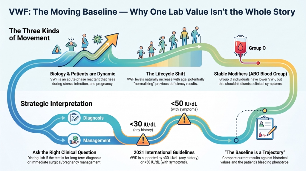
Why this spoke matters
One of the most common errors in evaluating von Willebrand disease is to treat a laboratory result as though it were a permanent characteristic of the patient.
It is not.
A VWF activity of 38 IU/dL during baseline health, 68 IU/dL during pregnancy, and 52 IU/dL several years later may all belong to the same person.
Assuming appropriate sample handling and valid assay performance, none of those values is necessarily wrong.
Each describes the biology of a particular moment.
The challenge is not that VWF changes. The challenge is that clinicians may mistake a changing measurement for a fixed biological trait.
Understanding normal VWF variation is therefore not simply about avoiding laboratory error.
It is about interpreting laboratory data within a living, changing biological system.
Every VWF value answers two questions
Every VWF measurement answers a biological question:
How much VWF is measurable today?
But diagnostic interpretation asks a second question:
Does today’s value represent the patient’s underlying hemostatic state?
Those questions are not always answered by the same number.
A laboratory result records today’s biology.
Diagnosis seeks to understand the patient’s more usual biology while also integrating:
- the bleeding phenotype
- family history
- prior VWF measurements
- the circumstances of sampling
- the relationship between VWF antigen, activity, and FVIII
- specialized or genetic testing when indicated
For diagnosis, measurements obtained during baseline health are often the most informative.
For immediate treatment, surgery, pregnancy, or replacement monitoring, however, the current value may be the clinically relevant one.
The same assay can therefore serve different purposes.
Interpretation begins by knowing which question is being asked.
The moving baseline
Many laboratory measurements vary over time.
VWF is particularly sensitive to physiologic state, age, endothelial activation, and clearance.
Its circulating level reflects the interaction of:
- synthesis and endothelial release
- clearance from plasma
- inherited variation
- ABO blood group
- age
- pregnancy
- acute inflammation or illness
- substantial physical or emotional stress
- assay and preanalytic factors
Some of these influences change VWF within the same person over time.
Others, such as ABO blood group and inherited clearance pathways, help establish the person’s underlying distribution of values.
The most useful way to think about baseline is therefore not as one perfect number.
Baseline is a physiologic range that can shift across the life course.
Or, more simply:
The baseline is not a point. It is a trajectory.
Three kinds of movement
The changing nature of VWF can be understood through three forms of movement:
- Biology moves
- The patient moves
- Interpretation moves
1. Biology varies
Stable determinants also matter
VWF is an acute-phase reactant.
Its concentration may rise during:
- infection
- inflammation
- acute bleeding
- trauma
- surgery
- strenuous exercise
- substantial emotional or physiologic stress
- pregnancy
The magnitude of these effects varies among individuals. Ordinary daily activity or minor emotional fluctuation does not automatically invalidate a result. But significant physiologic stress may transiently elevate VWF and obscure a lower underlying baseline.
Hormonal effects also require nuance.
Pregnancy reliably raises VWF and FVIII in most patients with quantitative VWF deficiency. Other endogenous or exogenous hormonal changes may influence levels, but their effects are generally less uniform and less predictable.
Normal physiology changes measured VWF.
But not all variation is dynamic.
Stable determinants also matter
ABO blood group is one of the strongest inherited determinants of VWF concentration. Group O individuals have lower average VWF levels than individuals with non-O blood groups, in part because of more rapid VWF clearance.
That fact provides biological context.
It does not make a low result clinically irrelevant.
Current international guidance does not require ABO-specific diagnostic thresholds. A low VWF value in a patient with abnormal bleeding should not be dismissed merely because the patient has group O blood.
ABO modifies the level. It does not replace clinical interpretation.
2. The patient moves
Patients move through different biological and clinical states.
The child reaches menarche.
The young adult becomes pregnant.
The previously healthy patient develops chronic inflammation.
The older adult develops malignancy, cardiovascular disease, renal dysfunction, or a need for antithrombotic therapy.
A patient may be tested:
- during health
- during active bleeding
- after surgery
- during infection
- during pregnancy
- after VWF replacement
- after desmopressin
- decades after the original diagnosis
Each circumstance changes the meaning of the sample.
The direction of change is not always predictable.
Inflammation may raise VWF antigen and activity. Aortic stenosis or mechanical circulatory support may produce acquired loss of high-molecular-weight multimers despite a normal or increased antigen level. Pregnancy may raise antigen substantially while a qualitative defect remains clinically relevant.
The patient changes even when the inherited diagnosis does not.
3. Interpretation moves
This is the movement clinicians most often overlook.
The same numeric value may carry different diagnostic implications depending on:
- bleeding history
- age
- prior measurements
- health state at sampling
- assay methodology
- subtype-specific findings
- local diagnostic terminology
A VWF activity of 45 IU/dL should not be interpreted in a vacuum.
Under the 2021 international guideline, a VWF level below 50 IU/dL in a patient with abnormal bleeding supports a diagnosis of type 1 VWD, while a level below 30 IU/dL supports the diagnosis regardless of bleeding history.1
At the same time, the label applied to a value between 30 and 50 IU/dL may differ across guidelines, institutions, and historical literature. Some clinicians and publications continue to use the term low VWF, especially when discussing partial quantitative deficiency.
The diagnostic threshold does not eliminate the need for context.
A value of 45 IU/dL may have different implications depending on whether it was obtained:
- in a symptomatic patient during baseline health
- during resolution of an inflammatory illness
- during pregnancy
- after a previously documented value below 30 IU/dL
- with discordant antigen and activity results suggesting qualitative dysfunction
- using an assay with recognized analytic limitations
The number may be the same. Its meaning may not be.
Apparent movement may come from the measurement system
Not every change in VWF reflects biology.
Apparent movement may also arise from:
- sample collection
- transport delay
- temperature exposure
- processing conditions
- laboratory-specific reference ranges
- analytic imprecision
- use of different VWF activity assays
- limitations of older ristocetin cofactor methods
- differences between platelet-dependent activity platforms
Preanalytic problems may produce falsely low or otherwise misleading results. Different activity assays may also yield clinically meaningful differences, particularly in patients with qualitative VWF abnormalities or sequence variants affecting ristocetin-dependent testing.
Serial values are most comparable when:
- samples are collected and processed appropriately
- the patient’s physiologic state is documented
- the same assay or analytically comparable assays are used
- antigen, activity, and FVIII are interpreted relationally rather than independently
Before concluding that the biology has moved, ask whether the measurement system has moved.
Repeat testing is interpretation, not repetition
Repeat testing is sometimes described as merely confirming an abnormal value.
Its deeper purpose is to ask:
Was the first result reproducible and representative?
Repeat testing can help evaluate:
- transient physiologic elevation
- persistent quantitative deficiency
- borderline results near a diagnostic threshold
- discordance between phenotype and laboratory findings
- assay-specific inconsistency
- suspected qualitative VWF dysfunction
But repeat testing is not magical.
Serial values may remain difficult to compare if:
- health states differ
- assay methods change
- preanalytic handling is inconsistent
- years have passed and age-related biology has shifted
- treatment was given before sampling
Statistical variation also matters. An unusually low or high result may move closer to the patient’s usual range when repeated, a phenomenon often described as regression toward the mean.
Repeat testing therefore does not automatically reveal the “true” value.
It provides additional observations from which the clinician can infer a more credible range.
Repeat testing refines interpretation rather than merely repeating measurement.
Aging illustrates the moving baseline
VWF commonly increases with age.
In longitudinal cohorts, many patients initially classified as having type 1 VWD or low VWF later moved into higher laboratory categories, including the normal range.2
Yet laboratory normalization did not clearly track with disappearance of the historical bleeding phenotype.
That does not prove that higher VWF has no clinical value.
It means that normalization by itself does not establish that the prior phenotype or future bleeding risk has resolved.
The important question is therefore not simply:
Has the number changed?
It is:
What does the new number mean in light of the original diagnosis, lifetime bleeding history, prior challenges, and current clinical setting?
Pregnancy illustrates the same principle
Pregnancy provides a particularly clear example of the moving baseline.
Platelet-dependent VWF activity and factor VIII activity (FVIII:C) usually rise substantially during gestation, especially in quantitative VWF deficiency.
These changes improve hemostatic reserve and are clinically important for delivery planning.
But a normal pregnancy-associated value does not necessarily represent the patient’s nonpregnant baseline.
Nor does it, by itself, negate a previously well-established diagnosis.
In type 2 VWD, antigen may rise while functional abnormalities persist. After delivery, VWF and FVIII fall toward baseline, sometimes rapidly, contributing to continued postpartum bleeding risk.
Pregnancy therefore teaches two different lessons:
- for diagnosis, pregnancy values may not represent baseline biology
- for management, current pregnancy values may be essential
The same result may be unrepresentative diagnostically and highly relevant therapeutically.
When the laboratory and the patient disagree
Discordance between laboratory findings and clinical phenotype should prompt curiosity, not automatic allegiance to either the number or the story.
Examples include:
- persistent bleeding despite apparently normal VWF
- markedly reduced VWF with little observed bleeding
- age-related normalization despite a convincing lifetime phenotype
- low antigen with preserved activity
- disproportionately low activity relative to antigen
- unexpectedly low FVIII
- new bleeding in a patient with previously stable inherited VWD
Possible explanations include:
- biologic variation
- limited exposure to hemostatic challenges
- assay or preanalytic problems
- qualitative VWD
- another inherited bleeding disorder
- medication effect
- an acquired VWF disorder
- an anatomic source of bleeding
- an inaccurate or incomplete original diagnosis
The goal is not to decide whether the laboratory or the patient is “right.”
The goal is to determine why they appear to disagree.
Discordance is not diagnostic failure. It is diagnostic information.
Clinical synthesis
It is an observation obtained at one moment within a changing biological and analytic system.
Interpretation requires attention to:
- the reason for testing
- the patient’s health state
- current and prior VWF values
- the bleeding phenotype
- stable modifiers such as ABO blood group
- age and pregnancy
- preanalytic and assay variability
- the relationship among antigen, activity, and FVIII
For diagnosis, ask whether the value reflects the patient’s usual biology.
For management, ask whether the value reflects the patient’s current hemostatic reserve.
The laboratory value is measured once. The patient has been changing for years.
Evidence anchor
| Evidence stream | What it shows | Why it matters | Main limitation |
|---|---|---|---|
| Physiologic and inherited variation | VWF varies over time with age, pregnancy, acute illness, inflammation, substantial stress, and strenuous exercise. Its baseline distribution is also influenced by stable determinants such as ABO blood group and inherited variation.3 | A single result may not represent the patient’s baseline state, while ABO-associated lowering should not be used alone to dismiss a clinically relevant result. | Magnitude of within-person effects varies; many data are observational. |
| Diagnostic thresholds and baseline-health testing | The 2021 international guideline supports type 1 VWD at VWF levels below 30 IU/dL regardless of bleeding, or below 50 IU/dL in a patient with abnormal bleeding. It emphasizes testing during baseline health because VWF is an acute-phase reactant.4 | Physiologic context matters, but it should not be used to neutralize a low result that meets diagnostic criteria in a symptomatic patient. | Conditional recommendations and very-low-certainty evidence inform some diagnostic questions. |
| Age-related change | Longitudinal type 1 VWD and low-VWF cohorts show increasing VWF with age and movement into higher laboratory ranges, while historical bleeding remained clinically relevant and did not clearly track with normalization.5 | Laboratory normalization does not by itself establish resolution of the prior phenotype or future bleeding risk. | Applies mainly to partial quantitative deficiency and should not be generalized to all VWD subtypes. |
| Preanalytic and assay variability | Sample handling, assay methodology, and platform-specific limitations may produce apparent changes or discordant VWF activity results.6 | Serial values should be compared using appropriately handled samples and analytically comparable methods whenever possible. | The clinical impact varies by assay, laboratory, subtype, and magnitude of discrepancy. |
| Contemporary synthesis | Modern diagnostic literature emphasizes VWF as a context-dependent measurement whose interpretation requires trajectory, phenotype, and sampling conditions.7 | Supports moving from threshold-only interpretation toward integrated clinical reasoning. | This is an interpretive synthesis rather than a validated diagnostic model. |
Guideline perspective: interpret the result in context
Based primarily on the ASH/ISTH/NHF/WFH 2021 diagnosis guideline and the 2024 British Society for Haematology laboratory guideline.
Shared guidance themes
- Interpret VWF antigen, platelet-dependent activity, and FVIII together rather than as isolated numbers.
- Perform diagnostic testing during baseline health whenever feasible.
- Repeat testing when the initial result may have been influenced by physiologic state, assay limitations, or persistent clinical suspicion.
- Do not use ABO blood group alone to dismiss a low VWF result or substitute ABO-specific diagnostic thresholds.
- Recognize that acute bleeding, inflammation, pregnancy, trauma, and substantial stress may raise measured VWF.
- Compare serial results using appropriately handled samples and analytically comparable assays whenever possible.
- Integrate laboratory findings with bleeding history and hemostatic challenges.
- Reconsider rather than automatically remove a previously confirmed type 1 VWD diagnosis when VWF normalizes with age, assuming the original diagnosis was sound.
- Investigate alternative or additional explanations when phenotype and laboratory findings remain discordant.
What guidelines do not eliminate
- uncertainty near diagnostic thresholds
- differences in nomenclature between type 1 VWD and low VWF
- variability among platelet-dependent activity assays
- difficulty determining whether one result reflects baseline physiology
- uncertainty about how age-related normalization changes future bleeding risk
- the need for clinical judgment when laboratory and phenotype diverge
Practical takeaway: Before interpreting a VWF value, ask why it was measured, whether the patient was at baseline health, how the result compares with prior values, which assay was used, and whether the number fits the bleeding phenotype.
Reflect & Apply Case
Two patients each have a platelet-dependent VWF activity of 46 IU/dL.
The first is a healthy 19-year-old evaluated during baseline health after lifelong epistaxis, easy bruising, and heavy menstrual bleeding.
The second is a 72-year-old recovering from pneumonia whose platelet-dependent VWF activity was 28 IU/dL three decades earlier.
The current numbers are identical.
But does the value provide the same estimate of baseline VWF in both patients?
Does it carry the same diagnostic meaning?
Does it reflect the same biological trajectory?
For the 19-year-old, a VWF level below 50 IU/dL in the setting of abnormal bleeding supports type 1 VWD under the 2021 international guideline, assuming appropriate testing and exclusion of relevant alternative explanations.
For the 72-year-old, age and pneumonia may both have raised the current value. The historical result below 30 IU/dL remains highly relevant if the original testing and diagnosis were valid.
Both patients may have type 1 VWD.
What differs is:
- the context of measurement
- the likely relationship to baseline
- the longitudinal trajectory
- the evidence supporting the diagnosis
- the clinical meaning of the current result
The laboratory values are identical. Their interpretation is not.
Diagnosis begins with measurement.
Interpretation begins with context.
And context begins with recognizing that the baseline itself moves.
Test your thinking
Quiz to follow