
Why prevalence does not equal disease burden
Note: The video and audio linked above were generated with the assistance of AI. Clinical accuracy has been reviewed, but no AI-generated content can be guaranteed to be fully error-free.
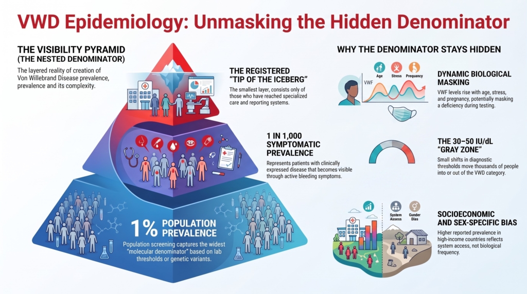
Why this spoke matters
Clinicians often hear that von Willebrand disease is “the most common inherited bleeding disorder.”
That statement is true, but incomplete.
Population-based studies have suggested VWD-like laboratory or clinical phenotypes near 1%, but clinically relevant VWD is much less frequent and highly dependent on the case definition used. Clinically symptomatic VWD is often estimated around 1 in 1,000. Registered or tertiary-care prevalence is lower still.1
The gap is not a statistical curiosity. It changes clinical reasoning.
Understanding the difference between laboratory prevalence, symptomatic prevalence, and reported prevalence helps prevent three errors:
- mistaking low VWF frequency for clinical disease burden
- dismissing VWD as common and therefore minor
- assuming that diagnosed cases represent all meaningful disease
Commonness does not imply triviality. Rarity does not imply severity.
Frequency and disease burden are related, but they are not the same thing.
This spoke explains why epidemiology in VWD is layered rather than singular.
The numerator problem: who counts as a case?
Prevalence depends on definition.
In VWD, definitions vary across studies. Some estimates rely mainly on laboratory thresholds. Others require bleeding symptoms. Others require bleeding history, low VWF levels, and familial inheritance.
The disease does not change.
The boundary changes.
A broad laboratory definition produces a large numerator. A stricter clinical definition produces a smaller numerator, but a population with higher average bleeding burden.
This matters because VWF levels are continuous. There is no natural biological cliff separating “normal” from “disease.” Thresholds are useful clinical tools, but they are not epidemiologic absolutes.
Two principles follow:
The broader the case definition, the higher the apparent prevalence.
The stricter the clinical threshold, the lower the prevalence but the higher the average disease burden.
The 30 to 50 IU/dL range is where this becomes most visible. In that zone, small shifts in definition can move many people into or out of the numerator. The 2021 ASH/ISTH/NHF/WFH diagnostic guideline classifies patients with abnormal bleeding and VWF levels below 50 IU/dL as type 1 VWD, whereas prior frameworks often separated “low VWF” from type 1 VWD. That change has clinical and epidemiologic consequences: it may improve access to care for symptomatic patients, but it may also increase diagnostic labeling in a biologically common gray zone.2
The number matters.
But the number is not the disease.
The denominator problem: who was actually looked for?
Every prevalence estimate has a denominator.
But in VWD, the denominator is often unstable.
Population screening asks: how many people meet a laboratory or epidemiologic definition?
Primary care asks: how many symptomatic individuals emerge in ordinary practice?
Registries ask: how many people have been diagnosed, reported, and retained in a health-system database?
These are not competing answers. They are different measurements of disease visibility.
A useful way to think about VWD epidemiology is as a set of nested populations:
- people with VWF levels or variants associated with VWD
- people with bleeding symptoms and low VWF
- people whose bleeding leads to testing
- people who receive a diagnosis
- people who are registered in a center, registry, or health system
Each layer is smaller than the one beneath it.
The hidden denominator is the population that exists biologically or clinically, but is never counted.
The hidden denominator: who is never seen?
Some people are never tested because they do not bleed or do not recognize their bleeding as abnormal. Others normalize symptoms within families, have symptoms attributed to non-hemostatic causes, or lack access to specialized coagulation testing.
Many become visible only after a trigger:
- dental extraction
- tonsillectomy
- surgery
- childbirth
- heavy menstrual bleeding
- recurrent iron deficiency
- family testing
- evaluation after a relative is diagnosed
A Canadian primary-care study estimated symptomatic VWD prevalence closer to 1 in 1,000, much lower than population-screening estimates near 1%.3
That difference is not a contradiction.
It is the hidden denominator.
Population screening captures laboratory or phenotypic possibility.
Clinical recognition captures expressed disease that becomes visible.
Both are real. Neither is complete.
Symptoms inflate the numerator
Bleeding symptoms are common.
Easy bruising, epistaxis, heavy menstrual bleeding, gum bleeding, and bleeding after procedures are frequent experiences. Most are not caused by VWD. Many are mild, situational, subjective, or related to local, hormonal, medication-related, anatomic, or non-hemostatic factors.
This creates a diagnostic tension:
- symptoms alone overestimate disease
- laboratory thresholds alone overestimate clinical burden
- family history may be absent, incomplete, or misleading
- bleeding history depends on memory, exposure, culture, and opportunity to bleed
Children add another layer of difficulty. Bruising and epistaxis are common in childhood, but children may not yet have faced major hemostatic challenges such as dental extraction, surgery, menarche, pregnancy, or childbirth.4
Structured bleeding assessment tools help make bleeding history more reproducible, but they do not by themselves define disease.
True clinical disease lies at the intersection of bleeding phenotype, VWF biology, inheritance, and context.
Detection is not neutral
Testing is never distributed evenly across a population.
People are tested because someone asks a question. That question may arise from bleeding, family history, surgery, pregnancy, iron deficiency, abnormal screening tests, or referral to hematology.
Referral-based cohorts therefore describe a selected denominator. They tell us about patients who reached clinical attention, not everyone with low VWF or VWF variants.
Global registration data make this especially clear. The World Federation of Hemophilia Annual Global Survey shows marked variation in VWD registration by geography and income level. Registration rates are far below expected population prevalence, especially in lower-resource settings.5
Stonebraker and colleagues similarly found that reported VWD prevalence varies substantially by national income classification, with higher reported prevalence in high-income countries and much lower reported prevalence in lower-income countries.6
This does not mean VWD biology is absent in low-registration regions.
It means the disease is less often made visible.
Reported prevalence is not true prevalence. It is true prevalence filtered through recognition, access, testing, diagnostic criteria, and reporting.
Prevalence is partly a property of health systems.
Mild disease drives variability
The variability in reported prevalence is driven especially by detection of type 1 VWD, low VWF, and milder bleeding phenotypes.
Type 3 VWD is rare but more consistently visible because the phenotype is severe, the laboratory abnormality is marked, and affected patients are more likely to reach specialized care. Even then, type 3 VWD may still be under-reported where diagnostic infrastructure is limited.
This creates a paradox:
Severe disease is rare but visible.
Mild disease is common but hidden.
In lower-income settings, type 3 VWD accounts for a larger proportion of registered VWD cases, suggesting that only the most severe phenotypes are consistently detected.7
That does not mean mild VWD is biologically uncommon in those regions.
It means mild VWD is epidemiologically quiet.
Biology makes prevalence conditional
VWD epidemiology is difficult because VWF biology is dynamic.
VWF levels vary with age, physiologic stress, exercise, inflammation, pregnancy, estrogen exposure, thyroid function, acute illness, blood group, and comorbid disease. Blood group O is associated with lower VWF levels than non-O blood groups. Pregnancy, inflammation, exercise, and acute stress may raise VWF levels and mask deficiency. Aging tends to increase VWF levels, especially in type 1 VWD or low VWF states.8
This matters epidemiologically because the measured variable is not fixed.
A patient tested during acute illness may appear normal. A pregnant patient with type 1 VWD may have physiologic correction of VWF and FVIII levels, whereas type 2 activity defects and type 3 severe deficiency often remain clinically relevant. An older adult with previously low VWF may now have levels above the diagnostic threshold.
When the measured biology moves, prevalence becomes conditional.
Prevalence depends not only on whom we test, but when we test them.
Overdiagnosis also changes the count
Epidemiology is not only about missed disease.
It is also about labels that persist after the evidence has changed.
Some patients carry a historical diagnosis of VWD based on older assays, borderline results, incomplete documentation, or childhood testing. Later in life, they may have normal VWF levels and little bleeding. This creates the opposite problem: not whether to diagnose VWD, but whether to reconsider, revise, or remove the diagnosis.9
This does not imply that the original diagnosis was incorrect. VWF levels can rise, assays evolve, and exposure histories may remain incomplete. Bleeding risk may persist despite normalization of VWF levels.
But the label itself affects epidemiology.
A diagnosis can be clinically useful, protective, and access-enabling.
It can also be burdensome, anxiety-producing, and inaccurate.
Good epidemiology must account for both underdiagnosis and overdiagnosis.
The sex-specific hidden denominator
VWD affects males and females at the genetic level because VWF is autosomal.
But clinical visibility is not sex-neutral.
Heavy menstrual bleeding, postpartum bleeding, iron deficiency, pregnancy, miscarriage management, gynecologic procedures, and childbirth make VWD more likely to become clinically visible in women and girls.
At the same time, these symptoms may be normalized, minimized, stigmatized, or attributed to gynecologic causes without hemostatic evaluation.
This creates two opposing forces:
- women may be more likely to come to attention because of reproductive bleeding
- women may also remain undiagnosed because reproductive bleeding is normalized
Global data show this tension. Females represent the majority of registered people with VWD globally, but in low-income countries males may predominate among registered cases, likely reflecting underrecognition of gynecologic bleeding and a skew toward severe disease detection.10
Thus, the hidden denominator is not only biological.
It is also cultural, clinical, and infrastructural.
Genetics reveals another denominator
Genetic data add another layer.
Large-scale genetic analyses suggest that VWF variants predicted to cause or contribute to VWD may be more common than clinical diagnoses imply. Some genetic epidemiology estimates exceed traditional clinical prevalence estimates.11
But molecular prevalence is not the same as clinical disease.
Within the genetic denominator are at least two populations:
- individuals with clinically meaningful but undiagnosed VWD
- individuals with low penetrance, variable expressivity, compensating modifiers, or insufficient hemostatic challenge to reveal bleeding
This distinction is essential.
Molecular prevalence describes biological possibility.
Clinical prevalence describes expressed disease.
Neither number cancels the other.
Genetics helps most when phenotype is specific
Genetic testing is increasingly useful in VWD, but its epidemiologic meaning depends on subtype.
For type 2 and type 3 VWD, genetic testing often helps confirm subtype, distinguish phenocopies, support family planning, and clarify difficult phenotypes. For type 1 VWD and low VWF, genetic testing is less straightforward because VWF levels are influenced by multiple loci and many variants have uncertain significance.12
Association between genotype and phenotype is usually cleaner in type 2 and type 3 VWD than in type 1 VWD or low VWF. That is why molecular counting is most clinically interpretable when the phenotype is specific.
A genetic variant is not the same as disease.
A low VWF level is not the same as disease.
A bleeding symptom is not the same as disease.
VWD emerges from the relationship among them.
Epidemiology as a function, not a number
Reported prevalence of VWD varies by method:
- population screening may suggest prevalence near 1%
- symptomatic primary-care estimates are closer to 1 in 1,000
- registered prevalence is much lower in many health systems
- tertiary-care prevalence is lower still
- type 3 VWD is rare, usually on the order of approximately 1 per million, though estimates vary by population and ascertainment
- genetic epidemiology may identify an even larger molecular denominator
Each estimate can be accurate within its method.
Each describes a different layer of visibility.
A useful synthesis is:
Prevalence = f(definition, detection strategy, population, biologic state, exposure, and health-system access)
In natural language: prevalence is not a fixed property of VWD. It is an output shaped by how the condition is defined, who is tested, why they are tested, what biological state they are in, and whether the health system is capable of recognizing them.
Clinical synthesis
When interpreting a prevalence claim about VWD, clinicians should translate it into six questions:
- What definition was used?
- Who was tested?
- What triggered testing?
- Were bleeding symptoms required?
- Under what physiologic conditions were measurements obtained?
- Who was never tested at all?
That final question is the hidden denominator.
Good clinicians treat epidemiology as context, not verdict.
Evidence anchor: why VWD prevalence is not one number
Summary derived from population studies, primary-care prevalence studies, global registry analyses, diagnostic guidelines, and expert reviews. The evidence consistently shows that VWD prevalence depends on what is counted: laboratory phenotype, symptomatic disease, diagnosed disease, registered disease, or genetic possibility.
| Evidence stream | What it shows | Why it matters | Main limitation |
|---|---|---|---|
| Population-based screening | Population studies have suggested VWD-like laboratory or clinical phenotypes near 1%, depending on criteria used.13 | These studies define the broad laboratory or phenotypic denominator. | They do not show that 1% of the population has clinically significant bleeding disease. |
| Symptomatic primary-care prevalence | Symptomatic VWD in primary care is often estimated closer to 1 in 1,000.14 | This estimate better reflects clinically expressed disease that becomes visible through symptoms. | It still misses symptomatic people who never present, normalize bleeding, or are not tested. |
| Registry and reported prevalence | Reported prevalence is much lower than population-screening estimates and varies substantially by country, income classification, and health-system infrastructure.15 | Registries measure what has been recognized, diagnosed, reported, and retained in a system. | They underestimate true disease when awareness, access, testing, or reporting is limited. |
| Severity-dependent visibility | Severe VWD, especially type 3, is rare but more visible; mild type 1 VWD and low VWF states are more common but often hidden.16 | Clinical cohorts overrepresent more severe disease because those patients are more likely to bleed, be tested, and reach specialty care. | Mild disease may be biologically present but epidemiologically quiet. |
| Sex-specific visibility | VWD is autosomal, but clinical visibility is shaped by menstruation, childbirth, postpartum bleeding, iron deficiency, stigma, and access to gynecologic evaluation.17 | Women may be more likely to come to attention because of reproductive bleeding, but also more likely to be missed when bleeding is normalized or stigmatized. | Registration patterns may reflect social and health-system visibility as much as biology. |
| Dynamic VWF biology | VWF levels vary with age, inflammation, pregnancy, stress, exercise, blood group, hormones, acute illness, and preanalytic conditions.18 | Prevalence depends not only on who is tested, but when and under what physiologic conditions testing occurs. | A single measurement may misclassify patients if obtained during a VWF-raising or VWF-lowering state. |
| Low VWF and type 1 gray zone | The 30–50 IU/dL range is heterogeneous and highly sensitive to diagnostic definition. Recent guidance classifies patients with abnormal bleeding and VWF below 50 IU/dL as type 1 VWD, while expert debate about low VWF and overdiagnosis continues.19 | Small definitional shifts can move many people into or out of the numerator. | Borderline values may represent disease, risk, modifier biology, or coincidental overlap with common bleeding symptoms. |
| Molecular denominator | Genetic analyses may identify VWF variants or predicted disease alleles more commonly than clinical diagnoses would suggest.20 | Genetics reveals a molecular denominator larger than the clinically recognized population. | Molecular possibility is not the same as expressed bleeding disease, especially in type 1 VWD and low VWF states. |
Interpretive note: These evidence streams point in the same direction: VWD prevalence is layered. Population screening, symptomatic prevalence, registry prevalence, and the molecular denominator each answer a different question. The central mistake is to treat them as competing estimates of the same thing. In VWD, prevalence is best understood as a measure of definition plus visibility, not biology alone.
Guideline perspective: prevalence as context, not verdict
Based on major diagnostic, laboratory, epidemiologic, and expert guidance, including ASH/ISTH/NHF/WFH diagnostic guidance, BSH laboratory guidance, global WFH registry analyses, and contemporary VWD reviews.
Shared guidance themes
- VWD diagnosis should integrate bleeding phenotype, VWF antigen, platelet-dependent VWF activity, FVIII, subtype-directed testing when needed, and family context.
- VWF testing should be interpreted in light of physiologic modifiers, including inflammation, pregnancy, stress, exercise, age, blood group, estrogen exposure, acute illness, and preanalytic variables.21
- Population-screening prevalence should not be equated with clinically significant disease burden.
- Registry prevalence should not be equated with true prevalence, because registration depends on awareness, access, testing, referral pathways, and reporting systems.22
- Mild VWD and low VWF states are especially vulnerable to underdiagnosis, overdiagnosis, and classification drift.
- Type 3 VWD is rare but more consistently visible; type 1 VWD and low VWF drive much of the variability in reported prevalence.
- Genetic testing is most clinically interpretable for type 2 and type 3 VWD; type 1 VWD and low VWF often require more cautious interpretation because of variable penetrance, modifier loci, and variants of uncertain significance.23
Where guidance and expert framing differ
Older diagnostic frameworks often treated VWF levels in the 30–50 IU/dL range as “low VWF,” emphasizing bleeding risk and diagnostic caution. The 2021 ASH/ISTH/NHF/WFH diagnostic guideline recommends diagnosing type 1 VWD in patients with abnormal bleeding and VWF below 0.50 IU/mL, reflecting a high value placed on avoiding missed diagnosis and improving access to care.24
The 2024 BSH laboratory guideline preserves caution around the 30–50 IU/dL range, emphasizing that associations among VWF level, bleeding, and VWF variants are weaker in this interval and that other hemostatic contributors may coexist.25
What guidance does not eliminate
- uncertainty in patients with borderline VWF levels
- uncertainty in children or adults without prior hemostatic challenges
- the possibility of underdiagnosis
- the possibility of overdiagnosis
- health-system bias in who gets tested and registered
- the distinction between assigning a label and planning safely for bleeding risk
Practical takeaway: Guidelines provide structure, not certainty. In VWD epidemiology, prevalence numbers are best treated as context. They tell clinicians what population was counted, what population was missed, and how much confidence to place in the number. They do not diagnose the patient in front of you.
Reflect & Apply Case
A 24-year-old woman is referred after prolonged bleeding following dental extraction. Her platelet-dependent VWF activity is mildly reduced at 42 IU/dL. She reports heavy menses since adolescence and recurrent iron deficiency, but she has never been evaluated for a bleeding disorder.
You review two sources:
- a population-screening study suggesting prevalence near 1%
- a primary-care study suggesting symptomatic prevalence closer to 1 in 1,000
Questions:
- Which estimate best reflects her clinical risk?
- What hidden denominator does each study miss?
- Would your interpretation change if testing occurred during pregnancy, inflammation, acute illness, or estrogen exposure?
- How would your reasoning change if she had a first-degree relative with diagnosed VWD?
- How would your reasoning change if repeat testing at baseline showed normal VWF levels?
- Would labeling her “type 1 VWD,” “low VWF,” or “bleeding with low VWF” change access to care, counseling, or treatment planning?
No prevalence estimate diagnoses her.
Population studies highlight how common low VWF biology may be. Symptomatic estimates emphasize that clinically meaningful disease is far less frequent. Registry data remind us that many affected individuals are never counted. Her risk depends on the relationship between bleeding phenotype, VWF level, family history, physiologic state, and future hemostatic challenges.
Her case also reminds us that even in well-resourced settings, many symptomatic individuals never reach testing.
In VWD, epidemiology does not give the answer.
It tells you which question you are asking.
Test your thinking
An interactive quiz for this topic is being developed and will be added soon.