
When hemostasis is disrupted by disease, flow, or clearance
Note: The video and audio linked above were generated with the assistance of AI. Clinical accuracy has been reviewed, but no AI-generated content can be guaranteed to be fully error-free.
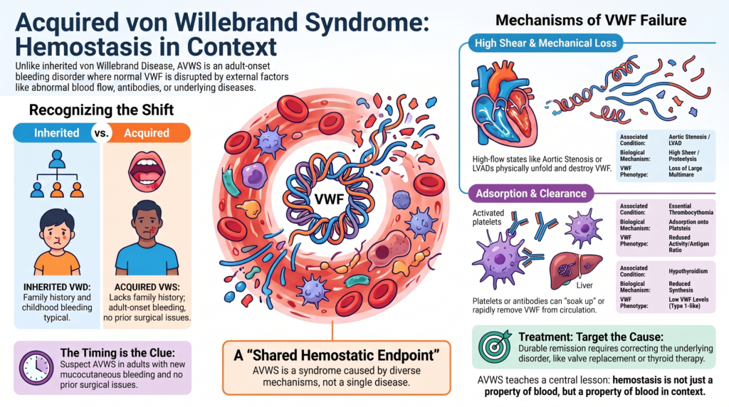
Why this spoke matters
Von Willebrand disease is usually taught as an inherited disorder. A patient is born with a quantitative or qualitative abnormality of von Willebrand factor. The bleeding history often begins early, the family history may help, and the laboratory abnormality reflects the biology of the VWF gene.
Acquired von Willebrand syndrome changes the frame.
Here, the patient may have normal VWF genes, normal childhood hemostasis, no family history of bleeding, and no previous surgical bleeding, and then later in life develop a VWD-like bleeding disorder.
The problem is not inherited production.
It is acquired loss, dysfunction, adsorption, accelerated clearance, excessive proteolysis, abnormal shear-dependent processing, or reduced synthesis of VWF.1
That is why acquired von Willebrand syndrome is so instructive. It shows that VWF biology is not fixed once VWF is made. VWF remains vulnerable to the conditions through which it circulates: flow can unfold it; transformed cells or expanded platelet burdens can adsorb or remove it; antibodies can accelerate its clearance or block its function; mechanical circulatory support can expose it to high shear and mechanical disruption; and hypothyroidism can produce a type 1-like AVWS pattern, probably through reduced VWF synthesis.2
Acquired von Willebrand syndrome teaches a central lesson: hemostasis is not only a property of blood. It is a property of blood in context.
The basic definition
Acquired von Willebrand syndrome is a bleeding disorder caused by an acquired quantitative, qualitative, or structural abnormality of VWF, producing clinical and laboratory features that resemble inherited VWD but arise secondary to another condition.3
The usual clues are:
- new mucocutaneous bleeding
- adult onset
- negative personal bleeding history
- negative family history
- an associated disorder
- laboratory findings that resemble inherited VWD, often with a type 2-like loss of high-molecular-weight multimers, although type 1-like and rarely more profound quantitative patterns can occur
The term “syndrome” is useful because AVWS is not one disease. It is a shared hemostatic endpoint reached by different mechanisms.
The clinical pattern
Patients often present with:
- epistaxis
- easy bruising
- gingival bleeding
- gastrointestinal bleeding
- iron deficiency anemia
- heavy menstrual bleeding
- postoperative bleeding
- bleeding after cardiac or invasive procedures
Bleeding is often mucosal because VWF is especially important where high shear and platelet-dependent adhesion dominate.4
The most important clinical clue is timing.
A patient who had dental extractions, childbirth, surgery, or trauma without abnormal bleeding, and then develops a VWD-like laboratory pattern later in life, should not be assumed to have newly discovered inherited VWD. That patient may have AVWS.
The major associated conditions
Commonly reported associations include:
- cardiovascular disease
- mechanical circulatory support
- myeloproliferative neoplasms
- lymphoproliferative disorders and plasma cell dyscrasias
- autoimmune disease
- solid tumors
- hypothyroidism
- selected medications and other rare conditions
Their relative frequency varies by cohort and referral setting. Older registry data emphasized lymphoproliferative disorders, while later cohorts and cardiac-device populations have increased recognition of cardiovascular and mechanical support-associated AVWS.5
The common mistake is to think of these as a list.
They are better understood as mechanisms.
Mechanism 1: high shear and loss of large multimers
The clearest example is aortic stenosis. As blood accelerates across a narrowed valve, VWF is exposed to high shear stress. This unfolds VWF and exposes the A2 domain, making high-molecular-weight multimers more susceptible to cleavage by ADAMTS13. The result is selective loss of the largest, most hemostatically active multimers.6
This is the biology behind Heyde syndrome:
- aortic stenosis
- loss of high-molecular-weight VWF multimers
- gastrointestinal bleeding, often from angiodysplasia
The same principle applies to other high-shear cardiovascular states, including some congenital heart lesions, hypertrophic obstructive cardiomyopathy, mitral regurgitation, and other structural lesions.7
The teaching point is powerful: hemostasis can improve not because hematology changed the blood, but because cardiology changed the flow.
After aortic valve replacement or TAVI, VWF multimer abnormalities may improve rapidly, sometimes within hours to days. The response may be incomplete if residual valve dysfunction, prosthesis-patient mismatch, or patient-specific VWF biology persists.8
Acquired and inherited mechanisms can also coexist. Severe bleeding in aortic stenosis should not automatically be attributed to shear-related AVWS alone. Aortic stenosis may unmask previously mild inherited VWD or VWF genetic susceptibility.9
Mechanism 2: mechanical circulatory support
Left ventricular assist devices, extracorporeal membrane oxygenation, and other mechanical circulatory support systems can produce AVWS. Here, shear-mediated unfolding and proteolysis may contribute, but direct mechanical disruption of VWF may also be important.10
Laboratory abnormalities may be very common, even near-universal in some series.
But bleeding is not universal.
Bleeding risk also depends on anticoagulation, antiplatelet therapy, device type, mucosal lesions, renal function, inflammation, and procedural exposures. AVWS is part of the bleeding biology; it is not the whole story.11
Mechanism 3: adsorption onto platelets or malignant cells
In myeloproliferative neoplasms, especially essential thrombocythemia and polycythemia vera with marked thrombocytosis, VWF may be adsorbed onto activated or expanded platelet surfaces. The largest multimers are preferentially lost, and the laboratory phenotype can resemble type 2A VWD:
- normal or mildly reduced VWF antigen
- reduced VWF activity
- low activity-to-antigen ratio
- loss of high-molecular-weight multimers
This explains the paradox of essential thrombocythemia. The same patient may be at risk for thrombosis and bleeding. The platelet count is high, but platelet-dependent hemostasis may be impaired because the VWF multimers needed for high-shear adhesion have been depleted.12
This is why AVWS matters when considering aspirin in patients with extreme thrombocytosis.
The risk is not simply “too many platelets.” It is too many platelets in a system that has consumed or removed the VWF structures needed for normal primary hemostasis.13
Mechanism 4: antibodies and accelerated clearance
In lymphoproliferative disorders and plasma cell dyscrasias, AVWS often reflects immune-mediated clearance or functional interference. The patient may have MGUS, Waldenström macroglobulinemia, multiple myeloma, lymphoma, or another B-cell disorder. Anti-VWF antibodies may be detectable, but testing is imperfect; absence of a demonstrable inhibitor does not exclude immune-mediated AVWS.14
This matters therapeutically.
Some patients respond poorly or briefly to desmopressin or VWF concentrates because the infused or released VWF is rapidly cleared. In selected IgG-mediated or monoclonal gammopathy-associated AVWS, high-dose intravenous immunoglobulin may prolong VWF survival and provide a more sustained hemostatic response. IVIG is not a general solution for shear-related AVWS. In IgM-associated states, especially Waldenström macroglobulinemia, plasmapheresis and treatment of the underlying clone may be more useful.15
Mechanism 5: reduced synthesis
Hypothyroidism is a classic example of AVWS caused primarily by reduced VWF synthesis. The phenotype often resembles type 1 VWD. Bleeding is usually mild and mucocutaneous, but surgical or clinically significant bleeding can occur. Correction of hypothyroidism may normalize VWF levels and resolve the bleeding tendency.16
This mechanism is conceptually different from shear-mediated AVWS. The problem is not destruction of large multimers; it is insufficient production. That distinction changes both testing and treatment.
Laboratory pattern
No single laboratory test reliably proves or excludes AVWS. The diagnosis depends on clinical context, VWF antigen and activity, activity-to-antigen ratios, factor VIII, collagen binding where available, multimer analysis, and evaluation for an underlying disorder.17
A practical evaluation includes:
- screening tests: CBC, platelet count, PT, aPTT
- VWF-specific tests: VWF antigen, platelet-dependent VWF activity, factor VIII activity
- pattern-defining tests: activity-to-antigen ratio, collagen-binding-to-antigen ratio, multimer analysis
- mechanism-refining tests: VWF propeptide-to-antigen ratio, selected inhibitor or antibody studies, and tests for associated disorders
Common patterns include:
- low VWF activity
- low or normal VWF antigen
- reduced VWF activity-to-antigen ratio
- low VWF collagen-binding-to-antigen ratio
- loss of high-molecular-weight multimers
- reduced factor VIII activity (FVIII:C) in some patients
- normal PT
- normal or prolonged aPTT depending on factor VIII level
But the laboratory pattern is not the diagnosis.
The diagnosis is relational. It requires linking the bleeding phenotype, age of onset, family history, VWF laboratory pattern, associated disease, mechanistic plausibility, and response to treatment of the underlying disorder.
A practical diagnostic question
The central question is not simply, “Does this patient have low VWF?” The better question is, “Why does this patient have a VWD-like pattern now?” That question forces the clinician to look for aortic stenosis, LVAD or ECMO, extreme thrombocytosis, monoclonal gammopathy, Waldenström macroglobulinemia, myeloma or lymphoma, autoimmune disease, hypothyroidism, solid tumor, drug exposure, or previously unrecognized inherited VWD.
AVWS should not become a diagnostic shortcut.
It should become a diagnostic discipline.
Treatment principle
Treatment has three goals:
- control active bleeding
- prevent bleeding during procedures
- treat or remove the underlying cause
The most durable treatment is usually treatment of the cause. Valve replacement can correct shear-mediated AVWS. Device removal can resolve mechanical circulatory support-associated AVWS. Cytoreduction can improve AVWS in myeloproliferative neoplasms. Clone-directed therapy can improve AVWS in lymphoproliferative disease. Thyroid replacement can correct hypothyroidism-associated AVWS.
Replacement may bridge the patient through bleeding, but remission usually requires addressing the biology that is removing, damaging, or suppressing VWF.18
Hemostatic tools
Immediate hemostasis may include:
- tranexamic acid for mucosal bleeding
- desmopressin in selected patients
- VWF-containing concentrates
- factor VIII/VWF concentrates when factor VIII is low
- local endoscopic, surgical, or procedural control
Mechanism-directed therapy may include:
- high-dose IVIG in selected immune-mediated or monoclonal gammopathy-associated AVWS
- plasmapheresis in selected IgM paraprotein states
- cytoreduction in extreme thrombocytosis
- valve intervention in clinically significant aortic stenosis-associated AVWS
- device removal, exchange, or optimization when feasible
- clone-directed therapy for lymphoproliferative or plasma cell disease
- thyroid replacement in hypothyroidism-associated AVWS
- recombinant factor VIIa in refractory bleeding when standard measures fail
The choice depends on mechanism. Desmopressin may help if releasable endothelial stores are present and clearance is not too rapid. VWF concentrates may help in bleeding or procedures, but responses can be short-lived when clearance is accelerated. IVIG is most useful in selected immune-mediated or monoclonal gammopathy-associated AVWS. Plasmapheresis may be helpful in IgM paraprotein states. Cytoreduction is central in extreme thrombocytosis. Valve intervention is central in severe aortic stenosis with clinically significant AVWS.19
Why desmopressin is not automatically benign
Desmopressin is familiar from inherited VWD, but AVWS patients are often older and may have cardiovascular disease. In cardiovascular AVWS, desmopressin may be ineffective, transient, or risky depending on the patient’s comorbidities. For major surgery or critical-site bleeding, it should not be relied upon as sole therapy. When used, response should be measured because AVWS responses can be unpredictable.20
Angiodysplasia and VWF
Gastrointestinal bleeding in AVWS is not only a mechanical bleeding problem. Loss of high-molecular-weight VWF multimers may impair platelet adhesion at fragile mucosal vascular beds. VWF may also participate in vascular integrity and angiogenesis, which may help explain the association between VWF abnormalities and angiodysplasia.
This angiogenesis link is biologically plausible and supported by experimental and clinical literature, but it should be framed as a contributing mechanism rather than a complete explanation for every case of gastrointestinal bleeding.21
Where clinicians get misled
VWS is easy to miss because each specialty sees only part of the syndrome:
- cardiology sees aortic stenosis
- gastroenterology sees angiodysplasia
- hematology sees low VWF activity
- oncology sees MGUS or Waldenström macroglobulinemia
- intensive care sees ECMO bleeding
- primary care sees iron deficiency anemia
The diagnosis emerges only when the pieces are connected. Another trap is assuming that absence of childhood bleeding excludes all VWF-related disease. It excludes typical inherited VWD as the full explanation. It does not exclude AVWS, nor does it exclude mild inherited VWD unmasked by acquired disease.
The central lesson
Inherited VWD asks: What did the patient inherit?
Acquired von Willebrand syndrome asks: What is happening to VWF now?
That shift is the heart of the topic. VWF is not simply a plasma level; it is a circulating structure. It responds to shear, binds cells, and is cleaved, cleared, stored, released, and sometimes destroyed. AVWS makes that biology visible. It turns VWF from a laboratory number into a moving participant in disease.
Bleeding can begin not because the patient’s genome changed, but because the patient’s circulation, clone, immune system, device, or endocrine state changed around it.
Clinical synthesis
Acquired von Willebrand syndrome is not a diagnosis of low VWF. It is a diagnosis of why VWF has become ineffective. The same laboratory pattern may arise from high shear, mechanical circulatory support, extreme thrombocytosis, immune-mediated clearance, paraproteins, or reduced synthesis. Identifying the mechanism is not an academic exercise; it determines treatment.
The diagnosis is strongest when adult-onset bleeding, the absence of a personal or family bleeding history, compatible laboratory findings, and an associated disorder all point in the same direction. It is weakest when a single abnormal VWF assay is interpreted without clinical context.
The most effective treatment often does not come from hematology alone. Hemostasis may improve because a valve is replaced, a platelet count is lowered, a plasma cell clone is treated, a paraprotein is removed, or hypothyroidism is corrected. Hemostatic therapy can bridge the patient through bleeding, but durable remission usually requires correcting the process that made VWF fail.
The clinician’s task is therefore not simply to recognize AVWS, but to ask a more fundamental question: what is happening to VWF in this patient now?
Evidence anchor: why mechanism matters more than the VWF level in AVWS
Summary derived from AVWS registries, cardiovascular AVWS studies, myeloproliferative and lymphoproliferative literature, hypothyroidism reviews, and expert guidance. The evidence consistently shows that acquired von Willebrand syndrome is not a single disease but a shared bleeding phenotype produced by different mechanisms. The same low VWF activity may reflect shear-mediated multimer loss, accelerated clearance, adsorption onto platelets or malignant cells, reduced synthesis, or mechanical disruption. Identifying the mechanism is therefore central to diagnosis, prognosis, and treatment.
| Evidence stream | What it shows | Why it matters | Main limitation |
|---|---|---|---|
| Registry and cohort data | AVWS is associated with cardiovascular disease, lymphoproliferative disorders, myeloproliferative neoplasms, autoimmune disease, solid tumors, hypothyroidism, and other conditions, although the relative frequency varies by cohort and referral setting.22 | Adult-onset mucocutaneous bleeding in a patient with an associated disorder should prompt consideration of AVWS rather than late recognition of inherited VWD. | Registry data are subject to referral and ascertainment bias, and disease frequencies differ across clinical populations. |
| High-shear cardiovascular studies | Severe aortic stenosis and other high-shear lesions produce loss of high-molecular-weight VWF multimers, and correction of the hemodynamic lesion often restores VWF structure and improves bleeding.23 | Flow can create a VWD-like disorder without an inherited VWF defect. Durable treatment may require correcting the underlying lesion rather than simply replacing VWF. | Laboratory evidence of AVWS is more common than clinically significant bleeding. |
| Mechanical circulatory support studies | LVADs and ECMO commonly produce high-shear-associated loss of high-molecular-weight multimers, although bleeding risk is modified by anticoagulation, device characteristics, mucosal lesions, and other factors.24 | A multimer abnormality explains only part of bleeding risk in device-supported patients. | Most evidence is observational and device-specific. |
| Myeloproliferative neoplasm studies | Extreme thrombocytosis can remove high-molecular-weight VWF multimers and produce a type 2-like AVWS pattern, explaining why bleeding and thrombosis may coexist in ET and PV.25 | Bleeding risk cannot be inferred from the platelet count alone; AVWS should influence decisions about antiplatelet therapy. | Platelet thresholds are imperfect predictors of bleeding. |
| Immune-mediated and paraprotein-associated AVWS | Paraproteins and antibodies may accelerate VWF clearance or interfere with function. Selected IgG-mediated cases respond to IVIG, whereas IgM-associated disease may require plasmapheresis and treatment of the underlying clone.26 | Mechanism determines therapy. A short-lived response to VWF replacement should prompt consideration of accelerated clearance. | Antibody assays have limited sensitivity, and immune mechanisms are heterogeneous. |
| Hypothyroidism studies | Overt hypothyroidism can produce a usually mild type 1-like AVWS pattern, probably through reduced VWF synthesis, and may improve with thyroid replacement.27 | Not all AVWS is caused by shear or clearance. Reduced synthesis represents a distinct biological mechanism and treatment strategy. | Evidence consists largely of case reports and observational studies. |
| Diagnostic testing | VWF antigen, platelet-dependent VWF activity, activity-to-antigen ratios, FVIII, collagen binding, multimer analysis, and selected propeptide or antibody studies each interrogate different aspects of VWF biology.28 | No single laboratory test reliably proves or excludes AVWS. Diagnosis depends on integrating laboratory findings with clinical context and the associated disease. | Specialized testing may not be widely available, and VWF assays remain susceptible to physiologic and preanalytic variability. |
Interpretive note: These evidence streams converge on a single conclusion: AVWS is fundamentally a mechanism-based diagnosis. The laboratory pattern identifies that VWF function is abnormal, but it does not explain why. The strongest diagnosis emerges when the bleeding phenotype, timing, VWF studies, and associated disorder all point toward the same biological mechanism. The key clinical question is therefore not simply How low is the VWF? but What is making VWF ineffective in this patient now?
Guideline perspective: AVWS as a mechanism-matched diagnosis
Based on major AVWS reviews, treatment guidance, VWD diagnostic guidance, and laboratory guidance. Unlike inherited VWD, AVWS has fewer high-certainty trial data and fewer formal mechanism-specific guideline recommendations. Most practical guidance is therefore built from registries, case series, expert reviews, laboratory principles, and extrapolation from inherited VWD management.
Shared guidance themes
- Suspect AVWS in patients with new mucocutaneous bleeding, especially when bleeding begins in adulthood and there is no personal or family history of abnormal bleeding.
- Search actively for associated disorders, including aortic stenosis or other high-shear cardiovascular lesions, LVAD or ECMO support, extreme thrombocytosis, monoclonal gammopathy, lymphoproliferative disease, plasma cell dyscrasia, autoimmune disease, solid tumor, hypothyroidism, and selected drugs.29
- Do not rely on one laboratory test. VWF antigen, platelet-dependent activity, FVIII, activity-to-antigen ratios, collagen binding, and multimer analysis provide complementary information.30
- Interpret the VWF panel through mechanism: shear-related AVWS, MPN-associated AVWS, immune/paraprotein-mediated AVWS, hypothyroidism-associated AVWS, and device-associated AVWS are not managed the same way.
- Measure response when using desmopressin or VWF concentrate, because responses may be short-lived or absent in accelerated-clearance states.31
- Treat active bleeding and procedural risk with hemostatic tools when needed, including local measures, antifibrinolytics, desmopressin, VWF-containing concentrates, IVIG, plasmapheresis, or bypassing therapy depending on context.
- Treat the underlying driver whenever possible: valve intervention for clinically significant shear-mediated AVWS, device removal or optimization when feasible, cytoreduction for extreme thrombocytosis, clone-directed therapy for lymphoproliferative or plasma cell disease, plasmapheresis for selected IgM paraprotein states, and thyroid replacement for hypothyroidism-associated AVWS.
- Manage complex cases through a hemostasis center or multidisciplinary team, particularly when cardiac disease, mechanical circulatory support, monoclonal gammopathy, major surgery, or competing thrombosis risk is present.
Where guidance requires judgment
- Laboratory AVWS does not always equal clinically important bleeding, especially in aortic stenosis, LVAD support, or other settings where multimer abnormalities may be common.
- AVWS and inherited VWD are not mutually exclusive; severe bleeding in aortic stenosis or another acquired state may unmask previously mild inherited VWD or VWF susceptibility.32
- IVIG is not a general AVWS treatment. It is most relevant to selected immune-mediated or monoclonal gammopathy-associated AVWS, especially IgG-mediated forms.
- Desmopressin is not automatically benign in AVWS, because many patients are older, have cardiovascular disease, or have rapid VWF clearance. It should not be relied on as sole therapy for major surgery or critical-site bleeding.33
- In MPNs with extreme thrombocytosis, aspirin decisions should account for bleeding history and AVWS testing, not platelet count alone.
- In mechanical circulatory support, VWF multimer loss is often only one part of bleeding risk; anticoagulation, antiplatelet therapy, device factors, renal dysfunction, angiodysplasia, and procedures also matter.
Practical takeaway
Guidance for AVWS is less about applying a universal VWD treatment algorithm and more about matching the mechanism. The core clinical question is not simply, “How low is the VWF?” It is, “What is happening to VWF now?” Once that question is answered, treatment can be directed both at the bleeding episode and at the disease, device, flow state, clone, platelet burden, or endocrine disorder that made VWF fail.
Reflect & Apply
A 78-year-old man is referred for recurrent iron deficiency anemia and intermittent melena. Colonoscopy shows multiple colonic angiodysplastic lesions. He reports no childhood bleeding, no excessive bleeding after dental extractions or an inguinal hernia repair performed 25 years ago, and no family history of bleeding disorders. Examination reveals a harsh systolic ejection murmur. Echocardiography demonstrates severe calcific aortic stenosis.
Laboratory studies show:
- VWF antigen: mildly reduced
- Platelet-dependent VWF activity: disproportionately reduced
- VWF activity-to-antigen ratio: decreased
- Multimer analysis: loss of high-molecular-weight multimers
The immediate temptation is to diagnose “von Willebrand disease” and prescribe VWF replacement.
Pause.
The more useful question is:
What has changed?
This patient had decades of apparently normal hemostasis before developing bleeding. His laboratory pattern reflects impaired VWF function, but the underlying problem is not defective VWF synthesis or an inherited VWF mutation. It is abnormal blood flow across a severely stenotic aortic valve, producing a high-shear environment that unfolds VWF and promotes loss of the largest multimers.
Recognizing that mechanism changes management. VWF concentrate or desmopressin may temporarily improve hemostasis during active bleeding or invasive procedures, but durable improvement is most likely to come from correcting the valve lesion. At the same time, unusually severe bleeding should prompt consideration that acquired shear-mediated AVWS and previously unrecognized inherited VWD can coexist.
Clinical pearl: In AVWS, the laboratory pattern tells you that VWF is failing. The clinical task is to determine why it is failing. Mechanism, not the VWF level alone, guides definitive management.
Test your thinking
An interactive quiz for this topic is being developed and will be added soon.