
Why instability near thresholds is expected, and why repetition strengthens diagnosis
Note: The video and audio linked above were generated with the assistance of AI. Clinical accuracy has been reviewed, but no AI-generated content can be guaranteed to be fully error-free.
Why this spoke matters
The preceding spokes establish why a single measurement cannot define von Willebrand disease.
This spoke asks what that means when you are holding a laboratory report in your hands.
VWD is not diagnosed by one number, but by a reproducible clinicolaboratory pattern.
That pattern includes:
- bleeding phenotype
- VWF antigen
- platelet-dependent VWF activity
- FVIII activity
- family context
- physiologic context
- assay method
- time
VWF levels fluctuate. Assays vary. Specimens can be mishandled. Testing may occur during illness, pregnancy, stress, acute bleeding, postoperative recovery, or other states that do not represent baseline biology.
If we ignore this instability, we misclassify patients.
We may underdiagnose those whose VWF is transiently elevated.
We may overdiagnose those whose borderline abnormality reflects noise, context, or another hemostatic problem.
Repeat testing is not hesitation.
In borderline, discordant, or nonbaseline settings, it is disciplined diagnosis.
In urgent bleeding, urgent procedural planning, or clearly severe deficiency, management may need to proceed while confirmation and subtyping continue.
How to read this essay
The previous diagnostic essays explained:
- how to localize the bleeding problem
- what the first-line VWF panel measures
- when specialized second-line testing is needed
- how to interpret VWF assays relationally
This essay asks a different question:
When should you trust a laboratory result—and when should you question it?
VWF is biologically dynamic. Assays differ. Specimen handling matters. Near diagnostic thresholds, a single value may not represent baseline biology.
Repeat testing is not about repeating a number. It is about confirming baseline biology.
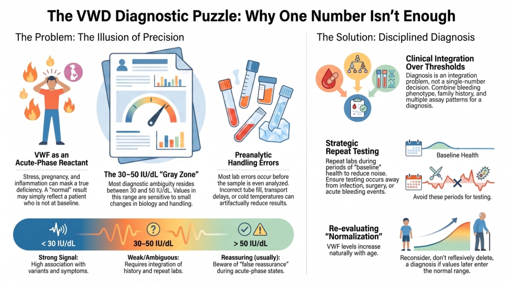
The illusion of precision
VWF levels are reported as exact numbers:
34 IU/dL
47 IU/dL
29 IU/dL
They look definitive.
They are not.
Plasma VWF spans a wide physiologic range in the general population. Values are influenced by inflammation, infection, acute bleeding, stress, exercise, pregnancy, surgery, aging, thyroid status, estrogen exposure, ABO blood group, and comorbid disease.
A single draw captures a moment, not necessarily baseline biology.
When a value is profoundly abnormal, instability matters less. A VWF activity of 8 IU/dL in a patient with a convincing bleeding phenotype is not the same interpretive problem as a VWF activity of 48 IU/dL.
Near thresholds, small movements matter.
A VWF activity of 48 IU/dL that repeats at 53 IU/dL may cross a reporting boundary without representing a meaningful biologic change.
The label can move more than the biology.
The 30–50 IU/dL zone
Below approximately 30 IU/dL, reduced VWF activity is more consistently associated with bleeding symptoms, VWF variants, and familial segregation.
Between 30 and 50 IU/dL, certainty dissolves.
This is where most diagnostic ambiguity resides.
Some individuals bleed.
Some do not.
Some have a VWF-centered bleeding tendency.
Some have another hemostatic modifier.
Some normalize with age.
Some remain low.
This is not diagnostic failure.
It is the biology of a continuous protein being forced into categorical boxes.1
The important point for this spoke is not to re-litigate the entire low VWF versus type 1 VWD debate. That belongs elsewhere in the module.
The point here is narrower:
Near a threshold, classification becomes sensitive to small changes in biology, sample handling, assay method, and timing.
A value below 30 IU/dL carries stronger diagnostic signal.
A value between 30 and 50 IU/dL requires integration.
That integration includes bleeding history, family history, repeat values, assay pattern, and the clinical setting in which testing was performed.
Near thresholds:
- small assay variation can alter classification
- transient physiologic elevation can mask low baseline VWF
- transient reduction or specimen artifact can exaggerate abnormality
- other causes of mucocutaneous bleeding must remain in view
Caution in this zone is not indecision.
It is proportional reasoning.
Preanalytic reality
Most laboratory errors occur before the analyzer runs.
That matters in VWD because the diagnostic signal is often small.
Specimens can be affected by:
- difficult phlebotomy
- incorrect tube fill
- clotting or hemolysis
- transport delay
- inappropriate temperature
- delayed processing
- freeze-thaw effects
- inadequate post-thaw mixing
- testing during acute-phase states
Some distortions are predictable.
Whole blood for VWF testing should generally be transported at ambient temperature, not on ice. Cold transport and refrigeration can artifactually reduce measured VWF and FVIII activity over time. Delays between collection and processing add noise at exactly the moment we are trying to decide whether a borderline value is real.2
Preanalytic problems can push the diagnosis in either direction.
They can make a normal person look like type 1 VWD.
They can make a type 1 VWD patient look more like type 2 VWD.
They can make a borderline result look categorical.
This is why repeat testing is most valuable when the first sample was drawn in a biologically or technically noisy setting:
- during infection or inflammation
- soon after surgery
- after vigorous exercise
- during pregnancy or postpartum transition
- during acute bleeding
- after transport or processing concerns
- when the result conflicts with the phenotype
If the clinical stakes are meaningful, confirmation on a second occasion is not redundancy.
It is quality control.
The problem of false reassurance
VWF is an acute-phase reactant.
That means the problem is not only false low results.
It is also false reassurance.
A patient with baseline low VWF may test “normal” during inflammation, pregnancy, physiologic stress, acute bleeding, or postoperative recovery. The laboratory report may look reassuring precisely because the patient is not at baseline.
The interpretive trap is simple:
The number is normal, so the disease is absent.
The better question is:
Was the patient’s biology normal when the sample was drawn?
For VWD, “normal” is not only a reference range.
It is a context.
When first-level VWF/FVIII tests are normal, VWD is unlikely. But repeat testing may be appropriate if clinical suspicion is high or if testing occurred during an acute-phase, pregnancy, inflammatory, or otherwise nonbaseline state.3
APTT and PFA are not escape hatches
A normal APTT does not exclude VWD.
FVIII may be normal in many patients with VWD, especially in milder quantitative deficiency. Even when FVIII is reduced, the APTT reagent may not be sensitive enough to detect the abnormality.
Likewise, platelet function analyzer closure times may support concern when clearly abnormal, especially in more severe VWD or some type 2 variants. But PFA testing can be normal in type 1 VWD and low VWF, and it is affected by platelet count, hematocrit, and other variables.
These tests provide context but do not replace the core VWD panel.
The core diagnostic panel remains:
- VWF antigen
- platelet-dependent VWF activity
- FVIII activity
Additional testing is guided by the pattern, not ordered reflexively without a question.4
Assay change is not biologic change
VWF testing has evolved.
Older ristocetin cofactor assays are variable. Newer platelet-binding assays, including VWF:GPIbM and VWF:GPIbR, and collagen-binding assays provide related but nonidentical information.
This matters because a patient’s apparent trajectory may reflect method change rather than biology.
The VWF ristocetin cofactor assay depends on ristocetin-induced interaction between VWF and platelet GPIb. It is useful, but it has well-recognized limitations, including inter- and intra-laboratory variability and susceptibility to ristocetin-binding sequence variants that may lower the assay result without reflecting impaired VWF function in vivo. Newer VWF:GPIbM assays avoid ristocetin and may have better precision, but no static assay perfectly reproduces VWF function under physiologic shear.5
Ratios also require caution.
Activity-to-antigen ratios are powerful, but method-dependent. A ratio near a cutoff should be interpreted with the local assay, clinical phenotype, and second-level testing in mind.6
This creates a second kind of instability.
Not biology.
Methodology.
A patient tested in 2008 and retested in 2026 may not be undergoing the same laboratory interrogation. The comparison may still be useful, but it is not always clean.
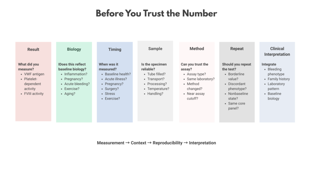
When interpreting change over time, ask:
- Was the same laboratory used?
- Was the same activity assay used?
- Were antigen, activity, and FVIII measured together?
- Was the patient at baseline on both occasions?
- Is the observed change large enough to matter clinically?
A trend is only as good as the comparability of its measurements.
Subtyping instability
Subtyping VWD requires more than a low value.
It requires pattern recognition.
Qualitative variants often depend on:
- activity-to-antigen ratios
- FVIII-to-VWF relationships
- collagen-binding results
- multimer analysis
- RIPA or genetic testing in selected cases
- VWF:FVIIIB testing when type 2N is suspected
Assigning a subtype from incomplete data invites misclassification.
Repeat evaluation may be needed when:
- activity-to-antigen ratios are borderline
- assays are discordant
- FVIII is disproportionately low
- multimers and ratios do not align
- thrombocytopenia raises concern for type 2B or platelet-type VWD
- phenotype and laboratory findings diverge
- prior subtyping was based on older or limited testing
Subtyping should be earned.
It should follow confirmation, not precede it.7
The problem of normalization
VWF levels increase with age.
Patients previously labeled as having type 1 VWD may later show values within the laboratory reference range. This is not rare. It is one of the central diagnostic conundrums in VWD.
What does normalization mean?
It does not automatically mean:
- the original diagnosis was wrong
- the patient no longer bleeds
- the patient is cured
- the label should be erased without review
It means the biology has changed.
The ASH/ISTH/NHF/WFH diagnosis guideline uses careful language here: the diagnosis should be reconsidered, not simply removed, when a patient with previously confirmed type 1 VWD later has normalized VWF levels.8
Reconsideration means returning to the full pattern:
- original bleeding history
- prior VWF values
- current VWF values
- assay methods used over time
- family history
- procedural bleeding history
- response to prior hemostatic therapy
The key is not whether the clinician can delete a diagnosis.
The key is whether the clinician can responsibly revise the patient’s risk narrative.
This “normalization problem” has already appeared elsewhere in this module as a mismatch problem and a life-stage problem. Here it is a laboratory interpretation problem:
What do you do with a prior label when the number no longer cooperates?
The answer is not to cling to the label.
The answer is not to discard it reflexively.
The answer is to recalibrate.
Repeat testing is not always the same question
Repeat testing can serve several purposes.
Sometimes it asks:
Is the abnormality reproducible?
Sometimes it asks:
Was the first sample distorted by illness, pregnancy, stress, or handling?
Sometimes it asks:
Has the patient’s VWF biology changed with age?
Sometimes it asks:
Has the subtype been assigned correctly?
Sometimes it asks:
Will this patient respond to desmopressin?
These are different questions.
They should not be collapsed into one vague instruction to “repeat labs.”
A diagnostic repeat should be performed during baseline health, with careful sample handling, and with the same core panel so the results can be compared.
A therapeutic repeat or desmopressin trial asks a different question: can the patient generate a clinically useful rise in VWF and FVIII, and how long does that response last?
Both are forms of repetition.
But they answer different problems.
When repetition is most valuable
Repeat testing is especially appropriate when:
- VWF values fall in the 30–50 IU/dL range
- testing occurred during illness, inflammation, pregnancy, acute bleeding, or postoperative recovery
- the historical diagnosis conflicts with the current phenotype
- assay methodology has changed
- VWF activity, antigen, FVIII, and bleeding history do not align
- subtype assignment depends on borderline ratios
- family history raises suspicion despite equivocal results
- a prior label was made with incomplete or outdated testing
- the decision will affect surgery, pregnancy, dental procedures, or patient counseling
In practice, repeat testing is most informative when it is structured.
The goal is not simply to order more labs.
The goal is to reduce noise.
A disciplined repeat strategy means:
- test during baseline health whenever possible
- avoid obvious acute-phase states
- repeat the same core panel
- use a laboratory experienced in VWF testing
- interpret activity, antigen, FVIII, and ratios together
- pursue second-line testing only when the first-line pattern justifies it
- document the context of the draw
Profound deficiency with a severe phenotype rarely requires repeated confirmation for diagnostic confidence.
The uncertainty lies in the gray zone.
The disciplined stance
A thoughtful clinician does not ask only:
“Is this VWD or not?”
They ask:
- Is this value stable?
- Is it reproducible?
- Is it biologically plausible?
- Was the patient at baseline?
- Was the specimen handled properly?
- Does the phenotype support it?
- Does the family history support it?
- Could another hemostatic disorder explain the bleeding?
- Would the label change counseling or management?
Diagnosis in VWD is an integration problem, not a threshold decision.
Repeat testing strengthens the signal and reduces the noise.
How we explain this matters.
“We repeat because the test is unreliable” undermines trust.
“We repeat because VWF naturally fluctuates, and we are trying to understand your baseline biology” protects trust.
Repetition is not weakness.
It is respect for biology.
Clinical synthesis
Treat borderline results as provisional until reproduced.
Assume physiologic and preanalytic variability unless conditions were clearly stable.
Exercise greatest caution near thresholds, where labels can flip without meaningful biologic change.
Do not close the case solely because a repeat value crosses above 50 IU/dL if the bleeding phenotype and family history remain compelling.
Use low VWF thoughtfully when disease category is uncertain.
Revisit historical diagnoses when values normalize, without assuming the original label was either correct or incorrect.
Confirm the foundation before subtyping.
When a prior label is reconsidered, replace it with a clear risk statement, not with silence.
Evidence anchor: why repeat testing is part of diagnosis, not a failure of diagnosis
Summary derived from diagnostic guidelines, laboratory guidance, assay reviews, low VWF studies, and expert reviews. The evidence consistently shows that VWF values are biologically variable, assay-dependent, and especially vulnerable to misclassification near diagnostic thresholds.
| Evidence stream | What it shows | Why it matters | Main limitation |
|---|---|---|---|
| Continuous VWF distribution | VWF levels are broadly distributed in the population. There is no natural biological cliff separating “normal” from “low.”9 | Thresholds are useful clinical tools, but they cannot carry the diagnosis alone. | A value near a cutoff may reflect disease biology, risk biology, physiologic variation, or coincidence. |
| Threshold strength | VWF activity below about 30 IU/dL is more consistently associated with bleeding symptoms, VWF variants, and familial segregation than values between 30 and 50 IU/dL.10 | Very low VWF carries stronger diagnostic signal. Borderline low VWF requires integration. | Even low values must be interpreted with phenotype, family history, assay method, and clinical context. |
| Low VWF gray zone | The 30–50 IU/dL range is heterogeneous. Some patients bleed significantly, but bleeding risk often does not correlate tightly with residual VWF level, and VWF variants are less consistently identified.11 | This explains why borderline results should be treated as provisional signals, not automatic labels. | The terminology remains contested: “low VWF,” type 1 VWD, possible VWD, and bleeding disorder of unknown cause may overlap in practice. |
| Preanalytic handling | Collection, citrate fill, transport temperature, processing delay, freeze-thaw handling, and post-thaw mixing can alter VWF and FVIII results.12 | Apparent abnormality may reflect specimen handling rather than patient biology. | Real-world handling details may be unavailable when old results are reviewed. |
| Acute-phase biology | VWF rises with inflammation, infection, stress, exercise, pregnancy, surgery, acute bleeding, and other physiologic states.13 | A normal result may falsely reassure if testing occurred during a VWF-raising state. | It is often difficult to define a perfectly baseline state, especially around pregnancy, surgery, or active bleeding. |
| Assay variability | VWF:RCo, VWF:GPIbM, VWF:GPIbR, VWF:CB, multimer analysis, and activity-to-antigen ratios provide related but nonidentical information.14 | A changed number may reflect a changed method, not changed biology. Ratios near cutoffs are especially method-dependent. | Different laboratories and assay platforms are not always directly comparable. |
| Normalization over time | VWF levels can increase with age, and previously low values may later fall within the reference range.15 | Historical VWD labels may need reassessment, but normalization does not automatically erase bleeding risk. | Evidence remains limited on how best to predict future bleeding after VWF normalization. |
| Repeat testing and reproducibility | Diagnostic confidence improves when borderline or unexpected abnormalities are confirmed under baseline conditions using a comparable core panel.16 | Repeat testing reduces noise and clarifies whether the result reflects baseline biology. | Repeat testing should not delay necessary hemostatic management in urgent bleeding, severe deficiency, or time-sensitive procedural planning. |
Interpretive note: These evidence streams point in the same direction: VWF testing is powerful, but it is not self-interpreting. A single value is most informative when it is far from a threshold, collected at baseline, handled properly, and consistent with the bleeding phenotype. Near thresholds, diagnosis depends on reproducibility plus context, not the number alone.
Guidance perspective: repeat testing as structure, not indecision
Based on major diagnostic and laboratory guidance, including ASH/ISTH/NHF/WFH diagnostic guidance, BSH/UKHCDO laboratory guidance, contemporary diagnostic reviews, assay-method reviews, and low VWF literature.
Shared guidance themes
- VWD diagnosis should integrate bleeding phenotype, VWF antigen, platelet-dependent VWF activity, FVIII activity, family context, assay method, and timing.17
- Testing should be interpreted in light of physiologic modifiers, including inflammation, infection, acute bleeding, pregnancy, stress, exercise, surgery, age, estrogen exposure, thyroid status, blood group, and comorbid disease.18
- Borderline VWF values should be confirmed when possible, especially if testing occurred during a nonbaseline state or if laboratory results and bleeding history are discordant.19
- VWF testing should use a core panel of VWF antigen, platelet-dependent VWF activity, and FVIII activity. Normal APTT, PT, platelet count, or PFA results do not exclude mild VWD.20
- VWF activity assays are not interchangeable. Activity-to-antigen ratios are useful, especially for suspected type 2 VWD, but their interpretation depends on the assay method and local laboratory performance.21
- Subtyping should follow a confirmed pattern. Borderline ratios, discordant assays, disproportionate FVIII reduction, thrombocytopenia, or suspected type 2 variants may require second-level testing such as VWF:CB, multimer analysis, RIPA or genetic testing, and VWF:FVIIIB testing.22
- When VWF levels normalize with age, guidance favors reconsidering the diagnosis rather than automatically removing it. Prior bleeding history, prior VWF values, family context, assay method, and procedural risk remain relevant.23
Where guidance and expert framing differ
Older frameworks often treated VWF values in the 30–50 IU/dL range as low VWF, emphasizing bleeding risk, diagnostic caution, and the danger of overmedicalizing borderline laboratory results. The 2021 ASH/ISTH/NHF/WFH diagnostic guideline recommends diagnosing type 1 VWD in patients with abnormal bleeding and VWF below 50 IU/dL, reflecting a high value placed on avoiding missed diagnosis and improving access to care.24
The 2024 BSH/UKHCDO laboratory guideline preserves caution in the 30–50 IU/dL range, emphasizing that the associations among VWF level, bleeding, and VWF variants are weaker in this interval, that other hemostatic contributors may coexist, and that abnormal results should be confirmed under appropriate laboratory conditions.25
These differences do not undermine the central lesson of this spoke. They reinforce it: thresholds organize decision-making, but they do not replace judgment.
What guidance does not eliminate
- uncertainty in patients with VWF values near 30–50 IU/dL
- uncertainty when testing occurs during illness, pregnancy, acute bleeding, or inflammation
- uncertainty in children or adults without prior hemostatic challenges
- uncertainty when prior results used older or different activity assays
- the possibility of underdiagnosis when VWF is transiently elevated
- the possibility of overdiagnosis when borderline VWF overlaps with common bleeding symptoms
- the need for hemostatic management before diagnostic certainty is complete in urgent or high-stakes settings
Practical takeaway: Guidelines provide structure, not certainty. In VWD laboratory interpretation, repeat testing is best understood as a way to identify baseline biology, confirm reproducibility, and protect patients from labels that are either too quick or too late. The goal is not to repeat endlessly. The goal is to know when repetition clarifies the signal and when clinical action must proceed despite remaining uncertainty.
Reflect & Apply Case
A 28-year-old woman is referred after delayed postpartum hemorrhage and a history of heavy menstrual bleeding.
Her first VWF activity is 44 IU/dL, with antigen 49 IU/dL, drawn two weeks after a respiratory infection.
Repeat testing three months later, when she feels well, shows VWF activity 53 IU/dL and antigen 58 IU/dL.
Her sister reports heavy menstrual bleeding but has never been tested.
Questions:
- Which result best reflects baseline biology, and why?
- What is the diagnostic risk of treating the higher value as “normal” and closing the case?
- What would you repeat next, and under what conditions, to reduce uncertainty rather than create more noise?
- How does family history change your threshold for labeling, counseling, and future procedural planning?
- How would you explain the result to the patient without making the laboratory seem arbitrary?
Test your thinking
An interactive quiz for this topic is being developed and will be added soon.