
Why bleeding protection and thrombosis risk are inseparable
Note: The video and audio linked above were generated with the assistance of AI. Clinical accuracy has been reviewed, but no AI-generated content can be guaranteed to be fully error-free.
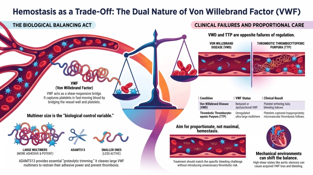
Why this spoke matters
Hemostasis is often taught as protection against bleeding.
But its deeper function is to prevent both bleeding and thrombosis.
At the same time.
The system must solve two problems:
stop bleeding when a vessel is injured
avoid clotting when a vessel is intact
Von Willebrand factor sits at the center of this compromise.
Too little effective VWF produces bleeding.
Too much adhesive VWF activity, or too little regulation of ultra-large VWF, can promote thrombosis.1
The same molecule that helps prevent hemorrhage can threaten vascular patency if insufficiently restrained.
That is the trade-off.
Understanding VWD therefore requires more than asking:
Why does this patient bleed?
It also requires asking:
What does the normal system risk in order to prevent bleeding?
The trade-off frame
Every survival system carries cost.
A system that responds too weakly fails during injury.
A system that responds too strongly injures the host.
Hemostasis is no exception.
In a closed, pressurized, flowing circulation, vascular injury must be sealed quickly.
But blood must also remain fluid across miles of intact endothelium.
This creates a narrow operating range.
Too little adhesion, and platelets cannot establish an early hemostatic surface.
Too much adhesion, and platelets accumulate where they should not.
Too little coagulation, and fibrin cannot stabilize injury.
Too much coagulation, and thrombosis follows.
The biology of VWF reflects this tension.
It is built for rapid action.
It is also built for restraint.
Why VWF is powerful
VWF solves a difficult physical problem.
In fast-moving blood, platelets must be captured before they are swept downstream.
VWF helps by acting as a multimeric bridge between injured vessel wall and platelet GPIbα.2
Its largest multimers are especially effective for platelet adhesion and aggregation under shear because they provide multivalent binding capacity and greater force responsiveness.3
That power is clinically visible in VWD.
When VWF is absent, reduced, structurally abnormal, or functionally impaired, bleeding appears where early platelet capture matters most:
- mucosal surfaces
- epistaxis
- heavy menstrual bleeding
- oral bleeding
- procedural sites
- postpartum tissue beds
- superficial wounds
VWD reveals what VWF normally protects.
Why power creates danger
The same properties that make VWF effective also make it risky.
Large multimers are hemostatically potent because they bind platelets well.
Ultra-large VWF multimers released from endothelial Weibel-Palade bodies are especially adhesive.
If they remain insufficiently regulated, they can capture platelets under flow and contribute to microvascular thrombosis.
The most hemostatically effective forms of VWF are also the forms that require the tightest regulation.4
Otherwise, the boundary between hemostasis and thrombosis begins to collapse.
ADAMTS13 as regulator
ADAMTS13 is often introduced as the protease deficient in thrombotic thrombocytopenic purpura.
That is clinically true.
But conceptually, ADAMTS13 is also a regulator of the hemostatic trade-off.
It limits the adhesive potential of VWF by cleaving unfolded VWF multimers.
The logic is elegant.
Endothelial cells release ultra-large, highly adhesive VWF.
Flow and tethering transmit force through VWF.
Force exposes the A2 domain.
ADAMTS13 cleaves.
Multimer size is reduced.
Adhesive power is restrained.5
Hemostasis is therefore not a static balance of “more clotting” or “less clotting.”
It is a dynamic cycle:
- release
- elongation
- platelet capture
- proteolytic trimming
- clearance
- renewed reserve
Excessive susceptibility of VWF to shear-dependent proteolysis, or extreme shear that accelerates loss of high-molecular-weight multimers, can produce bleeding.
Severe loss of ADAMTS13-mediated regulation allows ultra-large multimers to persist and promotes thrombosis.6
The same axis runs in both directions.
VWD and TTP as opposite lessons
VWD and TTP are often taught in different compartments.
One is a bleeding disorder.
The other is a thrombotic microangiopathy.
But VWF biology connects them.
In VWD, effective VWF function is insufficient.
Platelet tethering fails where it is needed.
Bleeding follows.
In TTP, ultra-large VWF multimers are insufficiently regulated because ADAMTS13 activity is severely deficient.
Platelets are captured where they should not be.
Microvascular thrombosis follows.
These are not the same disease.
But they reveal opposite edges of the same hemostatic constraint.
Too little functional VWF exposes the bleeding risk of flow.
Too much unregulated VWF exposes the thrombotic risk of adhesion.
Multimer size as a balancing variable
Multimer size is not a laboratory curiosity.
It is a biological control variable.
Small multimers are less adhesive.
Large multimers are more adhesive.
Ultra-large multimers are potentially thrombogenic unless regulated.
This explains why VWF multimer distribution matters clinically.
Loss of high-molecular-weight multimers can impair hemostasis, even when antigen is measurable.
Persistence of ultra-large multimers can support platelet-rich thrombosis, even when coagulation factors are not the primary problem.
Multimer size therefore sits between bleeding and thrombosis.
It is not simply “good” or “bad.”
It is useful in the right place, at the right time, in the right amount, under the right regulation.
Acquired VWF defects reveal the trade-off
Acquired von Willebrand syndrome offers another window into the same biology.
In high-shear states, especially severe aortic stenosis, VWF can become excessively unfolded and more susceptible to proteolysis.
High-molecular-weight multimers are lost.
The result may be bleeding, often from gastrointestinal angiodysplasia or other mucosal surfaces.7
Mechanical circulatory support can also disturb platelet and VWF biology through nonphysiologic shear, although mechanisms vary by device and context.8
Here the problem is not inherited absence of VWF.
It is mechanical overprocessing of VWF.
The same force-responsive system that normally permits regulation becomes pathologic when the mechanical environment is extreme.
This is why VWF cannot be understood as a static plasma concentration.
Its function depends on flow.
Its risk depends on context.
Treatment moves the patient along the curve
The trade-off matters clinically because treatment is not biologically neutral.
Desmopressin releases endogenous VWF.
VWF concentrates replace or augment VWF.
Antifibrinolytics stabilize clots locally.
Each intervention can be appropriate.
The issue is not that VWD therapy is inherently unsafe.
The issue is that the intensity, duration, and target of hemostatic correction should match the bleeding challenge and the patient’s thrombotic and treatment-related risks.9
In many patients with VWD, especially those with mucosal bleeding or procedure-related risk, the goal is not maximal correction.
The goal is proportional correction.
Enough hemostatic support to prevent bleeding.
Not so much that unnecessary thrombotic risk, treatment burden, or medicalization is introduced.
This is especially important in:
- older patients
- patients with cardiovascular disease
- patients receiving perioperative replacement
- patients with inflammatory states
- patients with limited mobility
- patients with acquired modifiers
- patients whose bleeding risk is uncertain
Standard therapy is appropriate when the bleeding challenge warrants it.
The clinical judgment lies in the target, duration, context, and monitoring.
The goal is not more hemostasis.
The goal is appropriate hemostasis.
Why mild reduction may be tolerated
Mild reductions in VWF activity may be tolerated across much of ordinary life because hemostasis is redundant.
Platelets, vessel-wall factors, fibrin formation, local vascular tone, and fibrinolytic balance all contribute to bleeding control.
This helps explain why some individuals with low VWF do not bleed substantially.
It also helps explain why risk may appear only during hemostatic stress:
- menarche
- childbirth
- dental extraction
- surgery
- trauma
- anticoagulant exposure
- aging-related vascular disease
A mild deficit may be invisible until reserve is challenged.
That does not make it meaningless.
It makes it conditional.10
The evolutionary frame is useful here, but should be used carefully.
We should not claim that low VWF was selected for without strong evidence.
A more defensible statement is this:
the hemostatic system contains enough redundancy that partial reductions in VWF may be clinically silent in some contexts, while severe deficiency or specific stressors reveal vulnerability.
Bleeding and thrombosis are not opposites
Clinicians often think of bleeding and thrombosis as opposite disorders.
But they are better understood as opposite failures of regulation.
Too little hemostatic activity at the site of injury produces bleeding.
Too much hemostatic activity in the wrong place produces thrombosis.
The same patient may even face both risks at different times.
An older patient with VWD may have bleeding risk from mucosal disease or procedures, while also having cardiovascular disease, immobility, inflammation, or replacement-related thrombotic concern.
A patient with acquired VWF loss from aortic stenosis may bleed from angiodysplasia while also living with a vascular disease that carries its own risks.
The categories “bleeding patient” and “thrombosis patient” can therefore mislead.
The biology is shared.
The balance shifts.
The ethical dimension of trade-off
Trade-offs are not only biological.
They are clinical and ethical.
Undertreatment can cause harm:
- postoperative bleeding
- postpartum hemorrhage
- iron deficiency
- painful or prolonged mucosal bleeding
- reduced quality of life
- loss of trust
Overtreatment can also cause harm:11
- hyponatremia after desmopressin
- unnecessary concentrate exposure
- thrombotic concern in selected settings
- cost
- surveillance burden
- activity restriction
- medical identity without benefit
Good care does not simply mean acting.
It means calibrating.
Sometimes treatment is essential.
Sometimes local or adjunctive therapy is enough.
Sometimes observation with a clear plan is the most rational choice.
Sometimes the ethical act is not to escalate.
This framework makes restraint visible as care, not neglect.
A clinical mental model
When making a VWD treatment decision, ask:
- what bleeding risk are we trying to reduce?
- what hemostatic challenge is coming?
- what mechanism explains this patient’s risk?
- what treatment changes that mechanism?
- what new risks does treatment introduce?
- what would count as success?
- what would make us de-escalate?
This is the practical expression of hemostasis as trade-off.
It moves the clinician away from a reflex:
low VWF → treat
and toward a strategy:
patient + phenotype + mechanism + challenge + risk tolerance → proportional plan
What the trade-off lens adds
The trade-off lens does not replace classification.
It deepens it.
Type 1 VWD raises questions about quantity, reserve, and stress.
Type 2 VWD raises questions about function, binding, multimers, and treatment response.
Type 3 VWD raises questions about severe deficiency, replacement, inhibitors, and lifelong strategy.
Acquired VWF defects raise questions about mechanical environments and comorbid disease.
Therapy raises questions about correction, duration, monitoring, and unintended effects.
All of these are trade-off questions.
How much hemostatic support is enough?
How much is too much?
What risk are we reducing?
What risk are we creating?
Clinical synthesis
Hemostasis is a regulated compromise.
VWF protects against bleeding by enabling platelet capture under flow.
The same properties that make VWF effective, including large multimer size, shear responsiveness, platelet binding, and endothelial release, create thrombotic potential when insufficiently regulated.
ADAMTS13 restrains that potential.
High-shear states can push the system toward acquired VWF loss and bleeding.
Severe ADAMTS13 deficiency can push the system toward microvascular thrombosis.
VWD reveals one edge of the balance.
TTP reveals another.
Treatment moves patients along the same curve.
That is why VWD care requires judgment.
Evidence anchor: why hemostasis is a regulated compromise
Summary derived from VWF mechanobiology, ADAMTS13 studies, multimer analyses, acquired von Willebrand syndrome, and contemporary treatment reviews. The evidence shows that the same features that make VWF effective in preventing bleeding also create thrombotic potential when insufficiently regulated.
| Evidence stream | What it shows | Why it matters | Main limitation |
|---|---|---|---|
| VWF-mediated platelet capture | VWF supports platelet tethering and adhesion under flow, particularly where high shear makes ordinary platelet adhesion difficult.12 | VWF protects against bleeding by solving a physical problem created by flowing blood. | VWF is one part of hemostasis; vessel wall responses, platelets, coagulation, and fibrinolysis also contribute. |
| Multimer size and adhesive potency | High-molecular-weight and ultra-large VWF multimers provide greater multivalent platelet-binding capacity and are especially effective under shear.13 | The forms of VWF that are most effective for hemostasis also require the tightest regulation. | Multimer size does not determine phenotype by itself; binding function, clearance, flow, and clinical context also matter. |
| ADAMTS13 regulation | Force exposes the VWF A2 domain, allowing ADAMTS13 to cleave VWF and limit multimer size and thrombus growth.14 | ADAMTS13 does not simply switch VWF off. It calibrates adhesive potential by regulating multimer size. | The spatial and temporal regulation of ADAMTS13 in vivo remains incompletely understood. |
| VWD and TTP as opposite failures | Reduced or dysfunctional VWF impairs platelet capture and causes bleeding, whereas severe ADAMTS13 deficiency permits persistence of highly adhesive VWF and platelet-rich microvascular thrombosis.15 | Bleeding and thrombosis can be understood as failures at opposite edges of a shared regulatory system. | VWD and TTP are distinct disorders and should not be treated as simple mirror images. |
| Excessive shear and acquired VWF loss | Severe aortic stenosis can cause loss of high-molecular-weight VWF multimers, impaired primary hemostasis, and bleeding that improves after valve replacement.16 | The mechanical environment can move the VWF system toward bleeding even without an inherited VWF defect. | Aortic stenosis is a specific acquired model and does not represent every high-shear state. |
| Mild quantitative reduction | VWF is a continuous trait, and mild reductions may be clinically silent until hemostatic reserve is challenged.17 | Risk depends on phenotype and challenge, not solely on the baseline laboratory value. | Mild bleeding symptoms are common in the general population, so association does not always prove causation. |
Interpretive note: These evidence streams support the same conclusion: hemostasis is not maximized adhesion. It is regulated adhesion. VWF must be sufficiently active to capture platelets where injury occurs, but sufficiently restrained to preserve flow elsewhere. Multimer size, mechanical force, ADAMTS13 activity, and clinical context determine where the system sits between bleeding and thrombosis.
Guidance perspective: treatment should aim for proportionate hemostasis
Based on contemporary VWD treatment reviews and perioperative management guidance. Recommendations do not use the phrase “hemostasis as trade-off” explicitly, but they consistently support individualized treatment matched to bleeding phenotype, VWD type, treatment response, procedural challenge, comorbidity, and treatment-related risk.
Shared guidance themes
- Treatment should be directed at the clinical bleeding risk or anticipated hemostatic challenge, not at the baseline VWF value alone.
- Desmopressin is appropriate only when the patient is likely to respond and when its safety profile is acceptable.
- VWF replacement is indicated when endogenous release is inadequate, unreliable, or unsafe, but dose and duration should be matched to the procedure and monitored response.
- Antifibrinolytics and local measures can reduce the amount of systemic therapy needed, particularly for mucosal and dental bleeding.
- Major surgery generally requires planned perioperative support and monitoring rather than a single treatment decision.
- Patient age, cardiovascular disease, immobility, inflammation, renal function, and the possibility of FVIII accumulation may influence the intensity and duration of replacement.
- Desmopressin requires attention to fluid restriction and hyponatremia risk.
- Standard VWD therapy is appropriate when indicated; the concern is not treatment itself, but treatment that is unnecessarily intense, prolonged, or poorly matched to the patient and challenge.18
How guidance translates into trade-off thinking
| Clinical question | Guidance-based interpretation | Practical implication |
|---|---|---|
| What bleeding risk are we treating? | Therapy should be linked to a specific phenotype, procedure, or anticipated challenge. | Avoid treating a laboratory value without defining the clinical objective. |
| How much correction is needed? | Targets depend on the severity and duration of the challenge. | Minor mucosal bleeding, dental procedures, and major surgery should not receive identical plans. |
| Which therapy best fits the mechanism? | Desmopressin, VWF replacement, antifibrinolytics, hormonal therapy, and local measures address different problems. | Use the least burdensome effective combination rather than defaulting automatically to maximal systemic therapy. |
| How long should support continue? | Duration should reflect ongoing bleeding risk, wound healing, and measured response. | Reassess rather than continuing treatment solely because it was started. |
| What risks does treatment introduce? | Hyponatremia, excessive FVIII levels, thrombotic concern in selected patients, cost, and treatment burden may matter. | Incorporate comorbidity, monitoring, and stopping criteria into the initial plan. |
| What counts as success? | Success is prevention or control of clinically meaningful bleeding, not simply normalization of every laboratory value. | Define the endpoint before treatment begins. |
What guidance does not support
- treating every mildly reduced VWF value
- assuming that a normal post-treatment level guarantees adequate clinical coverage
- using the same plan for every procedure
- continuing replacement without reassessing duration and response
- withholding indicated therapy solely because the patient has cardiovascular risk
- equating restraint with inaction
Practical takeaway
The goal is neither undertreatment nor maximal correction.
The goal is proportionate hemostasis:
- enough support for the actual bleeding challenge
- the right therapy for the underlying mechanism
- the shortest effective duration
- monitoring appropriate to the patient’s comorbidities and treatment response
- a clear definition of success and a plan for de-escalation
Guidance provides the structure. Clinical judgment determines where the patient should sit on the curve between bleeding risk and treatment-related risk.
Reflect & Apply Case
A 72-year-old man with type 1 VWD is scheduled for major abdominal surgery.
He has a history of postoperative oozing after dental extraction.
His baseline VWF antigen is 38 IU/dL.
VWF activity is 34 IU/dL.
He also has coronary artery disease, chronic kidney disease, and limited mobility.
Questions for reflection:
- What bleeding risk is treatment trying to reduce?
- What thrombotic or treatment-related risks might increase if VWF is overcorrected?
- Why is “normalize the VWF level” an incomplete goal?
- How might adjunctive therapy, duration of replacement, and monitoring reflect trade-off thinking?
- What would count as enough hemostatic support?
- What would make you de-escalate?
This case illustrates the central lesson:
VWD management is not simply correction of deficiency.
It is calibration of risk.
Hemostasis protects life because it balances competing dangers.
Good clinical care does the same work deliberately.
Test your thinking
An interactive quiz for this topic is being developed and will be added soon.