
When bleeding persists despite treatment
Note: The video and audio linked above were generated with the assistance of AI. Clinical accuracy has been reviewed, but no AI-generated content can be guaranteed to be fully error-free.
Why this spoke matters
Most VWD treatment works.
Desmopressin can mobilize endogenous VWF. VWF concentrates can replace missing or dysfunctional VWF. Antifibrinolytics can protect mucosal clots. Hormonal therapy can reduce menstrual bleeding. Local measures can stabilize exposed surfaces. Prophylaxis can reduce recurrent bleeding in selected patients.
But sometimes bleeding continues.
The instinct is to do more of the same:
more factor, more doses, longer coverage, higher targets.
Sometimes that is correct.
But not always.
In VWD, persistent bleeding may mean that the hemostatic target was too low.
It may also mean that the target was not the problem.
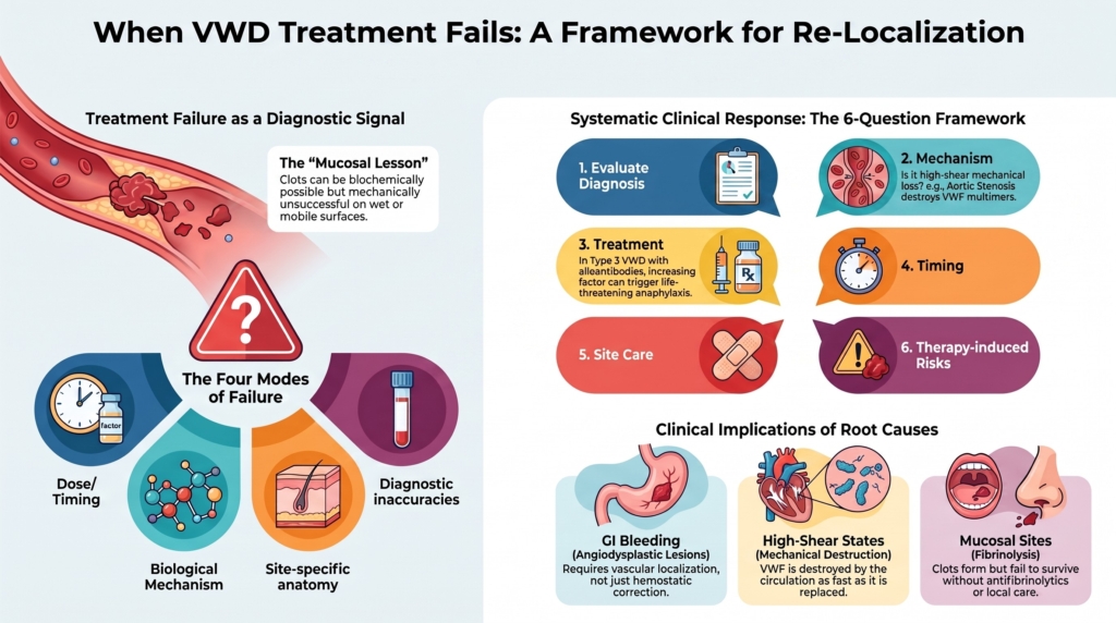
Treatment failure is therefore a diagnostic event.
It asks the clinician to re-localize the bleeding.
Treatment failure is not one thing
“Treatment failure” can mean several different problems.
Dose or timing failure
- not enough VWF
- not enough FVIII
- adequate peak but poor durability
- coverage that ended before the bleeding risk ended
Mechanism failure
- product did not match the VWD subtype
- desmopressin response was short-lived
- FVIII kinetics were wrong
- acquired high-shear VWF loss persisted
- anti-VWF alloantibodies developed
Site failure
- mucosal fibrinolysis was not controlled
- the bleeding site required local intervention
- angiodysplasia persisted
- a surgical, gynecologic, dental, ENT, or GI source was untreated
Diagnostic failure
- another bleeding disorder was present
- medications contributed
- the patient did not have VWD as the main explanation
These are different failures.
They require different responses.
The first question: what failed?
The first question should not be:
What should we add?
It should be:
What part of the hemostatic plan failed?
A useful failure analysis asks whether the problem lies in the diagnosis, the mechanism, the treatment, the timing, the site, or the new risks created by therapy.
This is not delay.
It is disciplined escalation.
Failure after desmopressin
Desmopressin can fail for predictable reasons.
There may be little releasable VWF. The released VWF may be dysfunctional. The response may be short-lived. Tachyphylaxis may limit repeated dosing. The patient may have type 3 VWD. The patient may have type 2B VWD, where desmopressin can worsen platelet binding and thrombocytopenia. The clinical challenge may require sustained coverage that desmopressin cannot provide.
A good one-hour rise does not guarantee durable protection.
A trial may show response but not suitability.
This is why the desmopressin trial should be interpreted as a response curve, not a yes-or-no result.
If DDAVP “fails,” the next step is not simply to declare the drug useless. Ask what kind of failure occurred:
- failure to release VWF
- release of VWF that cleared quickly
- response too brief for the challenge
- missing antifibrinolytic or local support
- wrong subtype or incomplete diagnosis
The response determines the next step.
Failure after replacement therapy
VWF replacement can also fail.
This is more unsettling because replacement feels definitive.
But replacement therapy corrects only part of the problem.
It can supply VWF. It can help stabilize FVIII. It can improve platelet-dependent hemostasis.
It cannot necessarily eliminate an angiodysplastic lesion, fix an unrecognized surgical source, correct a platelet function disorder, overcome an alloantibody safely, or prevent bleeding if the product is cleared too quickly, dosed too infrequently, or mismatched to the clinical timeline.
Persistent bleeding after replacement should prompt a structured review:
- were platelet-dependent VWF activity levels adequate?
- were FVIII levels adequate?
- was there FVIII accumulation?
- was the duration appropriate?
- was monitoring timely?
- was the bleeding site mucosal and fibrinolytic?
- was tranexamic acid or local therapy used?
- was endoscopic, surgical, gynecologic, cardiovascular, or dental treatment needed?
Replacement is powerful.
But it is not magic.
Mucosal bleeding: the clot may form and still fail
Mucosal bleeding is a common reason treatment seems disappointing.
VWF correction may allow clot formation.
But mucosal surfaces challenge clot survival.
The nose is exposed and easily re-injured. The mouth is wet and mechanically active. The uterus is cyclically remodeling. The postpartum uterus is healing while hemostatic physiology is changing. The GI tract may contain fragile vascular malformations.
In these settings, failure may mean that the clot was not protected.
Antifibrinolytics, topical therapy, packing, suturing, cautery, endoscopic therapy, hormonal therapy, or gynecologic intervention may matter as much as additional VWF.
This is the central mucosal lesson:
a clot can be biochemically possible and mechanically unsuccessful.
Gastrointestinal bleeding and angiodysplasia
Recurrent gastrointestinal bleeding is one of the hardest problems in VWD.
It is overrepresented in severe VWD, especially type 2A and type 3 disease, and in acquired VWF disorders. It is often associated with angiodysplasia. It may be recurrent, obscure, transfusion-requiring, and difficult to localize.
VWF replacement may reduce bleeding.
Prophylaxis may reduce recurrence.
But GI bleeding may continue despite apparently adequate VWF activity.
That is because the problem is not only hemostatic.
It is also vascular.
VWF biology appears to intersect with angiogenesis and vascular integrity, which may contribute to the association between VWD and angiodysplasia, although the mechanisms remain incompletely defined.1
A patient with recurrent GI bleeding therefore needs more than dose escalation.
They need localization.
Why GI bleeding resists simple correction
GI bleeding may persist because the lesion remains present.
Angiodysplastic lesions may be multiple, intermittent, inaccessible, or missed on endoscopy. VWF trough levels may be inadequate. The product may not provide sustained hemostatic coverage. FVIII may rise excessively with repeated VWF/FVIII dosing. High-shear acquired VWF loss may persist. Antiplatelet or anticoagulant therapy may be necessary. Anemia, renal disease, aortic stenosis, or other vascular modifiers may reduce physiologic reserve.
The “failed treatment” may be a successful hemostatic correction applied to an ongoing vascular lesion.
That distinction matters.
If the lesion persists, the bleeding may recur.
What to do with refractory GI bleeding
The first step is to confirm the mechanism and site as much as possible.
Consider:
- repeat endoscopic evaluation
- capsule endoscopy or deep enteroscopy when appropriate
- assessment for angiodysplasia
- review of VWD type and multimer pattern
- evaluation for acquired VWS
- review of aortic stenosis, LVAD, or other high-shear states
- review of antiplatelet or anticoagulant therapy
- assessment of iron deficiency and transfusion burden
- review of VWF product, dose, trough, and FVIII kinetics
VWF concentrate prophylaxis may reduce recurrent bleeding in selected patients, but optimal dosing is uncertain. One expert approach suggests starting VWF concentrate prophylaxis at 50 VWF IU/kg twice weekly for recurrent GI bleeding and escalating to three times weekly if bleeding is not controlled, but this should be understood as expert strategy rather than universal rule.2
Reported salvage approaches include thalidomide, octreotide, atorvastatin, tamoxifen, danazol, and bevacizumab in selected refractory cases, but evidence comes largely from case reports and small series, and none should be considered routine VWD therapy.3
These approaches should be presented modestly.
They are attempts to treat a difficult vascular-hemostatic phenotype when standard approaches are insufficient.
Acquired VWF loss: when the circulation keeps breaking the molecule
Sometimes treatment fails because the patient is not simply deficient.
The circulation is actively destroying the most hemostatically effective VWF forms.
Severe aortic stenosis is the classic example.
High shear across the stenotic valve can unfold VWF and promote loss of high-molecular-weight multimers, producing an acquired type 2A-like VWF defect.
This may contribute to bleeding, especially from gastrointestinal angiodysplasia, in what is often called Heyde syndrome.
In a study of patients with aortic stenosis, skin or mucosal bleeding occurred in 21% of patients with severe stenosis, and platelet-function abnormalities under high shear, reduced VWF collagen-binding activity, loss of largest multimers, or combinations of these were present in 67% to 92%. These abnormalities correlated with stenosis severity and improved after valve replacement when prosthesis mismatch was absent.4
The lesson is crucial.
If the mechanical lesion persists, replacement may be temporary.
The definitive treatment may be cardiovascular.
Mechanical circulatory support and other high-shear states
Mechanical circulatory support is another recognized high-shear setting associated with acquired VWF abnormalities, often with loss of high-molecular-weight multimers and GI bleeding.
In this setting, bleeding is not simply “VWD-like.”
It is VWF biology reshaped by the device.
Treatment may require cardiology collaboration, device assessment, speed optimization when possible, GI evaluation, antithrombotic adjustment, VWF-directed therapy in selected cases, iron and transfusion support, and management of angiodysplasia.
This is not inherited VWD management copied onto a device patient.
It is acquired VWF failure under mechanical stress.
Anti-VWF alloantibodies
Anti-VWF alloantibodies are rare.
But when present, they radically change treatment.
They occur mainly in type 3 VWD, especially in patients with large deletions or other severe null genotypes.
They may cause loss of response to VWF concentrates.
More importantly, patients may develop life-threatening anaphylactic reactions to VWF-containing products.5
This is one of the clearest examples where “more replacement” can be dangerous.
If anti-VWF alloantibodies are suspected, VWF-containing concentrates should not simply be continued.
Specialized management is required.
Consider anti-VWF alloantibodies when a patient with type 3 VWD has poor recovery after VWF infusion, unexpectedly short VWF survival, bleeding despite apparently appropriate dosing, infusion reactions, anaphylaxis, or a history of a large VWF gene deletion or severe null genotype.
Reported management approaches include recombinant FVIII, recombinant activated factor VII, and, in rare anecdotal reports, off-label emicizumab prophylaxis for type 3 VWD with alloantibodies.6
These are difficult, rare scenarios.
They should not be generalized to ordinary VWD care.
But they deserve a home in the module because they represent true treatment failure.
When FVIII becomes part of the failure
VWD treatment can fail because FVIII is too low.
It can also become complicated because FVIII becomes too high.
With repeated VWF/FVIII concentrate dosing, FVIII may accumulate. This is especially relevant in surgery, prophylaxis, older patients, cancer surgery, orthopedic surgery, immobility, and cardiovascular disease.
VWF-only products may be attractive in selected settings where repeated dosing is expected and FVIII accumulation is a concern.7
This is not treatment failure in the usual sense.
It is a reminder that “successful correction” can create a second problem.
In VWD, the goal is not maximal hemostasis.
It is proportional hemostasis.
Antiplatelet and anticoagulant therapy
Another difficult scenario occurs when a patient with VWD needs antiplatelet or anticoagulant therapy.
This may happen with atrial fibrillation, venous thromboembolism, coronary artery disease, stroke prevention, mechanical valves, or other cardiovascular disease.
The 2021 ASH/ISTH/NHF/WFH management guideline suggests giving necessary antiplatelet or anticoagulant therapy rather than withholding it in patients with VWD and cardiovascular disease, with individualized reassessment of bleeding risk. It also notes that patients with severe bleeding phenotypes may require VWF concentrate prophylaxis while receiving these therapies.8
This matters because failure can be conceptual.
The clinician may try to avoid thrombosis therapy because the patient bleeds.
But VWD does not remove thrombotic risk.
Sometimes the solution is not choosing bleeding care over cardiovascular care.
It is building a hemostatic plan that permits appropriate cardiovascular care.
When the diagnosis is incomplete
Treatment can fail because the diagnosis is incomplete.
The patient may have a platelet function disorder, platelet-type VWD, connective tissue disorder, hereditary hemorrhagic telangiectasia, acquired VWF syndrome, gynecologic pathology, liver disease, renal disease, medication-associated bleeding, unrecognized anticoagulant or antiplatelet exposure, NSAID use, SSRI or SNRI exposure, supplements affecting hemostasis, a local anatomic lesion, or malignancy.
This is especially important when bleeding is severe but VWF studies are only mildly abnormal.
A borderline VWF level can be real and still not be the whole explanation.
When the phenotype is worse than the VWF pattern predicts, the correct response is not simply more VWD therapy.
It is reopening localization.
The “wrong site” problem
Sometimes treatment fails because the bleeding site has not been treated.
Nasal bleeding may need nasal care, cautery, humidification, or ENT evaluation. Dental bleeding may need sutures, packing, topical antifibrinolytics, and careful dental technique. Heavy menstrual bleeding may need gynecologic assessment and reproductive-context treatment. GI bleeding may need endoscopic and vascular evaluation. Postoperative bleeding may need surgical assessment. Postpartum bleeding may need obstetric evaluation for uterine atony, retained products, laceration, infection, and delayed hemostatic decline.
VWF therapy can improve hemostasis.
It cannot replace site-specific care.
The site is part of the mechanism.
A framework for treatment failure
When bleeding persists, ask six questions.
1. Was the diagnosis right?
Does the phenotype fit VWD, or is another disorder present?
2. Was the mechanism right?
Is this quantitative deficiency, qualitative dysfunction, clearance, acquired high-shear loss, inhibitor, or local lesion?
3. Was the treatment right?
Was the correct combination of DDAVP, VWF replacement, antifibrinolytic, hormonal therapy, local therapy, or other intervention used?
4. Was the timing right?
Was coverage early enough, high enough, and long enough?
5. Was the site treated?
Did the local anatomy require procedural, endoscopic, gynecologic, ENT, dental, cardiovascular, or surgical management?
6. What new risk did treatment create?
Hyponatremia, FVIII accumulation, thrombosis, anaphylaxis, venous access burden, or overtreatment?
This framework prevents reflex escalation.
It turns failure into information.
When to refer or escalate care
Some failures require specialized centers.
Examples include:
- recurrent GI bleeding despite prophylaxis
- suspected angiodysplasia with transfusion dependence
- type 3 VWD with suspected alloantibody
- infusion reaction or anaphylaxis after VWF concentrate
- bleeding despite adequate measured VWF and FVIII
- need for antithrombotic therapy in severe VWD
- major surgery in severe type 2 or type 3 VWD
- possible acquired VWF syndrome from aortic stenosis or mechanical circulatory support
- uncertain diagnosis with severe phenotype
These are not routine management problems.
They are multidisciplinary problems.
The right team may include hematology, gastroenterology, cardiology, gynecology, anesthesia, surgery, transfusion medicine, laboratory medicine, and specialized bleeding-disorder nursing.
Clinical synthesis
When treatment fails in VWD, do not begin with more.
Begin with localization.
Failure may reflect inadequate VWF correction. But it may also reflect short duration, mucosal fibrinolysis, local anatomy, angiodysplasia, acquired high-shear VWF loss, alloantibodies, FVIII kinetics, antithrombotic needs, or an incomplete diagnosis.
Recurrent gastrointestinal bleeding is especially challenging because it is often both hemostatic and vascular.
Anti-VWF alloantibodies are rare but critical because VWF-containing products may provoke anaphylaxis.
High-shear states such as aortic stenosis or mechanical circulatory support can keep removing the most effective VWF multimers until the mechanical problem is addressed.
Persistent bleeding should not be interpreted as a simple failure of will, dose, or compliance.
It is a signal.
The question is not only:
How do we stop the bleeding?
It is:
What does the bleeding tell us we have not yet understood?
Evidence anchor: why treatment failure should prompt re-localization
Summary derived from VWD treatment guidelines, severe VWD reviews, acquired VWF literature, refractory GI bleeding discussions, and expert treatment reviews. The evidence consistently shows that persistent bleeding in VWD may reflect inadequate hemostatic correction, but it may also reflect the wrong mechanism, wrong site, acquired VWF loss, vascular lesions, alloantibodies, medication effects, or competing thrombotic needs.
| Evidence stream | What it shows | Why it matters | Main limitation |
|---|---|---|---|
| DDAVP response and durability | Desmopressin response may be absent, unsafe, short-lived, or inadequate for the clinical challenge. A peak response does not guarantee sustained coverage.9 | DDAVP “failure” should be interpreted as a response-pattern problem, not simply a yes/no drug judgment. | Definitions of adequate response vary by challenge and study. |
| Replacement failure | VWF replacement can correct VWF activity and FVIII support, but bleeding may persist if duration, trough levels, FVIII kinetics, local clot protection, or site-specific therapy are inadequate.10 | Adequate factor exposure does not exclude an untreated local or vascular problem. | Laboratory targets are surrogates for tissue-level hemostasis. |
| Mucosal bleeding biology | Mucosal bleeding may persist because clots form but fail to survive in wet, mobile, fibrinolytic, or mechanically stressed surfaces.11 | Antifibrinolytics, local measures, dental, ENT, gynecologic, endoscopic, or surgical interventions may be as important as additional VWF. | Evidence is site-specific and often based on expert practice. |
| GI bleeding and angiodysplasia | GI bleeding, especially angiodysplasia-associated bleeding, is overrepresented in severe VWD and acquired VWF disorders and may recur despite acute correction.12 | Recurrent GI bleeding is often both hemostatic and vascular; dose escalation alone may miss the lesion. | Optimal prophylaxis dosing and salvage strategies remain uncertain. |
| Acquired high-shear VWF loss | Severe aortic stenosis can cause acquired loss of high-molecular-weight VWF multimers and bleeding abnormalities that correlate with stenosis severity and improve after valve replacement when prosthesis mismatch is absent.13 | If the mechanical lesion persists, VWF replacement may be temporary; cardiovascular intervention may be hemostatic therapy. | Data are strongest for aortic stenosis; other high-shear settings require context-specific evidence. |
| Anti-VWF alloantibodies | Rare anti-VWF alloantibodies, mainly in type 3 VWD, may cause poor recovery, short survival, persistent bleeding, infusion reactions, and anaphylaxis to VWF-containing products.14 | This is a setting where more VWF replacement may be dangerous. | Management evidence is limited and requires expert-center care. |
| Antithrombotic therapy | Guidelines suggest giving necessary antiplatelet or anticoagulant therapy in VWD patients with cardiovascular disease, with individualized reassessment and prophylaxis in selected severe bleeding phenotypes.15 | VWD does not eliminate thrombosis risk; bleeding care may need to support cardiovascular care. | Evidence is limited and decisions are highly individualized. |
| Incomplete diagnosis | Disproportionate bleeding may reflect another or additional diagnosis, including platelet disorders, acquired VWS, hereditary hemorrhagic telangiectasia, connective tissue disease, medications, organ disease, gynecologic pathology, local lesions, or malignancy.16 | A borderline VWF level can be real and still not be the whole explanation. | Re-evaluation can be complex and may require specialized testing. |
Interpretive note: These evidence streams point in the same direction: persistent bleeding is not automatically proof that the patient needs more factor. It may mean the wrong mechanism, wrong duration, wrong site, wrong product, wrong diagnosis, or wrong system problem has been targeted.
Practical takeaway: When treatment fails in VWD, do not begin with more. Begin with localization.
Guideline perspective: refractory bleeding requires reassessment, not reflex escalation
Based on ASH/ISTH/NHF/WFH VWD diagnosis and management guidance, supported by severe VWD reviews, acquired VWS literature, and expert treatment reviews.
Shared guidance themes
- Persistent bleeding should prompt reassessment of diagnosis, VWD subtype, VWF activity, FVIII activity, treatment timing, and clinical context.17
- DDAVP should not be treated as adequate simply because a peak response occurs. Suitability depends on magnitude, durability, safety, VWD subtype, and the hemostatic challenge.18
- Replacement therapy should be monitored with attention to both platelet-dependent VWF activity and factor VIII activity (FVIII:C), particularly during repeated dosing, surgery, prophylaxis, severe disease, or thrombotic-risk settings.19
- Recurrent GI bleeding should trigger evaluation for angiodysplasia, acquired VWS, high-shear physiology, medication effects, iron deficiency, transfusion burden, and need for gastroenterology or endoscopic intervention.20
- GI prophylaxis dosing remains uncertain. Expert approaches may be used in selected refractory patients, but should not be presented as universal dosing rules.21
- Salvage approaches for refractory angiodysplasia-associated bleeding, including antiangiogenic or vascular-directed therapies, should be reserved for selected cases and discussed as limited-evidence strategies rather than routine VWD therapy.22
- Suspected anti-VWF alloantibodies in type 3 VWD require expert-center management, especially when poor recovery, short survival, infusion reaction, or anaphylaxis occurs.23
- Patients with VWD and cardiovascular disease should generally receive necessary antiplatelet or anticoagulant therapy, with individualized bleeding-risk reassessment and prophylaxis in selected severe bleeding phenotypes.24
- When bleeding phenotype is disproportionate to VWF findings, clinicians should consider additional diagnoses, medications, acquired VWS, local lesions, gynecologic disease, organ dysfunction, or vascular disorders.25
What guidelines do not eliminate
- uncertainty in refractory GI bleeding
- limited evidence for antiangiogenic and salvage therapies
- the need to distinguish acute correction from prevention of recurrence
- the need to treat local anatomy and not only plasma levels
- the risk of FVIII accumulation during repeated VWF/FVIII dosing
- the danger of VWF concentrate exposure in suspected alloantibody-associated anaphylaxis
- the need to balance bleeding and thrombosis in cardiovascular disease
- the need for multidisciplinary referral when standard therapy fails
Practical takeaway: Guidelines provide treatment tools, but refractory bleeding requires re-localization. The clinician must ask whether the problem is VWF level, FVIII kinetics, product choice, timing, mucosal clot survival, vascular lesion, high-shear acquired VWF loss, inhibitor, medication exposure, or incomplete diagnosis.
Reflect & Apply Case
A 72-year-old patient with type 2A VWD has recurrent melena from suspected small-bowel angiodysplasia.
He has required three hospitalizations in the past year.
He receives VWF/FVIII concentrate during bleeding episodes.
His platelet-dependent VWF activity rises appropriately after infusion.
His factor VIII activity (FVIII:C) becomes high with repeated dosing.
Bleeding stops transiently, then recurs weeks later.
He also has moderate-to-severe aortic stenosis.
Questions for reflection:
- Has replacement therapy failed, or has episodic treatment failed?
- What does the appropriate post-infusion VWF rise tell you?
- Why might bleeding recur despite adequate acute correction?
- How does angiodysplasia change the treatment problem?
- How might aortic stenosis contribute to the VWF phenotype?
- What would you reassess before simply increasing the VWF dose?
- What specialists should be involved?
- What would count as success: fewer bleeds, fewer hospitalizations, lower transfusion burden, improved iron status, or lesion control?
Expert synthesis: This patient’s acute replacement response appears biologically adequate, but episodic treatment is failing to prevent recurrence. The likely problem is not simply too little VWF. It is recurrent vascular-site bleeding, possible angiodysplasia, FVIII accumulation with repeated VWF/FVIII dosing, and a high-shear cardiac lesion that may contribute to acquired VWF dysfunction. Management should prioritize re-localization, GI and cardiology collaboration, consideration of prophylaxis or product change with FVIII monitoring, and attention to the aortic stenosis as a potentially hemostatic problem. Success would be measured by fewer hospitalizations, fewer transfusions, improved iron status, reduced recurrence, and better control of the vascular source, not simply by post-infusion VWF levels.
This case illustrates the central lesson:
treatment failure in VWD is not the end of reasoning.
It is the moment when reasoning matters most.
Test your thinking
An interactive quiz for this topic is being developed and will be added soon.