
Four ways function can fail
Note: The video and audio linked above were generated with the assistance of AI. Clinical accuracy has been reviewed, but no AI-generated content can be guaranteed to be fully error-free.
Why this spoke matters
Type 2 VWD is where classification becomes clinically useful.
In type 1 VWD, the main problem is quantity: there is not enough VWF. In type 2 VWD, the problem is function. There may be VWF in plasma, and the VWF antigen level may be only modestly reduced or even near-normal. But the protein does not perform one of its essential jobs.
That distinction changes everything:
- how the laboratory pattern is interpreted
- how the activity-to-antigen ratio is used
- when multimer analysis matters
- when RIPA matters
- when FVIII is the key clue
- when genetic testing helps
- when desmopressin is useful, risky, or insufficient
- how procedural plans are made
Type 2 VWD is not one disorder. It is a family of functional failures.
The central idea
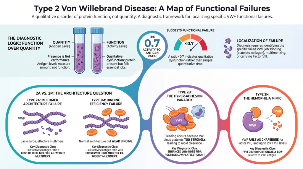
VWF has several clinically important jobs. It must bind platelet glycoprotein Ib under conditions that permit platelet tethering. It must bind collagen at sites of vascular injury. It must multimerize into large forms that are especially effective in high-shear hemostasis. It must carry and stabilize factor VIII.
Type 2 VWD occurs when one of these functions is disproportionately impaired. The protein may be present, but presence is not performance.
That is the core lesson.
Why antigen alone is not enough
VWF antigen measures how much VWF protein is present. That is useful, but it cannot tell whether the VWF works. A patient with type 2 VWD may have a VWF antigen level that is modestly reduced, normal, or near-normal. If only antigen is measured, the diagnosis may be missed.
That is why the initial VWD panel requires at least:
- VWF antigen
- platelet-dependent VWF activity
- FVIII activity
Additional assays, including collagen binding, multimer analysis, RIPA, FVIII-binding assays, and genetic testing, localize the functional defect when the initial pattern suggests type 2 VWD.1
Type 2 VWD teaches a diagnostic rule: amount is not the same as function.
The activity-to-antigen ratio
The activity-to-antigen ratio is one of the most important clues to type 2 VWD. In type 1 VWD, antigen and activity usually fall together, so the ratio is preserved. In many type 2 variants, activity is disproportionately low relative to antigen, producing a low ratio.
The 2021 ASH/ISTH/NHF/WFH diagnostic guideline suggests using a platelet-dependent activity-to-antigen ratio cutoff of less than 0.7, rather than less than 0.5, to confirm type 2 VWD in patients with an abnormal initial VWD screen.2
This shortcut mainly helps with type 2A, type 2B, and type 2M. Type 2N is different: platelet-dependent VWF activity may be relatively preserved, and the clue is FVIII that is disproportionately low relative to VWF.3
The ratio does not finish the diagnosis. It points to the next question: which function has failed?
A map of type 2 VWD
Type 2 VWD is traditionally divided into four subtypes:
- type 2A: loss or marked reduction of high-molecular-weight multimers
- type 2B: gain-of-function binding of VWF to platelet GPIbα
- type 2M: impaired platelet or collagen binding without selective loss of high-molecular-weight multimers
- type 2N: impaired binding of VWF to FVIII
The letters are not arbitrary labels. They are a functional map.
| Subtype | What fails? | Typical multimer pattern | Key clue |
|---|---|---|---|
| Type 2A | effective multimer architecture | loss of high-molecular-weight multimers | low activity-to-antigen ratio plus HMW multimer loss |
| Type 2B | excessive platelet binding | often loss of HMW multimers, but not always | enhanced low-dose RIPA; possible thrombocytopenia |
| Type 2M | platelet or collagen binding | HMW multimers preserved | low activity-to-antigen ratio without HMW multimer loss, or selective collagen-binding defect |
| Type 2N | FVIII carriage | usually normal | FVIII disproportionately low relative to VWF |
Type 2A: when large multimers are lost
Type 2A VWD is characterized by impaired VWF-dependent platelet adhesion caused by loss or marked reduction of high-molecular-weight multimers. Large multimers matter because they are the most hemostatically effective forms of VWF, especially under high shear stress.
In type 2A, VWF antigen may be present, but the most adhesive architecture is missing. The activity-to-antigen ratio is usually reduced. Collagen-binding may also be disproportionately reduced, particularly when the collagen-binding assay is sensitive to high-molecular-weight multimers. Multimer analysis shows loss of the larger forms.4
The clinical logic is straightforward: VWF is present, but the forms most able to build a platelet plug are depleted.
Why type 2A happens
Type 2A is not one molecular story. It is a final common phenotype: too few large multimers. That phenotype can arise through several mechanisms:
- defective multimer assembly
- impaired intracellular transport or secretion
- increased susceptibility to ADAMTS13-mediated proteolysis
- increased VWF clearance
- combinations of these processes
Historically described type 2A patterns, including IIA, IIC, IID, and IIE, reflect different molecular routes to the same functional endpoint. A2-domain variants often increase ADAMTS13 susceptibility, D3-domain variants often impair multimerization, D1-D2 propeptide variants can impair multimer assembly, and CK-domain variants can disrupt dimerization.5
This is an important principle for the whole VWD module: a subtype can name a functional phenotype while containing multiple biological mechanisms.
Type 2A bleeding phenotype
Type 2A often produces mucocutaneous bleeding, including epistaxis, bruising, gum bleeding, heavy menstrual bleeding, post-dental bleeding, and postoperative bleeding. Gastrointestinal bleeding, particularly from angiodysplasia, is especially important in patients with loss of high-molecular-weight multimers.
Several cohorts suggest that type 2A may carry a greater bleeding burden on average than type 2M, particularly for gastrointestinal bleeding, although individual phenotype remains variable. This difference may not be fully explained by VWF or FVIII level alone. The qualitative defect itself, especially loss of the most effective multimers, matters.6
Severity in VWD is not simply how much antigen is present. It is whether the right form of VWF is available at the right moment.
Type 2B: when binding is too strong
Type 2B VWD is conceptually different. The problem is not weak platelet binding. It is excessive platelet binding.
Type 2B is caused by gain-of-function VWF variants, usually in the A1 domain, that increase binding to platelet GPIbα. This can lead to spontaneous or exaggerated VWF-platelet interaction, clearance of VWF-platelet complexes, loss of high-molecular-weight multimers, and thrombocytopenia.7
This is the paradox that makes type 2B memorable: the phenotype is bleeding, but the molecular behavior is hyperadhesion. Too much binding produces loss. Too much interaction produces deficiency.
Type 2B is heterogeneous
The classic type 2B pattern includes:
- enhanced low-dose RIPA
- low activity-to-antigen ratio
- loss of high-molecular-weight multimers
- mild, intermittent, or persistent thrombocytopenia
- worsening thrombocytopenia during stress, pregnancy, surgery, infection, or after desmopressin
But not every patient follows the classic pattern. Some patients with type 2B have preserved large multimers, normal platelet counts, and less typical laboratory profiles. Enhanced VWF-platelet interaction remains the defining functional abnormality, but multimer pattern and platelet count may vary.8
This matters because type 2B should not be excluded simply because one expected feature is absent. The diagnostic question is not only “are the multimers missing?” It is also “is platelet binding pathologically increased?”
Type 2B and platelet-type VWD
Type 2B can be confused with platelet-type VWD. The phenotypes overlap because both involve increased VWF-platelet interaction. In type 2B, the abnormality is in VWF. In platelet-type VWD, the abnormality is in platelet GPIbα, usually due to GP1BA variants.
Enhanced low-dose RIPA is therefore not specific for type 2B VWD. When the phenotype suggests increased VWF-platelet interaction, platelet-type VWD must be excluded by specialized RIPA mixing studies and/or targeted genetic testing.9
Type 2B teaches a diagnostic warning: the same phenotype can arise from opposite sides of an interaction.
Why type 2B matters clinically
Type 2B matters because it changes treatment. Desmopressin is generally avoided in type 2B VWD because it can release abnormal VWF, intensify VWF–platelet binding, and worsen thrombocytopenia. Rare minor-use exceptions should be specialist-directed and based on a careful understanding of the patient’s phenotype and prior response.10
In many clinically significant type 2B situations, treatment relies on VWF-containing concentrate plus adjunctive measures such as tranexamic acid when appropriate. The goal is not simply to raise VWF antigen. It is to provide hemostatic VWF without worsening the VWF-platelet interaction.
Type 2M: when binding is weak but multimers remain
Type 2M VWD is often harder to teach because the architecture looks relatively preserved. The “M” refers to multimers: type 2M describes decreased VWF-dependent platelet adhesion, or in some cases collagen binding, without selective loss of high-molecular-weight multimers.
The VWF multimers are present, but function is impaired. Classically, this involves reduced platelet GPIb binding with preserved multimers. Some type 2M variants primarily impair collagen binding rather than platelet GPIb binding, especially A3-domain variants. These may require VWF collagen-binding assays and/or genetic testing for correct classification.11
Type 2M teaches a subtle but important principle: normal architecture does not guarantee normal binding.
Why type 2M is often missed
Type 2M is a diagnostic trap. If the laboratory panel includes only VWF antigen, platelet-dependent activity, and FVIII, type 2A and type 2M may look similar because both can show a low activity-to-antigen ratio. Without multimer analysis or collagen-binding assessment, type 2M may be misclassified as type 1 or type 2A.
This underrecognition is not trivial. It affects diagnosis, family counseling, interpretation of bleeding risk, and treatment planning. Reviews and external quality assessment studies have repeatedly emphasized that type 2M is frequently misidentified, partly because many laboratories do not have access to the full testing panel needed to distinguish it from type 2A.12
The key distinction is this:
- type 2A: function is impaired because large multimers are missing.
- type 2M: function is impaired despite preserved large multimers.
That is why type 2M belongs in a reasoning-based module. It asks the clinician not just to see that function is abnormal, but to localize why.
Why collagen-binding defects matter
VWF must bind both platelets and subendothelial matrix. The classic platelet-dependent activity assay focuses on platelet binding, but VWF also binds collagen through specific domains that can be selectively affected.
A3-domain variants may cause reduced collagen binding with relatively preserved platelet-dependent activity, antigen, and multimer pattern. In these cases, the usual platelet-binding assay may not fully explain the bleeding phenotype. Collagen-binding assays can therefore provide important functional information, especially when the clinical picture suggests VWD but the standard activity pattern is incomplete.13
This is another type 2 lesson: VWF has more than one functional surface.
Type 2N: when FVIII carriage fails
Type 2N VWD is unique enough to deserve its own essay, but it belongs on the type 2 map. In type 2N, VWF cannot bind FVIII normally. Platelet adhesion and collagen binding may be relatively preserved. VWF antigen may be normal or only mildly reduced. The multimer pattern is usually normal.
The striking abnormality is low FVIII, often out of proportion to the VWF antigen level. This can mimic mild or moderate hemophilia A in males, or hemophilia A carrier status in females.14
Type 2N is conceptually powerful because it reminds us that VWF is not only a platelet-adhesion protein. It is also the chaperone for FVIII.
Type 2N as the exception to the ratio shortcut
The activity-to-antigen ratio is central for many type 2 variants, but type 2N does not necessarily announce itself through low platelet-dependent activity relative to antigen. Instead, the clue is disproportionate FVIII reduction relative to VWF.
Type 2N is usually recessively inherited. A low FVIII-to-VWF antigen ratio should raise suspicion, especially when the inheritance pattern does not fit X-linked hemophilia A. Confirmation generally requires a VWF:FVIIIB assay and/or molecular testing of VWF.15
Type 2N asks a different functional question: can VWF carry FVIII?
Type 2N treatment implications
Type 2N also changes treatment logic. Because FVIII clearance is accelerated when FVIII cannot bind VWF, FVIII-only replacement may have a short half-life. When replacement therapy is required, VWF-containing concentrates are preferred because they provide functional VWF that stabilizes FVIII and supports sustained FVIII correction.16
Desmopressin may raise FVIII transiently in some patients, but it does not correct the underlying FVIII-binding defect. It should be used only when a test dose demonstrates adequate and sustained FVIII levels for the planned challenge.
The laboratory pattern is a localization tool
Type 2 VWD is best taught as laboratory localization. The tests are not isolated facts; they ask where VWF function has failed.
- Low activity-to-antigen ratio: qualitative VWF dysfunction, especially type 2A, 2B, or 2M.
- Loss of high-molecular-weight multimers: type 2A or classic type 2B.
- Enhanced low-dose RIPA: type 2B or platelet-type VWD.
- Preserved multimers with low platelet-dependent activity: type 2M.
- Disproportionately low FVIII: type 2N or hemophilia A.
- Low collagen binding with preserved platelet activity: collagen-binding defect, often within type 2M biology.
A type 2 workup is therefore not just confirmatory. It is anatomical in a functional sense: platelet surface, collagen surface, multimer structure, or FVIII-binding site.
RIPA: a test with a specific job
Ristocetin-induced platelet agglutination, or RIPA, is not a general screening test for VWD. Its most important role is to identify enhanced VWF-platelet interaction, especially in type 2B VWD and platelet-type VWD.
Low-dose ristocetin causes abnormal platelet agglutination when VWF-platelet binding is pathologically increased. That finding helps separate type 2B from type 2A and type 2M, but it does not by itself prove that the defect is in VWF rather than platelet GPIbα. Additional testing, including mixing studies or genetic testing, may be needed to distinguish type 2B VWD from platelet-type VWD.17
The question RIPA asks is narrow but important: is binding too strong?
Multimer analysis: structure becomes visible
Multimer analysis made type 2 VWD intelligible. It showed that not all VWF dysfunction is about total amount. Some patients lack the largest, most hemostatically effective multimers, explaining why antigen can be present but function impaired.
The multimer pattern helps distinguish:
- type 2A, where high-molecular-weight multimers are reduced or absent
- type 2B, where high-molecular-weight multimers are often reduced but may be preserved in some variants
- type 2M, where high-molecular-weight multimers are generally preserved
- type 2N, where multimers are usually normal
Multimer analysis is not just a confirmatory test. It is a structural lens. It lets clinicians see the architecture behind the activity result.
Genetics can clarify, but phenotype still leads
Genetic testing is increasingly useful in type 2 VWD. It can help:
- confirm type 2B VWD
- distinguish type 2B from platelet-type VWD
- distinguish type 2N VWD from hemophilia A
- support classification when phenotypic tests are ambiguous
- assist family counseling
- clarify genotype-phenotype patterns in type 2M and type 2A
Contemporary reviews emphasize that genetic testing is especially useful for confirming type 2 subtypes and type 3 VWD. But classification should still begin with the phenotype: what is the protein doing or failing to do?18
Genotype can clarify. Phenotype localizes the failure.
Bleeding phenotype in type 2 VWD
Type 2 VWD often produces mucocutaneous bleeding:
- epistaxis
- easy bruising
- oral bleeding
- heavy menstrual bleeding
- post-dental bleeding
- postoperative bleeding
- postpartum bleeding
The pattern varies by subtype. Patients with type 2A, type 2B, and type 2M often bleed like patients with disorders of primary hemostasis. GI bleeding may be especially important in patients with loss of high-molecular-weight multimers, particularly type 2A. Patients with type 2N may have a more hemophilia-like pattern because the dominant abnormality is low FVIII, with bleeding after surgery, trauma, tonsillectomy, dental extraction, or postpartum hemostatic challenge.19
Subtype helps predict risk. It does not replace the patient’s personal bleeding history.
Why type 2 can be more severe than type 1
A patient with type 2 VWD may have a VWF antigen level that looks less alarming than a patient with type 1 VWD. But the functional defect may be more clinically important.
Examples:
- A patient with type 2A may have VWF antigen present but lack large multimers.
- A patient with type 2B may have ongoing VWF-platelet binding, multimer loss, and thrombocytopenia.
- A patient with type 2M may have VWF architecture present but binding impaired.
- A patient with type 2N may have low FVIII and procedure-related bleeding.
This is why clinicians should not rank VWD severity by antigen alone. A qualitative defect can be more consequential than the amount suggests.
Treatment principles
Treatment in type 2 VWD depends on subtype, bleeding phenotype, baseline VWF and FVIII levels, prior response to therapy, and the hemostatic challenge. The major tools are:
- local measures
- tranexamic acid
- hormonal therapy for heavy menstrual bleeding
- desmopressin in selected patients
- VWF-containing concentrate when desmopressin is ineffective, risky, or insufficient
Desmopressin responsiveness in type 2 VWD is subtype- and variant-dependent. It should not be assumed. A trial is generally needed to assess magnitude and duration of response, except that type 2B is the major caution because of thrombocytopenia risk.20
For major bleeding, major surgery, high-risk sites, or patients with poor desmopressin response, VWF replacement therapy is usually required. Monitoring may need to include both VWF activity and FVIII because FVIII can accumulate with repeated VWF/FVIII concentrate dosing.21
The treatment lesson is the same as the diagnostic lesson: first localize the failure, then match therapy to the failure and the challenge.
Subtype-specific treatment implications
Type 2 classification is clinically useful because each subtype carries different treatment implications.
- Type 2A: desmopressin may help some patients, but response is often limited or short-lived; VWF concentrate is often needed for major bleeding or surgery.
- Type 2B: desmopressin is generally avoided because it may worsen platelet-VWF binding and thrombocytopenia.
- Type 2M: desmopressin may be useful in selected patients, especially when the response is adequate and sustained.
- Type 2N: desmopressin may transiently raise FVIII, but FVIII survival may be short because VWF still cannot bind it normally; VWF-containing concentrate is preferred when sustained FVIII correction is required.
This is why “type 2” alone is not enough. The subtype tells the clinician what kind of failure is being treated.
A practical diagnostic synthesis
When the initial VWD panel suggests type 2 VWD, the reasoning should move stepwise:
- Confirm discordance. Is platelet-dependent activity disproportionately low compared with antigen?
- Look at structure. Are high-molecular-weight multimers lost?
- Ask about excessive binding. Is low-dose RIPA enhanced?
- Ask about collagen. Is collagen binding disproportionately low?
- Ask about FVIII carriage. Is FVIII disproportionately low relative to VWF?
- Use genetics selectively. Does the phenotype need confirmation, counseling, or distinction from a mimic?
The goal is not to memorize four labels. The goal is to determine which VWF job has failed.
The bottom line
Type 2 VWD is not defined simply by how much VWF is present. It is defined by what VWF fails to do. Type 2A impairs effective multimer architecture, type 2B produces excessive platelet binding, type 2M impairs platelet or collagen binding despite generally preserved multimers, and type 2N impairs FVIII carriage.
The four subtypes can therefore be approached through four functional questions:
- Can VWF preserve its effective large-multimer architecture?
- Can VWF regulate platelet binding appropriately?
- Can VWF bind platelets or collagen effectively?
- Can VWF carry and stabilize FVIII?
The clinician’s task is to identify which job has failed.
Clinical synthesis
Type 2 VWD is qualitative VWD. The protein may be present, but one of its essential jobs has failed.
The central diagnostic task is to localize the failed function. Type 2A is primarily a problem of multimer architecture, type 2B is a problem of excessive platelet binding, type 2M is a problem of platelet or collagen binding despite preserved multimers, and type 2N is a problem of FVIII carriage.
Antigen alone is therefore insufficient. The clinician must interpret VWF antigen in relation to platelet-dependent activity, FVIII, multimer pattern, RIPA, collagen binding when relevant, and the bleeding phenotype.
The activity-to-antigen ratio is a useful clue for many type 2 variants, especially type 2A, 2B, and 2M, but it is not the whole map. Type 2N may have a normal platelet-dependent activity-to-antigen ratio because the failed job is FVIII binding, not platelet binding.
Treatment follows mechanism. Desmopressin may be useful in selected patients, but response must be demonstrated and sustained. It is generally avoided in type 2B because it may worsen VWF-platelet interaction and thrombocytopenia. VWF-containing replacement is often needed for major bleeding, major surgery, high-risk sites, or inadequate desmopressin response.
Type 2 VWD should not be taught as four labels to memorize. It should be taught as four questions: can VWF keep its large multimers, regulate platelet binding, bind platelets or collagen effectively, and carry FVIII?
The clinician’s task is to find the failed job.
Evidence anchor: why type 2 VWD is not one disease
| Evidence stream | What it shows | Why it matters | Main limitation |
|---|---|---|---|
| Functional classification | Type 2 VWD comprises qualitative defects in VWF function rather than simple quantitative deficiency. Platelet-dependent activity is often disproportionately reduced relative to antigen.22 | The diagnosis depends on identifying the failed function, not simply measuring the amount of VWF. | No single laboratory assay captures every aspect of VWF biology. |
| Multimer analysis | Type 2A typically shows loss or reduction of high-molecular-weight multimers, whereas type 2M generally preserves them.23 | Multimer analysis distinguishes loss of VWF architecture from impaired VWF binding. | Testing is specialized and not universally available. |
| Platelet-binding studies | Type 2B reflects increased VWF–platelet interaction, but thrombocytopenia and multimer loss are variable.24 | Enhanced low-dose RIPA should prompt consideration of type 2B or platelet-type VWD. | Phenotypic heterogeneity means no single feature is universally present. |
| Genotype-phenotype studies | Type 2M includes both platelet-binding and collagen-binding defects despite preserved multimers.25 | Normal multimer architecture does not guarantee normal VWF function. | Bleeding severity varies even within the same genotype. |
| FVIII-binding studies | Type 2N causes disproportionately low FVIII because VWF cannot bind and stabilize it, creating a biologic mimic of mild hemophilia A.26 | Low FVIII does not always indicate an F8 disorder. | Specialized FVIII-binding assays are not widely available. |
Interpretive note: Type 2 VWD is best understood as a family of functional disorders rather than a single disease. The laboratory task is not simply to measure how much VWF is present, but to determine which VWF function has failed—multimer architecture, platelet binding, collagen binding, or FVIII carriage.
Guideline perspective: how to use the type 2 label
Guidelines help structure the evaluation of type 2 VWD, but they do not replace mechanism-based reasoning. The 2021 ASH/ISTH/NHF/WFH diagnostic guideline recommends using the platelet-dependent VWF activity-to-antigen ratio to identify qualitative VWF defects and supports a cutoff of less than 0.7, rather than less than 0.5, to increase sensitivity for detecting type 2 VWD in patients with abnormal initial testing.27
Once qualitative VWF dysfunction is suspected, the goal is not simply to assign a subtype. It is to identify the failed function. Practical implications include:
- use multimer analysis to distinguish loss of VWF architecture (type 2A) from preserved architecture (type 2M)
- use low-dose RIPA when increased platelet binding suggests type 2B or platelet-type VWD
- consider collagen-binding assays when platelet-dependent activity alone does not explain the bleeding phenotype
- suspect type 2N when FVIII is disproportionately low relative to VWF antigen, even if the platelet-dependent activity-to-antigen ratio is normal
- use targeted genetic testing when phenotypic testing cannot confidently distinguish subtypes or when results will affect diagnosis, counseling, or management
Treatment should follow mechanism rather than subtype name alone. Desmopressin responsiveness varies among type 2 variants and should be demonstrated rather than assumed. It is generally avoided in type 2B because it may worsen VWF–platelet interaction and thrombocytopenia. Major bleeding and major surgery often require VWF-containing replacement therapy rather than desmopressin alone.28
The practical lesson is straightforward: the label “type 2 VWD” is only the beginning. Good clinical care depends on determining which VWF function has failed, because that mechanism predicts both the laboratory pattern and the treatment plan.
Reflect & Apply Case
A 32-year-old woman is evaluated for heavy menstrual bleeding, recurrent epistaxis, and prolonged bleeding after dental extraction.
Initial testing shows:
- VWF antigen: 64 IU/dL
- platelet-dependent VWF activity: 24 IU/dL
- FVIII activity: 58 IU/dL
- activity-to-antigen ratio: low
- platelet count: normal
Multimer analysis shows preserved high-molecular-weight multimers.
Questions for reflection:
- Why does this pattern suggest qualitative VWF dysfunction rather than simple quantitative deficiency?
- Why does the low activity-to-antigen ratio not automatically mean type 2A VWD?
- What does preserved high-molecular-weight multimer distribution suggest?
- Why should type 2M VWD be considered?
- What additional testing might help clarify whether the defect involves platelet binding, collagen binding, or another function?
- How would the treatment plan depend on bleeding phenotype, procedure risk, and desmopressin response?
- Why is “type 2 VWD” not specific enough as a treatment label?
Expert synthesis: This patient has VWF antigen that is only mildly reduced but platelet-dependent activity that is disproportionately low, supporting qualitative VWF dysfunction. The preserved high-molecular-weight multimers argue against classic type 2A and make type 2M an important consideration. Additional testing may include repeat platelet-dependent activity assay, collagen-binding assay, multimer review, and targeted genetic testing depending on local availability and phenotype. Management should be based on bleeding history, hemostatic challenge, and demonstrated response to desmopressin; VWF-containing replacement may be needed if response is inadequate or sustained correction is required.
This case illustrates the central lesson: in type 2 VWD, the question is not only how much VWF is present. It is which VWF job has failed.
Test your thinking
A short quiz on type 2 VWD.