Blood doping refers to deliberately increasing circulating red cell mass in an attempt to improve aerobic performance.
It can be accomplished by transfusing stored red cells or by stimulating erythropoiesis with agents such as erythropoietin. Both approaches are prohibited in competitive sport.1
But the practice is not based on athlete folklore.
Controlled studies have shown that increasing red cell mass can increase maximal oxygen uptake and improve endurance performance, including in trained athletes.2
So blood doping can work.
The more interesting question is:
Why?
At first the answer seems obvious.
More red cell mass means more hemoglobin.
More hemoglobin means greater oxygen carrying capacity.
But hemoglobin does not circulate freely in plasma.
It is packaged inside red blood cells.
That packaging has enormous advantages. It keeps hemoglobin contained, protects it from loss and toxicity, and allows the red cell to regulate its internal environment.
But it comes at a cost.
To add more hemoglobin to the circulation, we generally have to add more red cell volume.
As the fraction of blood occupied by red cells rises, hematocrit rises.
And as hematocrit rises, whole blood viscosity rises.3
So the physiological tradeoff is not simply:
more hemoglobin versus less hemoglobin.
It is:
more hemoglobin and therefore greater oxygen carrying capacity
versus
more red cell volume and therefore greater resistance to blood flow.
So increasing red cell volume creates a fundamental tradeoff:
greater oxygen carrying capacity versus greater resistance to blood flow.
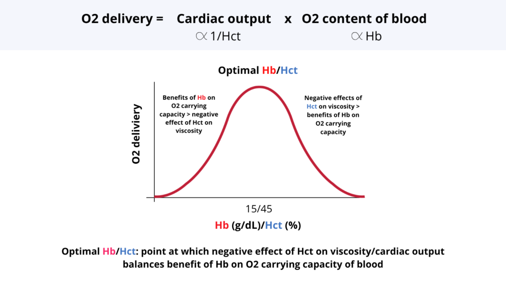
That tradeoff predicts that there should be a hematocrit at which oxygen delivery is maximal.
Indeed, conventional models place the optimal Hct roughly within the range normally found in humans. If that were the whole story, increasing Hct beyond normal should provide little additional benefit and might eventually reduce oxygen delivery.
At low Hct, oxygen delivery is limited by inadequate oxygen carrying capacity.
At sufficiently high Hct, the circulatory cost of increasing the red cell volume fraction begins to outweigh the gain in oxygen content.
The result is a bell shaped relationship between hematocrit and oxygen delivery.5
Somewhere near the top of that curve lies an optimal hematocrit.
Figure 1. The tradeoff that determines optimal hematocrit. Increasing Hct generally increases Hb and arterial oxygen content, but also increases blood viscosity, which tends to oppose blood flow. Oxygen delivery is maximal where the benefit of increasing oxygen carrying capacity is balanced by the increasing rheologic cost of a higher Hct.
Is the optimal hematocrit fixed?
The bell shaped curve is often drawn as though its peak were a property of hematocrit itself.
But look again at the equations.
Hct affects viscosity.
Viscosity affects resistance to flow.
But neither viscosity nor vascular resistance is fixed.
Blood is a non-Newtonian fluid: as shear rate increases, its apparent viscosity decreases as red cell aggregates disperse and cells deform and align with flow.
Vessel caliber also matters enormously:
R ∝ 1/r⁴
During exercise, resistance vessels supplying working skeletal muscle dilate, local blood flow rises markedly, shear conditions change, and the pressure flow relationship of the muscle bed is altered.67
Thus, exercise alters both sides of the rheologic equation: vessel caliber increases and the apparent viscosity of blood at a given Hct can decrease.
The flow cost associated with a given Hct therefore need not be the same during exercise as it is at rest.
If that cost is reduced, the balance between:
oxygen content gained
and
flow lost
shifts in favor of a higher Hct.
The prediction is straightforward:
The optimal Hct should shift to the right.
Does it?
An experiment that showed the peak moving rightward
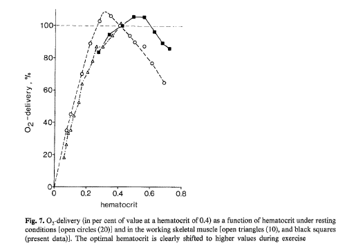
In 1979, Gaehtgens, Kreutz, and Albrecht studied isolated canine gastrocnemius muscle during rhythmic isotonic contraction.8
They perfused the preparation with blood spanning Hct values from 0.21 to 0.81 and measured blood flow, oxygen delivery, oxygen uptake, oxygen extraction, and contractile performance.
This allowed them to construct something that is extraordinarily difficult to obtain in humans:
oxygen delivery as a function of Hct during active skeletal muscle work.
Maximal oxygen delivery occurred at Hct values between approximately 0.50 and 0.60.
At lower Hct, oxygen delivery fell because oxygen carrying capacity was inadequate.
At extreme hemoconcentration, oxygen delivery again fell.
Most importantly, the investigators concluded that compared with resting skeletal muscle, the optimal Hct was shifted toward higher values during muscular work.
In graphical terms:
The peak shifted to the right.
Figure 2. Oxygen delivery as a function of Hct in resting and working canine skeletal muscle. The Hct associated with maximal oxygen delivery shifts to the right during muscle work. Gaehtgens P, Kreutz F, Albrecht KH. Optimal hematocrit for canine skeletal muscle during rhythmic isotonic exercise. European Journal of Applied Physiology and Occupational Physiology. 1979;41:27–39. doi:10.1007/BF00424466.
This is the central experimental observation: A rightward shift of the optimal Hct has been demonstrated experimentally in a working mammalian skeletal muscle bed.
There is, however, an important limitation.
This was an isolated peripheral muscle preparation under controlled perfusion conditions.
The original investigators themselves cautioned against simply extrapolating the findings to an intact exercising human, in whom blood volume, cardiac performance, reflex cardiovascular regulation, and multiple vascular beds interact.9
Why might working muscle tolerate a higher hematocrit?
The Gaehtgens study asked where oxygen delivery peaks across a wide range of Hct values during muscle work.
A separate canine study by Gustafsson, Appelgren, and Myrvold asked a different question: what happens to blood flow when acute polycythemia is imposed on working versus nonworking muscle?10
Rather than mapping oxygen delivery across a broad range of Hct values, the investigators compared the flow penalty produced by increasing Hct under the two conditions.
In working muscle, the reduction was only about 5% to 10% and was not statistically significant.
Thus, the same increase in red cell volume fraction carried a much smaller local flow penalty when muscle was contracting.
Indeed, red cell flow increased in working muscle after induction of polycythemia while decreasing in nonworking muscle.11
The two experiments therefore provide complementary evidence: Gaehtgens showed that the Hct associated with maximal oxygen delivery shifts to the right during muscle work; Gustafsson showed that the flow penalty imposed by a higher Hct is markedly attenuated in working muscle.
That finding helps explain the rightward shift.
But it would be a mistake to reduce the explanation to:
exercise → vasodilation → viscosity no longer matters.
The circulation is not passive plumbing.
The whole circulation complicates the story
The relationship between Hct and flow cannot be explained by viscosity alone.
In exercising dogs, investigators raised Hct from approximately 35% to 50% using methemoglobin-containing red cells, increasing Hct and viscosity without the usual increase in functional O₂-carrying capacity. Exercise cardiac output did not fall significantly. When methemoglobin was subsequently converted back to functional hemoglobin at essentially the same Hct and viscosity, arterial O₂ content increased and cardiac output fell.12
This experiment does not identify an optimal Hct. Rather, it shows that the intact circulation actively adjusts flow according to oxygen content as well as rheology.
Hct does not determine flow through viscosity alone.
That complexity is one reason a local Hct–oxygen delivery curve cannot simply be extrapolated to the whole human circulation.
EXPERIMENTAL LOGIC: SEPARATING VISCOSITY FROM O₂ CONTENT
The question: Does cardiac output respond primarily to the increased viscosity or to the increased O₂ content?
Some mammals perform natural blood doping
Comparative physiology provides another striking clue.
Horses and some athletic dogs store a substantial reserve of red cells in the spleen.
During intense exercise, sympathetic stimulation causes splenic contraction and releases those cells into the circulation.13
Functionally, this produces an acute endogenous autotransfusion.
In highly aerobic dogs, splenic contraction can markedly increase circulating Hct, blood volume, and oxygen carrying capacity, with systemic Hct rising from around 40% at rest toward 60% during exercise.14
Horses show an even more striking exercise associated increase in Hct, with values above 60% well described during strenuous exertion.15
Importantly, these natural blood dopers do not simply concentrate an unchanged quantity of blood.
They deploy a stored red cell reservoir into the active circulation.
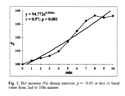
Figure X34. Hematocrit rises rapidly during exercise. Hct increased progressively during the first 10 minutes of exercise, reaching approximately 135% of the baseline value. The increase reflects the dynamic nature of Hct during exercise and should not be confused with the experimentally demonstrated rightward shift in the Hct that optimizes oxygen delivery.
Unlike athletic dogs and horses, humans do not use a large splenic erythrocyte reservoir to produce a major exercise induced expansion of circulating red cell mass. Human exercise can increase Hct modestly, but much of that change reflects plasma volume contraction rather than massive splenic release of red cells.16
The comparison should not be pushed too far.
Dogs, horses, and humans differ in splenic anatomy, blood volume regulation, red cell properties, and cardiovascular physiology.
But the broader principle is striking:
The circulating red cell fraction useful during maximal exercise need not be the same as the one maintained at rest.
Humans can move to the right artificially
Humans cannot physiologically mobilize a splenic red cell reservoir comparable to that of an athletic dog or horse.
But we can approximate part of the experiment artificially.
A classic study by Buick and colleagues examined 11 highly trained runners who underwent autologous reinfusion of approximately 900 mL of stored blood in a double blind study.18
The improvements were also present seven days later.
Thus, increasing red cell mass above baseline improved maximal exercise performance.
This tells us something important:
Over the range studied, the physiological benefit of additional red cell mass exceeded its cost.
Blood doping therefore demonstrates that a normal human Hct is not necessarily the point beyond which additional red cell mass ceases to provide an exercise benefit.
But it does not tell us precisely where the human exercise peak lies.
It does not map the descending limb.
And by itself it does not prove that the whole body exercise optimum is shifted to the right of the resting optimum.
There is another reason for caution.
Sixteen weeks after reinfusion, hematologic values had returned toward baseline, yet VO₂max remained above control values.20
The study therefore cannot be interpreted as a perfectly clean experiment isolating Hct, viscosity, and oxygen content.
More broadly, a systematic review identified 28 autologous transfusion studies and found that reinfusion generally improved performance during exercise lasting approximately 5 to 45 minutes and performed at about 70% to 100% of VO₂peak, including in well trained athletes. Only four studies, however, fulfilled the review’s highest quality criteria of placebo controlled, double blind crossover design.21
Blood doping probably works through more than one mechanism
Increasing functional hemoglobin and arterial oxygen content is clearly important.
But it may not be the whole story.
Böning, Maassen, and Pries emphasized that several other changes accompanying blood boosting may contribute to its effects, including changes in blood volume, cardiac performance, oxygen diffusion, red cell properties, buffering capacity, and vascular regulation.22
So a rightward shift of the optimal Hct should not be presented as the explanation for why blood doping works.
Rather, it provides one physiologically compelling explanation for why increasing Hct can remain beneficial during strenuous exercise despite the accompanying increase in viscosity.
What does theory predict?
Mathematical modeling provides another piece of the argument.
Any model of optimal Hct needs a constraint: otherwise the heart could simply generate ever more pressure or power to compensate for increasing viscosity.
Traditional models assume that blood is driven through the circulation by a fixed pressure. As Hct rises, viscosity and resistance rise, flow falls, and the calculated optimal Hct generally lies around 30–50%, close to the normal human range.23
But strenuous exercise is not a fixed-pressure experiment.
Sitina and colleagues therefore used a different model. Instead of holding perfusion pressure constant, they held cardiac mechanical power constant.
Cardiac power is approximately:
Power = pressure × flow
This allows pressure and flow to trade off while the total power generated by the heart remains fixed.
Under this assumption, the calculated optimal Hct shifted to the right, to approximately 50–70%.24
These numbers are model dependent and should not be interpreted as target Hcts for athletes.
The important point is simpler:
Change the conditions under which the circulation operates, and the optimal Hct can change.
How far to the right?
Not indefinitely.
The curve ultimately has a descending limb.
Schuler and colleagues manipulated Hct over a broad range in mice and measured maximal exercise performance.25
In wild type mice treated with a long acting erythropoietin analogue, maximal oxygen uptake peaked at approximately:
Hct was not altered in isolation. Blood volume and cardiovascular work changed along with it.
So the study demonstrates an exercise performance optimum in an intact mammal, but not a pure isolated effect of Hct.
The lesson remains:
Rightward does not mean endlessly rightward.
Could we prove the whole body shift in humans?
The ideal experiment is easy to imagine.
Take the same individuals.
Set their Hct sequentially across a wide range.
At each Hct, measure whole body oxygen delivery at rest.
Then repeat the measurements during maximal exercise.
Construct the two curves.
If the exercise peak lies farther to the right, the question is settled.
We do not have those paired human curves.
That is the central evidentiary boundary.
A rightward shift has been demonstrated in working skeletal muscle.27
Natural blood dopers markedly increase circulating red cell volume during exercise.28
Human blood boosting improves exercise performance over the ranges studied.29
And models predict a higher optimum under exercise relevant cardiovascular constraints.30
But the precise whole body human exercise optimum remains unknown.
EVIDENCE BOX
What has been directly demonstrated?
Working skeletal muscle
In isolated canine gastrocnemius, oxygen delivery during rhythmic contraction peaked at approximately Hct 0.50 to 0.60, and the investigators concluded that the optimal Hct was shifted to higher values relative to resting muscle.31
Whole animal exercise performance
Experimentally increasing Hct over a broad range in mice produced an exercise performance optimum, although Hct was not manipulated independently of other cardiovascular changes.32
Human performance
Autologous blood reinfusion can increase Hb, VO₂max, and endurance performance in trained athletes.33
What complicates the simple viscosity model?
Whole animal experiments show that cardiac output responds to functional oxygen content as well as to blood rheology.
Increased Hct and viscosity alone do not fully account for the hemodynamic response to polycythemia.34
What occurs naturally?
Horses and some athletic dogs acutely release splenic red cells during exercise, increasing circulating red cell volume and Hct.35efn_note]Dane DM, Hsia CCW, Wu EY, et al. Splenectomy impairs diffusive oxygen transport in the lung of dogs. J Appl Physiol. 2006;101:289-297. doi:10.1152/japplphysiol.01600.2005.[/efn_note]
What is inferred?
That the optimal whole body human Hct during intense exercise is shifted to the right of the optimal resting Hct.
What remains unknown?
The precise location of the human resting and exercise peaks, the magnitude of any systemic rightward shift, and how much that shift contributes to the performance benefit of blood doping.
So why does blood doping work?
The simplest answer is:
More functional hemoglobin means greater oxygen carrying capacity.
But hemoglobin has to be carried somewhere.
In blood, it is packaged inside red cells.
Increasing the amount of packaged hemoglobin therefore generally increases the red cell volume fraction and with it blood viscosity.
Blood doping works over the ranges studied because the benefit of additional oxygen carrying capacity can exceed the accompanying circulatory cost during strenuous exercise.36
Why can that balance remain favorable?
One reason may be that exercise changes the optimum itself.
In working skeletal muscle, the Hct associated with maximal oxygen delivery has been measured at a higher value than at rest.37
So the most defensible conclusion is:
A rightward shift of the exercise specific Hct optimum provides one compelling physiological explanation for why additional red cell mass can remain advantageous during strenuous exercise despite the accompanying rise in blood viscosity.
CLOSING THOUGHT
Packaging hemoglobin inside red cells allows blood to carry enormous amounts of oxygen. The price is viscosity. Exercise changes the balance between the two.