
When one name contains many diseases
Note: The video and audio linked above were generated with the assistance of AI. Clinical accuracy has been reviewed, but no AI-generated content can be guaranteed to be fully error-free.
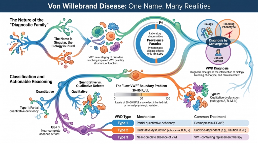
The name is singular.
The biology is plural.
“Von Willebrand disease” sounds like one disorder. It is better understood as a diagnostic family: a name applied to several related ways in which von Willebrand factor biology can fail.
Von Willebrand factor (VWF) is a multimeric glycoprotein produced primarily by endothelial cells and megakaryocytes. It supports platelet adhesion under shear, contributes to platelet accumulation at sites of vascular injury, and stabilizes factor VIII in the circulation. The disorders grouped under the VWD label arise when VWF quantity, structure, function, or survival is impaired.1
That distinction matters.
A disease name can create the impression of unity. It can suggest one mechanism, one phenotype, one diagnostic pathway, and one treatment strategy.
VWD offers none of those so neatly.
Some patients have too little VWF. Some produce VWF that is present but dysfunctional. Some clear it too quickly. Some lose the largest multimers. Some have increased platelet binding rather than impaired binding. Some cannot stabilize factor VIII. Some have almost no VWF at all.
These are related disorders, but they are not biologically identical.
The label is real.
The borders are not perfectly sharp.
The major types of VWD
VWD is classified into three major types.
Type 1 VWD is a partial quantitative deficiency of VWF. The protein is present and usually functions relatively normally, but there is not enough of it to provide a full hemostatic reserve.
Type 2 VWD is qualitative VWF dysfunction. The amount of VWF may be normal, reduced, or only mildly reduced, but the protein does not work normally. Type 2 is subdivided into 2A, 2B, 2M, and 2N, reflecting different functional defects in multimer structure, platelet binding, adhesive function, or factor VIII binding.
Type 3 VWD is near-complete absence of VWF. Because VWF is needed both for platelet-dependent hemostasis and for stabilizing factor VIII, type 3 disease can produce severe bleeding and may sometimes resemble a coagulation-factor deficiency.
This simple three-part structure is the entry point. The deeper lesson is that each type asks a different biological question: is there too little VWF, abnormal VWF, or almost no VWF at all?
Two different questions hide inside the name
VWD contains two distinct kinds of complexity.
The first is internal heterogeneity.
This asks:
How many diseases are inside the name?
The answer is: several.
The major categories correspond to genuinely different biological problems. Type 1 VWD describes partial quantitative deficiency. Type 2 VWD includes qualitative defects affecting multimer structure, platelet binding, adhesive function, or factor VIII binding. Type 3 VWD reflects near-complete absence of VWF.2
A patient with type 1 VWD may have reduced synthesis, impaired secretion, or accelerated clearance. A patient with type 2A VWD may lose high-molecular-weight multimers because of defective assembly, impaired secretion, increased proteolysis, or a combination of these mechanisms. A patient with type 2B VWD has excessive interaction between VWF and platelet GPIb. A patient with type 2M VWD has impaired platelet-dependent function despite relatively preserved multimer structure. A patient with type 2N VWD has defective binding of VWF to factor VIII.3
One name therefore contains multiple failure modes.
The second complexity is boundary uncertainty.
This asks:
Where does disease end and normal variation begin?
VWF levels vary continuously across the population. They are influenced by ABO blood group, age, pregnancy, hormonal state, inflammation, exercise, stress, thyroid function, and other biological modifiers.4
A mildly reduced value may indicate inherited bleeding risk.
It may also reflect normal variation.
Sometimes it reflects both.
These two problems are related, but they should not be confused. Internal heterogeneity concerns the number of mechanisms contained within the diagnosis. Boundary uncertainty concerns where clinicians draw the line between disease, susceptibility, and normal biology.
One question asks what is inside the category.
The other asks where the category should end.
A pre-molecular name
Erik von Willebrand described a hereditary bleeding disorder in 1926 in a family from the Åland Islands, decades before the responsible protein was identified.5
The name therefore arose before the biology was understood.
That history is important.
Many medical labels are created from phenotype first and mechanism later. Once molecular understanding arrives, the original name may remain even as the biology beneath it fragments into multiple subtypes.
“Von Willebrand disease” is one such name.
It began as a clinical pattern involving hereditary bleeding, mucocutaneous manifestations, affected women as well as men, and a phenotype distinct from classic hemophilia.
Later research showed that this pattern could arise through several different defects in VWF quantity, structure, function, survival, or interaction with other components of hemostasis.
The name survived.
Its meaning expanded.
This is not necessarily a flaw. A shared name can help clinicians recognize related disorders, organize testing, communicate risk, and build treatment strategies.
But the name can also conceal important differences.
A patient with type 1 VWD, a patient with type 2B VWD, and a patient with type 3 VWD do not simply occupy different points on a severity scale. They may have fundamentally different biology, inheritance, laboratory patterns, and treatment responses.
The linguistic category is broader than any one mechanism.
The disorder is common. Clinical disease is less so
The prevalence of VWD depends on what is being counted.
Population studies have identified reduced VWF levels or laboratory-defined VWD in approximately 0.6% to 1.3% of individuals, depending on the population studied and the criteria applied. Symptomatic disease is less common, often estimated at approximately 0.1%, or 1 in 1,000. Patients followed in specialized treatment centers represent a still smaller group, often estimated at approximately 0.01%, or 1 in 10,000.6
These are not three competing estimates of the same thing.
They describe three different constructs:
- A laboratory abnormality.
- Symptomatic bleeding disease.
- Referral-center disease.
A low laboratory value may identify biological vulnerability. A bleeding phenotype may identify clinically expressed disease. Referral-center populations identify patients whose diagnosis, severity, complications, or treatment needs bring them into specialist care.
Diagnostic thresholds create categories. Biology does not create cliffs.
The difference between 29 IU/dL and 31 IU/dL may matter diagnostically or administratively, but it does not create a new biological species.
The category helps clinicians act.
The underlying trait remains continuous.
What the name gains and loses
The name “von Willebrand disease” gains several things by gathering these disorders together. It creates a shared conceptual home for VWF-mediated bleeding, supports a common diagnostic framework, allows clinicians to reason from laboratory patterns toward mechanism, and connects phenotype, inheritance, molecular biology, and treatment. It also makes visible the central role of VWF in platelet adhesion under shear, platelet accumulation, and factor VIII stabilization.
But the name also loses information. It can obscure the difference between quantitative and qualitative disease, blur the distinction between inherited VWD and acquired von Willebrand syndrome, and make mildly reduced VWF appear more categorical than the biology warrants.
It can also encourage clinicians to treat a diagnostic label as a fixed truth rather than as a working explanation that must remain accountable to the patient’s history, laboratory pattern, physiologic context, and response to hemostatic challenge.
The diagnosis is therefore strongest when the whole pattern points in the same direction.
No single laboratory value, bleeding score, genetic result, or family history carries the diagnosis alone.
VWD emerges from convergence:
- A compatible bleeding phenotype.
- Evidence of reduced or dysfunctional VWF.
- Inheritance when present.
- Exclusion of better explanations.
- A clinical context in which the findings make biological sense.
This does not weaken the diagnosis.
It describes how the diagnosis works.
Classification makes the name actionable
Classification matters because the name alone is not enough.
A diagnosis of “VWD” does not yet tell the clinician whether the defect is quantitative or qualitative, whether high-molecular-weight multimers are preserved, whether platelet binding is impaired or increased, whether factor VIII binding is defective, or which treatment is most likely to succeed.
Subtype converts a broad name into a more useful biological explanation.
Type 1 VWD often responds to desmopressin, although a trial is needed to confirm that the response is adequate and sustained. Type 2B VWD requires caution because enhanced VWF-platelet binding may be associated with thrombocytopenia, and desmopressin may worsen the platelet count in some patients. Type 3 VWD usually requires VWF-containing replacement therapy because endogenous VWF is nearly absent.7
Type 2A VWD illustrates a different therapeutic problem. Loss of high-molecular-weight multimers may reduce the usefulness of simply increasing endogenous VWF, depending on the underlying variant and demonstrated treatment response. Type 2M shows why a preserved multimer pattern does not guarantee preserved function. Type 2N shows why a patient with disproportionately low factor VIII may require evaluation for defective VWF-FVIII binding rather than being assumed to have hemophilia A.
These distinctions show why classification is not merely descriptive.
It has diagnostic and therapeutic consequences.
The purpose of classification is not to end reasoning.
It is to make reasoning safer.
The mild quantitative borderland
The phrase low VWF captures the most contested boundary within the name.
For many years, patients with VWF levels between 30 and 50 IU/dL and abnormal bleeding were often described as having low VWF rather than type 1 VWD.
The 2021 ASH/ISTH/NHF/WFH diagnostic guideline recommended confirming type 1 VWD in patients with abnormal bleeding and VWF levels below 50 IU/dL. It also supported the diagnosis when VWF is below 30 IU/dL regardless of bleeding history.8
That framework reflects a clinical priority: reducing missed diagnoses and improving access to care.
It also exposes the tension built into the name.
A broader category may help patients whose bleeding has long been dismissed. It may also include some people whose mildly reduced VWF represents a bleeding risk factor rather than a discrete monogenic disease.9
The debate is therefore not only about terminology.
It is about what disease labels are for.
Are they intended to identify a molecular entity, predict bleeding, justify treatment, support access to care, or communicate uncertainty?
The answer is usually: all of these, imperfectly.
The boundary with acquired disease
Not every VWF abnormality belongs inside inherited VWD.
Acquired von Willebrand syndrome can produce similar laboratory and clinical patterns through mechanisms that arise later in life.
Severe aortic stenosis is the classic example. High shear unfolds VWF and increases its susceptibility to proteolytic cleavage, resulting in loss of high-molecular-weight multimers. The acquired abnormality may improve or normalize after valve replacement.10
The structural endpoint resembles inherited qualitative VWD.
The cause is different.
A patient with late-onset bleeding, no family history, and newly abnormal VWF testing may therefore require evaluation for an underlying cardiac, hematologic, immunologic, or mechanical process rather than automatic assignment of an inherited label.
The name “von Willebrand” can point in two directions:
- Toward an inherited disorder of VWF biology.
- Toward an acquired syndrome that mimics it.
The distinction matters because treatment must address both the bleeding and the cause.
A practical definition
In practice, “von Willebrand disease” names a group of inherited bleeding disorders in which abnormal VWF quantity or function provides a clinically meaningful explanation for bleeding.
That definition is intentionally practical.
It does not require every patient to share the same mutation.
It does not imply that every borderline laboratory value represents disease.
It does not pretend that categories are natural cliffs.
It asks whether the available evidence supports VWF-mediated bleeding strongly enough to guide care.
The diagnosis should remain accountable to the patient over time:
- Prior bleeding.
- Current and historical VWF measurements.
- Physiologic modifiers.
- Family history.
- Procedural history.
- Subtype-specific testing.
- Response to treatment.
The name is useful when it organizes reality.
It becomes misleading when it replaces it.
Clinical synthesis
“Von Willebrand disease” is not one mechanism.
It is a diagnostic family created at the intersection of phenotype, laboratory biology, inheritance, and clinical judgment.
The name contains real disorders.
It also contains uncertainty.
That is not a reason to abandon the name.
It is a reason to use it carefully.
The name is singular.
The biology is plural.
The best clinicians honor both truths.
Reflect & Apply
Patient A has VWF levels of 35 IU/dL, blood group O, mild lifelong epistaxis, no excessive bleeding after dental extraction, and no known affected relatives.
Patient B has current VWF levels within the reference range but lifelong heavy menstrual bleeding, postpartum hemorrhage, excessive bleeding after dental work, and several relatives with similar mucocutaneous bleeding.
Questions for reflection
- Which patient has the stronger current laboratory signal?
- Which patient has the stronger lifetime bleeding phenotype?
- What does blood group O contribute to the interpretation of Patient A?
- What would you want to know about the timing and clinical context of Patient B’s VWF measurements?
- What prior laboratory results, procedural records, or family information would most change your assessment?
- How should each patient be evaluated before major surgery?
- Does either profile prove or exclude VWD?
- What does this comparison reveal about the difference between a laboratory abnormality and a clinically meaningful disease construct?
Test your thinking
Apply the concepts from this essay in the companion interactive quiz.