
How one molecule connects vessel wall, platelets, and coagulation
Note: The video and audio linked above were generated with the assistance of AI. Clinical accuracy has been reviewed, but no AI-generated content can be guaranteed to be fully error-free.
Why this spoke matters
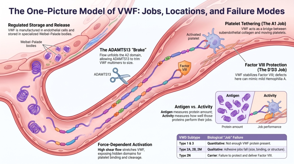
At a site of vascular injury, the VWF story can be seen in one picture.
Collagen is exposed.
VWF binds the injured surface.
Flow stretches VWF and changes its shape.
Platelets tether through GPIbα.
Additional platelets are recruited.
Factor VIII is protected and delivered into a hemostatic environment.
ADAMTS13 trims VWF when the A2 domain unfolds under force.
That is the essential image.
Von Willebrand factor is easy to reduce to laboratory values:
- VWF antigen
- VWF activity
- factor VIII activity
- multimer analysis
But VWF is not just a plasma number. It is a stored, secreted, multimeric, force-responsive protein whose function changes with location, size, conformation, binding partners, and flow.1
It connects the injured vessel wall, exposed collagen, flowing platelets, factor VIII, endothelial storage and release, shear stress, ADAMTS13-mediated size control, and plasma clearance.
This is why von Willebrand disease cannot be understood by memorizing subtypes alone. The subtypes are shorthand. The biology is the map.
The central question is simple:
Where is VWF, what is it trying to do there, and what happens when that job fails?
Location 1: the endothelial cell
VWF is synthesized primarily by endothelial cells and megakaryocytes.2
In endothelial cells, VWF is stored in Weibel-Palade bodies, specialized granules that allow rapid regulated release. In platelets, VWF is stored in alpha granules and can contribute locally after platelet activation.
This location matters clinically because plasma VWF is only one part of the system. Plasma testing samples circulating VWF, but not the full choreography of endothelial release, platelet activation, collagen exposure, flow, and local hemostatic surface formation.
VWF therefore has both a circulating life and an injury-site life.
Location 2: the storage granule
VWF begins as a pre-pro-protein, with a signal peptide, a large propeptide, and a mature VWF subunit. During intracellular processing, VWF undergoes folding, glycosylation, dimerization, multimerization, and propeptide cleavage.3
The assembly sequence matters.
In the endoplasmic reticulum, VWF subunits form tail-to-tail dimers through their C-terminal cystine-knot domains.
In the Golgi, those dimers are linked head-to-head into large multimers through N-terminal disulfide bonding.
The VWF propeptide is not passive. It supports multimerization and trafficking into regulated storage granules.4
This intracellular choreography matters clinically because defects at different steps can produce different VWD phenotypes. VWF may be reduced because it is poorly synthesized, poorly folded, poorly secreted, abnormally stored, incorrectly assembled, or cleared too quickly.
VWF is therefore not simply secreted. It is manufactured, folded, packed, released, stretched, trimmed, and cleared.
Location 3: the injured vessel wall
When a blood vessel is injured, subendothelial collagen is exposed.
VWF binds to collagen and becomes positioned at the injured surface. Under shear or elongational flow, VWF shifts from a compact conformation toward an extended form, exposing functional sites that support platelet tethering.5
VWF function is critically modulated by physical force.
Under high shear or elongational flow:
- VWF elongates
- platelet-binding sites become more available
- the A1 domain binds platelet GPIbα
- platelets tether and translocate along the surface
- platelet activation and aggregation follow
This is why VWF is especially important in high-shear vascular settings. Platelets cannot simply settle onto a damaged surface in rapidly flowing blood. They need a tether.
VWF provides that tether.
Job 1: tethering platelets
The first major job of VWF is to help platelets attach to injured vessel wall.
This is mediated largely through the interaction between the VWF A1 domain and platelet GPIbα. Under normal circulating conditions, this interaction is restrained. Under flow, conformational changes in VWF permit platelet tethering at sites where vascular injury has exposed adhesive surfaces.6
This job explains why VWF is central to mucocutaneous hemostasis.
It also explains why a patient may have VWF protein present but still bleed if that protein cannot bind platelets properly.
Job 2: recruiting platelets under flow
VWF is not just an adhesive patch. It is a multimeric scaffold.
Multimer size matters because the largest VWF multimers are disproportionately hemostatically active. They provide more binding sites, bind platelets and collagen more effectively, and respond more strongly to flow.7
Their advantage is most evident under high shear, where smaller multimers may fail to sustain platelet tethering.
This explains why loss of high-molecular-weight multimers can produce bleeding even when VWF antigen remains measurable.
A patient may have VWF in the plasma, but if the most functional multimers are missing, the molecule may not perform properly where it is most needed.
Job 3: carrying factor VIII
VWF also carries factor VIII.
Most circulating factor VIII is bound to VWF. This interaction protects factor VIII from premature degradation and clearance.8
When VWF is severely reduced or absent, factor VIII survival falls.
This is why severe VWF deficiency can resemble hemophilia A clinically and biologically. The primary defect is VWF, but the downstream consequence is reduced factor VIII.
A low factor VIII level does not always mean primary factor VIII deficiency. It may reflect a VWF problem.
In VWD, factor VIII is often best understood as a downstream readout of VWF biology.
The domain map: where jobs become failure points
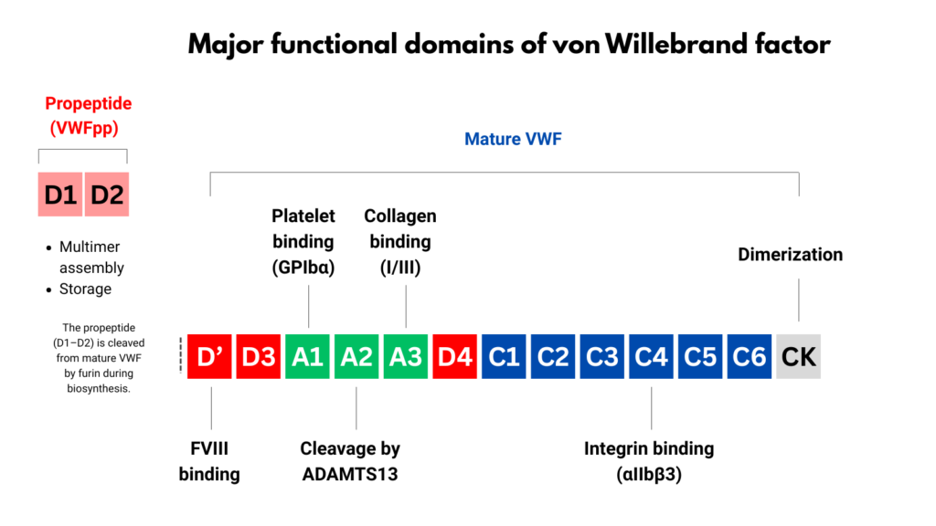
VWF domains are not simply structural. They are functional failure points.
The D′D3 region binds factor VIII.
The A1 domain binds platelet GPIbα and contributes to binding some collagen types.
The A2 domain contains the ADAMTS13 cleavage site.
The A3 domain contributes to binding collagen types I and III.
The C-terminal region, including the C4 domain, contains an RGD sequence that can interact with platelet integrin αIIbβ3.
The CK domain supports dimerization during biosynthesis.9
These relationships explain why different VWF defects produce different clinical and laboratory patterns.
A platelet-binding defect is not the same as a factor VIII-binding defect.
A collagen-binding defect is not the same as accelerated clearance.
Loss of high-molecular-weight multimers is not the same as reduced synthesis.
The label “VWD” contains multiple biological failures.
ADAMTS13: the built-in brake
VWF is powerful because it can recruit platelets under flow.
That power has to be controlled.
Physiologically, VWF multimer size is regulated by ADAMTS13, a metalloprotease that cleaves VWF in the A2 domain at the Tyr1605-Met1606 bond.10
But ADAMTS13 does not digest VWF at random.
The A2 cleavage site is hidden in resting VWF. It becomes available when VWF is stretched and the A2 domain unfolds under force.11
This produces an elegant biological balance:
The same flow-dependent biology that makes VWF adhesive also allows VWF to be trimmed.
Disruption of that balance can shift the system toward bleeding or thrombosis.
Too little effective VWF causes bleeding. Too much unregulated VWF activity can promote thrombosis.
Failure mode 1: too little VWF
VWF may be quantitatively reduced.
This can reflect:
- reduced synthesis
- impaired folding
- intracellular retention
- impaired secretion
- abnormal storage
- accelerated clearance
The result is a lower steady-state VWF level in plasma and, often, reduced VWF availability at sites of injury.
A low VWF level does not automatically mean reduced synthesis. It may reflect accelerated removal from the circulation.12
Failure mode 2: abnormal VWF structure
VWF may be present but assembled abnormally.
The most clinically important example is loss of high-molecular-weight multimers. In that setting, VWF antigen may be detectable, but adhesive function is disproportionately impaired.
This is the biological logic behind many type 2A-like patterns and some acquired high-shear states.
The problem is not simply how much VWF is present. It is whether the right forms of VWF are present.
Failure mode 3: impaired ligand binding
VWF may fail to bind one of its key partners.
It may bind platelets poorly.
It may bind collagen poorly.
It may bind factor VIII poorly.
Each defect produces a different physiologic problem.
Impaired platelet binding affects tethering and platelet recruitment.
Impaired collagen binding affects localization of VWF to injured vessel wall.
Impaired factor VIII binding reduces factor VIII survival and can mimic mild hemophilia A.
These are not interchangeable defects. They are different broken jobs.
Failure mode 4: dysregulated processing or clearance
VWF may be too susceptible to proteolysis under flow, or it may be cleared too rapidly from plasma.
In some settings, enhanced ADAMTS13-mediated cleavage contributes to loss of high-molecular-weight multimers. In other settings, clearance mechanisms determine the steady-state VWF level more than synthesis does.13
VWD can therefore arise from defects in synthesis, intracellular trafficking, storage, secretion, multimer assembly, ligand binding, regulated proteolysis, or clearance.14
Why antigen and activity can diverge
One of the most important clinical lessons in VWD is that VWF antigen and VWF activity are not the same thing.
VWF antigen asks: how much VWF protein is present?
VWF activity asks: how well does that VWF perform a particular function in the assay used?
A proportional reduction in antigen and activity suggests a quantitative problem.
A disproportionately reduced activity suggests qualitative dysfunction.
This is the conceptual basis for the activity-to-antigen ratio.
The ratio is not a mathematical trick. It is an attempt to distinguish amount from function.
For example, a patient with mucosal bleeding, normal VWF antigen, low platelet-dependent VWF activity, and preserved multimers does not have a simple amount problem. The pattern suggests a specific qualitative defect in platelet-dependent VWF function, depending on the assay and after excluding assay artifact.
Why subtype labels are shorthand
Classification systems are clinically useful, but they compress underlying biology into simplified categories.
Type 1 generally points toward partial quantitative deficiency.
Type 3 points toward near-complete or complete deficiency.
Type 2 points toward qualitative dysfunction.
But real patients do not always fit cleanly into conceptual boxes.
A variant may affect secretion, multimer assembly, platelet binding, collagen binding, factor VIII binding, ADAMTS13 susceptibility, and clearance in overlapping ways.
The goal is not simply to name the subtype. The goal is to understand which VWF job is failing, because that failure pattern influences bleeding risk, laboratory interpretation, and treatment.
A practical shorthand is:
- Type 1 or low VWF: amount problem, sometimes with altered kinetics
- Type 2A, 2B, or 2M: adhesive job problem involving structure, platelet binding, collagen binding, or multimer distribution
- Type 2N: factor VIII carrier job problem
- Type 3: near absence of both adhesive and carrier jobs
The subtype comes later.
The biology comes first.
The one-picture model
Return to the injury site.
Collagen is exposed. VWF binds the damaged surface. Flow stretches the molecule. Platelets tether through GPIbα. Platelet activation strengthens adhesion and aggregation. VWF protects factor VIII in the circulation, allowing FVIII to remain available for coagulation at the site of injury. ADAMTS13 trims VWF when force exposes the A2 cleavage site.
Hemostasis succeeds when these elements align in time and space.
Disease emerges when they do not.
Why this matters clinically
Understanding VWF as a molecule with jobs and locations changes how clinicians interpret VWD testing.
It explains why:
- VWF antigen and activity may diverge
- a normal or borderline VWF level does not always settle the question
- factor VIII may be low because VWF is low or dysfunctional
- multimer loss can produce disproportionate bleeding risk
- collagen-binding function must be specifically assessed when clinically relevant
- clearance can determine steady-state VWF level
- subtype labels are useful only when connected to mechanism
- laboratory results must be interpreted as relationships, not isolated values
VWD is not diagnosed by asking whether a single number is low. It is diagnosed by determining whether the bleeding history, laboratory values, and VWF biology form a coherent story.
Clinical synthesis
VWF is the hemostatic bridge between vessel wall injury and platelet adhesion, and between primary hemostasis and coagulation.
Its biology is shaped by where it is made, where it is stored, how it is released, how large its multimers are, how it responds to flow, how well it binds its partners, how ADAMTS13 trims it, and how rapidly it is cleared.
A useful mental model of VWD begins with four questions:
Where is the VWF problem?
How much VWF is present?
How well does it work?
Which hemostatic job is failing?
That is the one-picture model.
Evidence anchor: why VWF must be interpreted as a system, not a number
Summary derived from structural biology, mechanobiology, multimer studies, clearance literature, diagnostic reviews, and management guidance. The evidence consistently shows that VWF function depends on where the molecule is, how it is assembled, how large its multimers are, how it responds to flow, what it binds, how it is cleaved by ADAMTS13, and how rapidly it is cleared.
| Evidence stream | What it shows | Why it matters | Main limitation |
|---|---|---|---|
| Domain architecture | VWF domains map to distinct functions: D′D3 binds and stabilizes factor VIII; A1 mediates platelet GPIbα binding and contributes to collagen IV/VI binding; A2 contains the ADAMTS13 cleavage site; A3 binds collagen I/III; C4 contains the RGD motif for platelet integrin interaction; CK supports dimerization.15 | VWD subtypes are best understood as failures of specific structural functions, not simply as different degrees of “low VWF.” | Domain maps simplify a dynamic multimeric molecule and cannot fully capture conformation, shear, glycosylation, cellular release, or vascular context. |
| Biosynthesis and storage | VWF is synthesized as pre-pro-VWF, dimerizes in the endoplasmic reticulum through CK-domain interactions, multimerizes in the Golgi through N-terminal disulfide bonding, and is stored in endothelial Weibel-Palade bodies and platelet alpha granules.16 | Quantitative VWD can arise from defects in synthesis, folding, multimer assembly, storage, secretion, or clearance. Desmopressin response depends on stored and releasable endogenous VWF. | Most routine laboratory tests do not directly identify which biosynthetic step is abnormal. |
| Multimer biology | High-molecular-weight multimers are disproportionately effective in platelet adhesion and collagen interaction under shear.17 | Similar VWF antigen levels can have different meaning if multimer distribution differs. Loss of high-molecular-weight multimers helps explain type 2A VWD and acquired high-shear VWF loss. | Multimer analysis is technically specialized, may not be rapidly available, and must be interpreted with other assays. |
| Mechanobiology and shear | Shear and elongational flow can stretch VWF, promote platelet-binding function, and expose the A2 domain for ADAMTS13 cleavage. Single-molecule studies support force-dependent A2 unfolding as a prerequisite for efficient ADAMTS13 cleavage.18 | VWF is built for hemostasis in moving blood. Flow both activates VWF and marks it for trimming, explaining why VWF function cannot be fully inferred from static antigen measurements. | In vitro and single-molecule systems isolate mechanisms but do not fully reproduce the vascular microenvironment. |
| A1 platelet-binding phenotypes | Reduced A1-GPIbα interaction can produce type 2M-like platelet-binding defects, whereas increased A1-GPIbα binding causes type 2B VWD with VWF-platelet complex clearance, loss of high-molecular-weight multimers, and possible thrombocytopenia.19 | Both too little and too much platelet binding can cause bleeding. This distinction changes classification and influences desmopressin safety. | Platelet-binding assays are imperfect proxies for shear-dependent platelet capture in vivo. |
| A2-ADAMTS13 axis | ADAMTS13 regulates VWF multimer size by cleaving the A2 domain when the cleavage site is exposed by force. Excessive susceptibility to cleavage can contribute to loss of high-molecular-weight multimers; severe ADAMTS13 deficiency permits ultra-large VWF-mediated thrombosis.20 | VWD and TTP can be understood as opposite failures along the VWF-ADAMTS13 regulatory axis. A2 functions as a force-sensitive regulatory region that allows VWF size and activity to be edited under flow. | This axis explains many patterns but does not account for every modifier of bleeding or thrombosis risk. |
| High-shear acquired VWF loss | Severe aortic stenosis can cause acquired loss of the largest VWF multimers, reduced collagen-binding activity, and mucocutaneous or gastrointestinal bleeding, with improvement after valve replacement when shear is relieved.21 | A type 2A-like laboratory pattern can be acquired, not inherited. The same force-dependent biology explains inherited and acquired VWF dysfunction. | The presence of a high-shear lesion does not prove that all bleeding is VWF-mediated; clinical context remains essential. |
| Factor VIII carriage | The D′D3 region binds factor VIII and protects it from clearance. Type 2N VWD impairs factor VIII binding and can resemble mild hemophilia A.22 | Disproportionately low factor VIII should prompt consideration of type 2N VWD, especially when the inheritance pattern does not fit hemophilia A. | Factor VIII levels are influenced by multiple variables, so interpretation requires VWF antigen, activity, family pattern, and sometimes FVIII-binding or genetic testing. |
| Collagen-binding defects | VWF binds collagen I/III mainly through A3 and collagen IV/VI through A1. Some patients may have impaired collagen binding despite preserved antigen, platelet-dependent activity, and multimer distribution.23 | Collagen-binding assays can reveal selected type 2M mechanisms that platelet-binding assays may not specifically assess. | Collagen-binding testing varies by assay design and collagen type and is not uniformly available. |
| Clearance biology | Plasma VWF level reflects synthesis, secretion, proteolysis, and clearance. VWF clearance is influenced by glycosylation, macrophage uptake, lectin and scavenger receptors, ABO blood group, and VWF variants. The VWFpp/VWF:Ag ratio can help identify accelerated clearance.24 | A normal desmopressin peak may be misleading if VWF falls quickly. Duration of response matters for procedural planning. | VWFpp testing is not universally available, and clearance phenotypes may overlap with other quantitative defects. |
| Assay interpretation | VWF antigen, platelet-dependent VWF activity, VWF collagen binding, multimer analysis, factor VIII activity, VWFpp/VWF:Ag ratio, and genetic testing are different windows into VWF biology. Ristocetin-based assays may be affected by assay-specific limitations such as A1 polymorphisms.25 | Laboratory results should be interpreted as partial structural readouts, not direct measurements of the whole molecule in vivo. | No single assay captures VWF function in moving blood, and results must be integrated with bleeding phenotype. |
Interpretive note: These evidence streams point in the same direction: VWF structure-function reasoning is not ornamental. It is the basis for interpreting antigen-activity discordance, multimer patterns, factor VIII levels, desmopressin response, acquired high-shear VWF loss, and subtype classification. The goal is not to memorize the domain map. The goal is to ask which structural function has failed.
Guideline synthesis: applying structure–function reasoning in VWD
Use the “jobs, locations, and failure modes” model when interpreting VWD testing.
Ask not only:
How much VWF is present?
Also ask:
Where should this VWF work?
What should it bind?
What size is it?
How does it behave under force?
Is it being cleaved or cleared too quickly?
Does the laboratory pattern explain the bleeding phenotype?
Use structure-function reasoning when interpreting
- the lowest documented VWF antigen and platelet-dependent activity levels
- whether antigen and activity are proportionate or discordant
- whether high-molecular-weight multimers are present
- factor VIII level relative to VWF antigen
- platelet count, especially when type 2B is possible
- collagen-binding results, if available
- VWFpp/VWF:Ag ratio or desmopressin survival curve, if clearance is suspected
- whether testing occurred during stress, inflammation, pregnancy, acute bleeding, or another VWF-raising state
- whether the bleeding history fits the laboratory pattern
- whether the patient has faced meaningful hemostatic challenges
- whether a planned procedure requires a treatment plan even if the label remains uncertain
Avoid common interpretive errors
- treating VWF antigen as the whole diagnosis
- assuming normal antigen excludes qualitative VWD
- assuming low platelet-dependent activity always means type 1 VWD
- missing type 2N VWD in a patient labeled as mild hemophilia A
- missing type 2B VWD when thrombocytopenia and loss of high-molecular-weight multimers are present
- judging desmopressin response only by the peak value
- treating multimer analysis as optional trivia when type 2A or 2B is suspected
- interpreting ristocetin-based activity as if it perfectly reproduced shear-dependent VWF function
- assuming acquired VWF loss cannot mimic inherited type 2A patterns
- letting subtype terminology replace mechanism-based reasoning
Useful language
Instead of:
“Your VWF antigen is not that low.”
Try:
“Your VWF amount is only one part of the picture. We also need to know how well the VWF works, whether the largest multimers are present, and whether the bleeding history fits.”
Instead of:
“The activity is low, so this is type 1.”
Try:
“When activity is lower than antigen, we should think about a qualitative VWF problem, not just a quantitative deficiency.”
Instead of:
“The desmopressin trial worked.”
Try:
“The peak response was good. Now we need to know whether the response lasted long enough for the bleeding-risk situation.”
Instead of:
“This looks like mild hemophilia A.”
Try:
“Because factor VIII is disproportionately low, we should consider whether VWF is failing to bind and protect factor VIII, as in type 2N VWD.”
Instead of:
“The multimer result is just a specialty test.”
Try:
“The multimer pattern tells us whether the VWF present has the size distribution needed for platelet adhesion under shear.”
Instead of:
“The aortic stenosis is unrelated to the bleeding workup.”
Try:
“High shear can cause acquired loss of large VWF multimers, so the valve lesion may be part of the hemostatic explanation.”
Practical takeaway
Structure-function reasoning makes VWD safer to interpret.
The question is not only:
How much VWF is there?
It is:
What size is it?
What does it bind?
How does it behave under force?
Is it being cleaved or cleared too quickly?
Does the laboratory pattern explain the bleeding phenotype?
Does the mechanism predict the treatment plan?
The best VWD care interprets VWF as a system, not a number.
Reflect & Apply Case
A patient has normal VWF antigen, markedly reduced platelet-dependent VWF activity, and a normal multimer distribution.
Which VWF interaction is most likely impaired?
This pattern suggests impaired platelet-dependent VWF function, classically in the type 2M conceptual space, rather than a simple quantitative deficiency. The likely interaction is VWF A1 domain binding to platelet GPIbα, interpreted in the context of the specific activity assay and after excluding assay artifact.
In contrast, a proportional reduction in antigen and activity would suggest a quantitative defect.
The key lesson is that normal antigen does not guarantee normal function.
Test your thinking
A short quiz on VWF jobs, locations, and failure modes.