
What it means to live between normal variation and disease
Note: The video and audio linked above were generated with the assistance of AI. Clinical accuracy has been reviewed, but no AI-generated content can be guaranteed to be fully error-free.
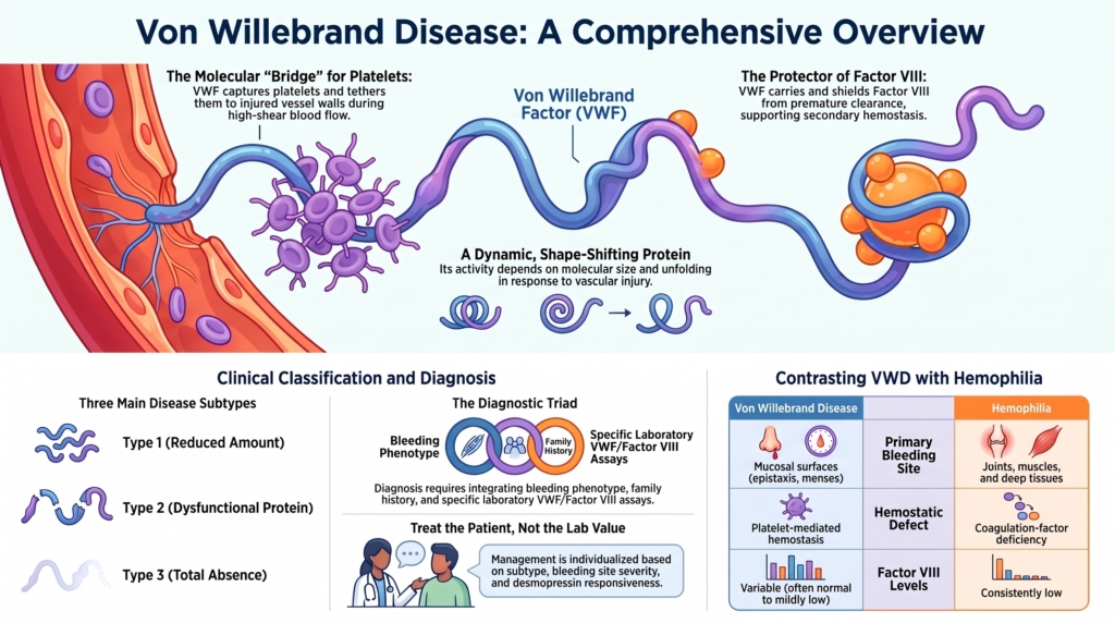
What is von Willebrand disease?
Von Willebrand disease (VWD) is the most common inherited bleeding disorder. Laboratory-defined reductions in VWF are found in approximately 1% of the population, while clinically significant VWD is much less common, affecting roughly 1 in 1,000 people. This difference illustrates a central feature of VWD: abnormal laboratory findings, bleeding risk, and clinically recognized disease overlap but are not equivalent. VWD results from a quantitative deficiency or qualitative dysfunction of von Willebrand factor, a large multimeric glycoprotein synthesized by endothelial cells and megakaryocytes and present in plasma, platelets, and the vascular subendothelium.
VWD is best understood as a family of disorders united by abnormal VWF biology.
That biology is unusually broad. VWF participates in platelet adhesion and aggregation, protects factor VIII from premature clearance, and responds dynamically to blood flow. Abnormalities of VWF can therefore disturb primary hemostasis and, in more severe forms, reduce factor VIII sufficiently to impair secondary hemostasis as well.
This helps explain why VWD varies so widely among patients. The disorder may present as mild bruising or nosebleeds, heavy menstrual bleeding, unexpected procedural bleeding, recurrent gastrointestinal hemorrhage, or, in its most severe forms, deep tissue and joint bleeding.
One protein, many connected functions
Unlike many coagulation proteins, VWF is not a static circulating factor. Its activity changes continuously in response to molecular size, blood flow, vascular injury, secretion, proteolysis, and clearance.
In the intact circulation, VWF generally remains folded and relatively inactive. When vascular injury exposes subendothelial collagen, VWF attaches to the damaged surface. Shear forces generated by flowing blood then alter its conformation, exposing sites that bind platelet glycoprotein Ib. This allows platelets to slow, tether, and adhere where direct platelet contact with the vessel wall would otherwise be difficult.
The largest VWF multimers contain the greatest number of binding sites and are therefore the most hemostatically active. Their size must be carefully regulated: multimers that are too small may not support effective platelet capture, while excessively large forms may promote pathological platelet adhesion.
At the same time, VWF serves as the carrier protein for factor VIII, shielding it from rapid clearance and preserving its contribution to thrombin generation.
These functions are not separate items on a list. They form an interconnected system linking the vessel wall, platelet recruitment, blood flow, and coagulation. A defect at any point in that system can produce a distinctive clinical and laboratory phenotype.
Why VWD differs from hemophilia
VWD and hemophilia are both inherited bleeding disorders, but they usually produce different patterns of bleeding because they affect different parts of hemostasis.
VWD primarily impairs platelet-mediated hemostasis at mucosal surfaces. Patients commonly experience epistaxis, easy bruising, oral bleeding, heavy menstrual bleeding, and prolonged bleeding after dental work, surgery, or childbirth.
Hemophilia, by contrast, is primarily a coagulation-factor deficiency and more often causes bleeding into joints, muscles, and other deep tissues.
The distinction is not absolute. In severe type 3 VWD, the near absence of VWF results in very low factor VIII levels because factor VIII is no longer protected in the circulation. These patients may develop hemarthroses and deep tissue bleeding resembling severe hemophilia. Type 2N VWD can also produce disproportionately low factor VIII levels because the abnormal VWF binds factor VIII poorly, creating a phenotype that may resemble mild hemophilia A. Unlike hemophilia A, type 2N VWD is usually inherited in an autosomal recessive pattern and may therefore affect women as well as men.
VWD therefore occupies an unusual position between disorders of primary and secondary hemostasis.
Not one disease, but many
The name VWD encompasses several biologically distinct disorders.
In type 1 VWD, the amount of circulating VWF is partially reduced, but the remaining protein is generally proportionate in its function.
In type 2 VWD, VWF is present but one or more of its functions are abnormal. The qualitative defect may involve multimer formation, platelet binding, collagen binding, or protection of factor VIII. These disorders are classified as types 2A, 2B, 2M, and 2N according to the dominant functional problem. Platelet-type VWD is a rare phenocopy of type 2B caused by gain-of-function variants in platelet GPIbα rather than VWF and requires separate testing to distinguish the two.
In type 3 VWD, VWF is virtually absent, producing the most severe quantitative deficiency.
This classification is more than taxonomy. Subtype classification localizes biology more reliably than it predicts bleeding severity. It may clarify inheritance, laboratory interpretation, desmopressin responsiveness, replacement strategy, and bleeding risk.
Yet classification does not capture everything. Patients with the same subtype may bleed differently, and some laboratory phenotypes do not fit neatly into a single category. The classification is a clinical map, not a complete description of the individual patient.
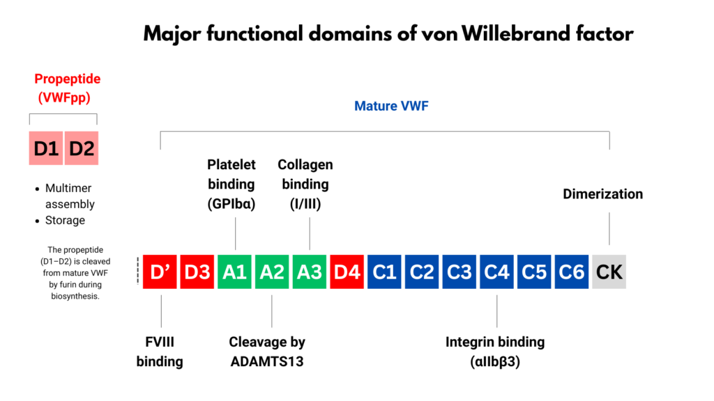
Table 1. Quantitative von Willebrand disease
| VWD subtypes | Type 1 | Type 1C | Type 3 |
|---|---|---|---|
| Mechanism | Partial quantitative deficiency of VWF | Accelerated plasma clearance due to VWF variants, commonly involving D3/A1 | Near-complete or complete quantitative deficiency of VWF |
| Principal domain(s) | Multiple domains (reduced quantity) | D3/A1 (clearance variants) | Multiple domains (absent or severely reduced VWF) |
| Laboratory pattern | • ↓ VWF:Ag • ↓ VWF activity (proportionate) • FVIII normal or slightly ↓ | • ↓ VWF:Ag • ↓ VWF activity (proportionate) • FVIII normal or slightly ↓ • Elevated VWFpp/VWF:Ag ratio | • Very low/undetectable VWF:Ag • Very low/undetectable VWF activity • FVIII very low • Multimers absent |
Table 2. Qualitative von Willebrand disease
| VWD subtypes | Type 2A | Type 2B | Type 2M | Type 2N |
|---|---|---|---|---|
| Mechanism | Loss of HMW multimers due to defective assembly or secretion and/or increased ADAMTS13 proteolysis | Gain of function: increased VWF affinity for platelet GPIbα, causing enhanced platelet binding | Defective platelet binding (and/or collagen binding) with normal multimers | Defective FVIII binding due to D′/D3 region variants |
| Principal domain(s) | A2 (ADAMTS13 cleavage region) ± others | A1 (GPIbα binding site) | A1 (GPIbα binding site) and/or A3 (collagen binding) | D′/D3 (FVIII binding site) |
| Laboratory pattern | • ↓/N VWF:Ag • ↓ VWF activity (disproportionate) • Loss of HMW multimers • FVIII normal or ↓ | • ↓/N VWF:Ag • Disproportionately ↓ activity • HMW multimers often reduced • Variable thrombocytopenia • Increased low-dose RIPA | • N/↓ VWF:Ag • Markedly ↓ VWF activity (disproportionate) • Multimers essentially preserved • FVIII normal or mildly ↓ | • N/↓ VWF:Ag • VWF activity N or ↓ (proportionate to Ag) • FVIII disproportionately ↓ • Multimers normal |
Diagnosis requires more than a laboratory value
At first glance, diagnosing VWD appears straightforward: measure the amount and function of VWF.
In practice, neither bleeding nor VWF exists in a simple binary state.
Laboratory assessment begins by comparing VWF quantity with function. VWF antigen measures the amount of circulating protein, while platelet-dependent VWF activity evaluates its ability to support platelet binding and is commonly measured using GPIbM or GPIbR assays. A disproportionately low activity relative to antigen, often reflected by an activity-to-antigen ratio below approximately 0.7, suggests a qualitative defect.
Low-dose ristocetin-induced platelet aggregation (RIPA) is a different test. It evaluates how the patient’s own platelet-rich plasma responds to low concentrations of ristocetin and is particularly useful when type 2B VWD or platelet-type VWD is suspected.
Factor VIII levels, multimer analysis, the VWF–factor VIII binding assay, and other specialized tests can help localize the abnormality further when indicated.
VWF levels vary continuously across the population. VWF behaves as an acute-phase reactant and may rise with stress, exercise, inflammation, pregnancy, acute bleeding, and other physiological or clinical conditions. Levels are also influenced by ABO blood group and age. A patient tested during illness or pregnancy may have a higher VWF level than at baseline. Improper sample collection, processing, or transportation can also distort results.
The bleeding history has limitations of its own. Bruising and nosebleeds are common in the general population. Children may not yet have encountered a major hemostatic challenge. Family history may be absent because of variable penetrance, small family size, unrecognized bleeding, or recessive inheritance.
Diagnosis therefore requires integration of three forms of evidence:
- the nature and severity of the bleeding phenotype
- the personal and family context
- laboratory assessment of VWF quantity, platelet-dependent activity, and factor VIII
This integration becomes especially important when VWF levels fall between approximately 30 and 50 IU/dL. Contemporary diagnostic guidelines support a diagnosis of type 1 VWD at levels below 30 IU/dL regardless of bleeding history, and at levels below 50 IU/dL in patients with abnormal bleeding. Some clinicians use the term low VWF for patients in the 30–50 IU/dL range, particularly when the relationship between the laboratory finding and bleeding phenotype remains uncertain. As a result, the same patient with a VWF level between 30 and 50 IU/dL and abnormal bleeding may be described as having type 1 VWD under current diagnostic criteria or as having low VWF in traditional clinical usage. This difference reflects both nomenclature and an unresolved biological question: whether mildly reduced VWF in this range represents mild type 1 VWD, a distinct low-VWF state, or a multifactorial bleeding risk that varies among patients.
Whatever terminology is used, a mildly reduced VWF antigen or platelet-dependent activity result may contribute to bleeding without fully explaining it, and another hemostatic or anatomical problem may coexist.
The goal is not simply to identify an abnormal laboratory result, but to determine whether abnormal VWF biology explains the patient’s bleeding.
Treatment is individualized
Management of VWD follows a closely related principle: treat the patient, not the laboratory value.
The appropriate strategy depends on the VWD subtype, the patient’s prior bleeding history, the site and magnitude of the hemostatic challenge, the expected duration of risk, the response to desmopressin, and relevant comorbidities.
Desmopressin can release stored endogenous VWF and factor VIII in responsive patients. VWF concentrates provide replacement when endogenous release is inadequate, too brief, or unsafe. Antifibrinolytic agents are particularly useful at mucosal surfaces, where fibrinolytic activity is high. Hormonal and gynecologic therapies may be central to the management of heavy menstrual bleeding, while local hemostatic measures can be crucial during dental and surgical procedures.
Some patients with severe and recurrent bleeding benefit from long-term prophylaxis. Others require treatment only during menstruation, pregnancy, trauma, surgery, or dental work.
The therapies are relatively few. The challenge lies in matching the right therapy, dose, timing, and duration to the patient and the clinical situation.
Living with VWD
For many patients, VWD is experienced less as continuous spontaneous bleeding than through life’s hemostatic challenges.
The disorder may become visible at menarche, after a dental extraction, during childbirth, following surgery, or after an injury. A person may appear entirely well between these events, yet bleed excessively when the hemostatic system is tested.
This pattern contributes to delayed recognition. Heavy menstrual bleeding may be normalized within families or dismissed as part of being a woman. Recurrent epistaxis and bruising may be regarded as minor. Procedural bleeding may be attributed to surgical technique. Years may pass before these apparently separate events are recognized as parts of the same disorder.
The burden also extends beyond visible bleeding. Heavy menstrual bleeding may lead to iron deficiency, fatigue, impaired concentration, and reduced quality of life. Anticipated procedures may require advance planning and coordination. Patients may carry uncertainty about when bleeding will occur, whether clinicians will understand their diagnosis, and whether appropriate treatment will be available.
VWD is therefore experienced through both bleeding and anticipation.
Why VWD continues to challenge clinicians
VWD sits at the boundary between normal variation and disease.
Its central protein is dynamic. Its laboratory measurements vary. Its symptoms overlap with common experiences in the general population. Its inheritance may be obvious, subtle, or absent. Its classification is useful but imperfect.
Important questions therefore remain:
- Where should the boundary between physiologic variation, low VWF, and VWD be drawn?
- How should laboratory results be interpreted when they do not match the bleeding phenotype?
- When does genetic testing clarify the diagnosis, and when does it add uncertainty?
- How should a historical diagnosis be reconsidered when VWF levels normalize with age?
- Which patients require episodic treatment, and which benefit from prophylaxis?
These are not failures of knowledge. They reflect the nature of a disorder in which molecular biology, laboratory measurement, bleeding history, and clinical context do not always align.
VWD requires judgment because no single value, symptom, or label can carry the diagnosis alone.
Looking ahead
The sections that follow examine VWF biology, disease classification, diagnostic reasoning, laboratory testing, treatment, pregnancy, surgery, acquired von Willebrand syndrome, patient experience, history, and evolution.
Each approaches VWD from a different direction, but all return to the same questions:
What is the biological problem?
How confidently can it be localized?
What does it mean for this patient, in this setting, at this moment?
Reflect & Apply
Von Willebrand disease lies at the intersection of vascular biology, hemostasis, genetics, laboratory medicine, and clinical care.
Pause and ask:
- When you hear “von Willebrand disease,” do you first think about bleeding symptoms, laboratory assays, or molecular biology?
- Which aspect of the disease feels most intuitive?
- Which aspect seems most uncertain?
- When the bleeding phenotype and laboratory findings disagree, which do you trust first, and why?
- Do you think of low VWF and type 1 VWD as separate entities, or as parts of a continuous biological spectrum?
The goal of this module is to build a framework for thinking about VWD.
Suggested Reading
1. Leebeek FWG, Eikenboom JCJ. Von Willebrand’s disease. N Engl J Med. 2016;375:2067–2080.
This review offers a clear, clinically focused overview of von Willebrand disease, covering how the disorder presents, how it is classified, and how it is diagnosed and treated in everyday practice. It provides an excellent starting point for understanding VWD as a family of related conditions rather than a single disease.
2. Seidizadeh O, Eikenboom JCJ, Denis CV, et al. Von Willebrand disease. Nat Rev Dis Primers. 2024;10:51.
This article brings the story of VWD up to date, explaining current ideas about how often it occurs, how different types arise from specific defects in von Willebrand factor, and why diagnosis can be difficult. It also discusses factors such as age, blood group, and exercise that naturally influence von Willebrand factor levels.
3. James PD, Connell NT, Ameer B, et al. ASH/ISTH/NHF/WFH 2021 guidelines on the diagnosis of von Willebrand disease. Blood Adv. 2021;5:280–300.
These international guidelines describe how clinicians should approach the diagnosis of VWD, including which bleeding symptoms to look for, which tests to order, and how to interpret borderline results. They are a useful reference for understanding why diagnosis depends on both laboratory values and careful clinical judgment.
4. Leebeek FWG, Susen S. Von Willebrand disease: clinical conundrums. Haemophilia. 2018;24(Suppl 6):37–43.
This review examines difficult clinical scenarios in the diagnosis and management of VWD, including limitations of diagnostic classification and challenging bleeding manifestations such as gastrointestinal bleeding. It also considers acquired VWF abnormalities in settings such as mechanical circulatory support, illustrating why individualized assessment is often required.
5. Sadler JE. Low von Willebrand factor: sometimes a risk factor and sometimes a disease. Hematology Am Soc Hematol Educ Program. 2009:106–112.
This piece explains why mildly reduced von Willebrand factor levels may represent normal variation in some people and a true bleeding disorder in others. It encourages readers to think carefully about where to draw the line between “risk factor” and “disease.”
6. Springer TA. Von Willebrand factor, Jedi knight of the bloodstream. Blood. 2014;124:1412–1425.
This article focuses on how von Willebrand factor works at a molecular level, describing how its shape changes under blood flow and how those changes control platelet binding and proteolysis. It provides a vivid explanation of why VWF sits “at the crossroads” of hemostasis and thrombosis.
7. Lenting PJ, Casari C, Christophe OD, Denis CV. Von Willebrand factor: the old, the new and the unknown. J Thromb Haemost. 2012;10:2428–2437.
This review summarizes what is known about the structure and multiple functions of von Willebrand factor, including its role as a carrier for factor VIII and its interactions with platelets and collagen. It also highlights emerging questions about non‑hemostatic roles such as inflammation and angiogenesis.
8. James P, Leebeek F, Casari C, Lillicrap D. Diagnosis and treatment of von Willebrand disease in 2024 and beyond. Haemophilia. 2024;30(Suppl 3):103–111.
This forward-looking review considers how the diagnosis and treatment of VWD may evolve beyond current practice. It discusses advances in laboratory and genetic characterization, persistent diagnostic uncertainties, and emerging approaches intended to improve individualized management.
Test your thinking
Apply the concepts from this essay in the companion interactive quiz.