
When VWF is absent and both arms of hemostasis fail
Note: The video and audio linked above were generated with the assistance of AI. Clinical accuracy has been reviewed, but no AI-generated content can be guaranteed to be fully error-free.
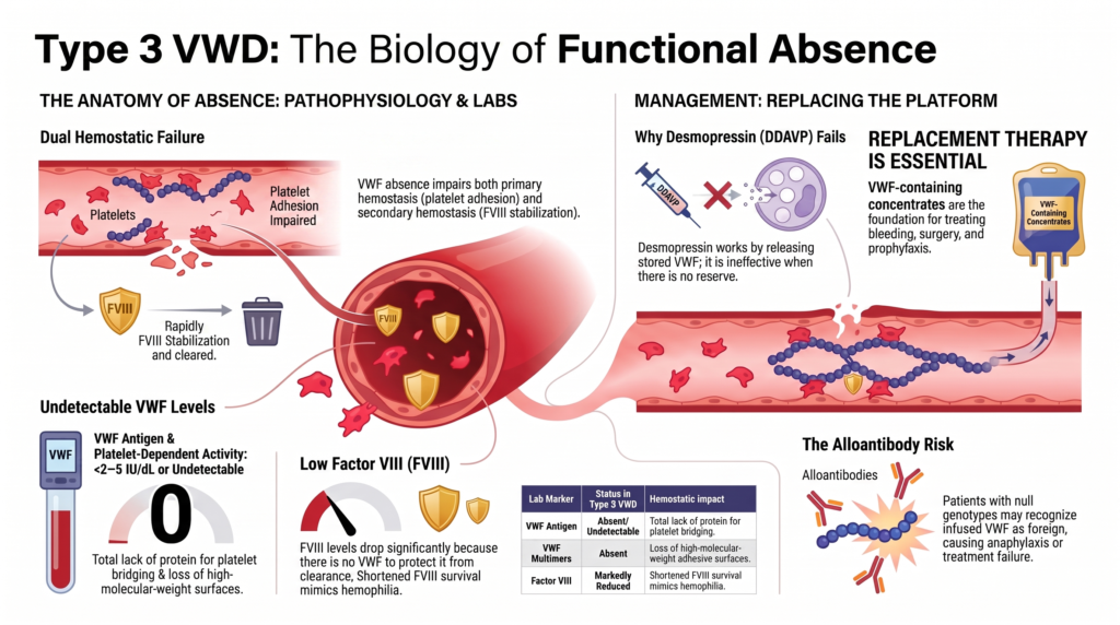
Why this spoke matters
Type 3 VWD is rare, but conceptually essential. In type 1 VWD, VWF is reduced. In type 2 VWD, VWF is dysfunctional. In type 3 VWD, VWF is absent or nearly absent.
That absence reveals the full biology of VWF. It shows why VWD can resemble both a platelet-type bleeding disorder and a coagulation factor deficiency, why mucosal bleeding and hemarthroses can coexist, and why replacement therapy becomes central when endogenous VWF is missing.
Type 3 VWD is not simply “more severe type 1.” It is the clearest demonstration that VWF is a bridge between primary and secondary hemostasis.
The basic definition
Type 3 VWD is the most severe quantitative form of VWD. It is characterized by absent or nearly absent VWF:
- VWF antigen is undetectable or virtually absent.
- VWF platelet-dependent activity is undetectable or virtually absent.
- Multimers are absent or nearly absent.
- FVIII is often markedly reduced because VWF is not available to bind, stabilize, and protect it.
The 2006 ISTH classification defines type 3 VWD as a virtual absence of VWF. In practice, type 3 is usually defined by undetectable or virtually absent VWF, often below assay detection limits or approximately less than 2 to 5 IU/dL depending on assay sensitivity.1
The key is not the exact number alone. The key is the functional absence of VWF.
A rare disease within a common disorder
VWD is often called the most common inherited bleeding disorder. That statement is true at the broad population level, but type 3 VWD is rare.
Published estimates are commonly in the range of approximately 0.5 to 4 cases per million, varying by geography, consanguinity, ascertainment, and diagnostic infrastructure.2
This creates an important teaching paradox: VWD is common, but type 3 VWD is rare. Commonness and severity are not the same axis.
Inheritance
Type 3 VWD is typically recessive. Affected patients often have biallelic pathogenic VWF variants, including null variants, large deletions, nonsense variants, frameshift variants, splice variants, or compound heterozygous combinations.
Heterozygous carriers may have low or borderline VWF levels, a mild type 1-like phenotype, or little bleeding. In some families, the heterozygous state is not clinically silent, which complicates counseling and reinforces the need to think beyond simple dominant/recessive labels.3
Type 3 VWD therefore has implications beyond the patient. It matters for siblings, reproductive counseling, partner testing in selected contexts, and prenatal or preimplantation discussions in families who desire them.
Why FVIII is low
FVIII is not usually low in type 3 VWD because the F8 gene is defective. FVIII is low because VWF is absent—not because the F8 gene is abnormal.
VWF normally binds FVIII and protects it from premature clearance. Without VWF, FVIII survival is shortened, and FVIII activity may fall into a hemophilia-like range.4
This is why type 3 VWD can produce both:
- mucocutaneous bleeding from impaired primary hemostasis
- joint, muscle, or soft-tissue bleeding from low FVIII
The phenotype is dual because VWF has dual hemostatic roles.
Why type 3 VWD can resemble hemophilia
Joint bleeding is not the classic teaching image of VWD. But in type 3 VWD, it matters.
Very low FVIII can produce hemarthroses and muscle bleeding, more reminiscent of moderate or severe hemophilia. Repeated joint bleeding can lead to arthropathy, pain, limited mobility, reduced activity, and reduced quality of life.5
Type 3 VWD is not hemophilia. The primary defect is absent VWF. But among VWD subtypes, type 3 comes closest to crossing the traditional boundary between platelet-type bleeding disorders and coagulation factor deficiencies.
The bleeding phenotype
Type 3 VWD can present early in life and has the broadest bleeding vocabulary of the inherited VWD subtypes.
Mucocutaneous bleeding may include:
- severe epistaxis
- oral cavity bleeding
- gingival bleeding
- easy bruising
- bleeding from minor wounds
- heavy menstrual bleeding
Hemostatic-challenge bleeding may include:
- post-dental bleeding
- postoperative bleeding
- post-traumatic bleeding
- postpartum hemorrhage
Severe or deeper bleeding may include:
- gastrointestinal bleeding
- hemarthroses
- muscle hematomas
- life-threatening bleeding
The mucocutaneous phenotype reflects absent VWF-mediated platelet adhesion. The joint and muscle phenotype reflects low FVIII. Not every patient has every manifestation, but the range is broader than in milder VWD.
Gastrointestinal bleeding
Gastrointestinal bleeding can be a major problem in type 3 VWD. It may be recurrent, difficult to localize, transfusion-requiring, and associated with angiodysplasia.
The problem is not always solved by simply correcting a level. Angiodysplasia represents a vascular lesion interacting with a hemostatic defect. VWF biology may also intersect with angiogenesis and vascular integrity, which may help explain why GI angiodysplasia is such a difficult phenotype in severe VWD.6
Type 3 VWD therefore belongs not only in classification. It belongs in the “when treatment fails” conversation.
Heavy menstrual bleeding, pregnancy, and delivery
For patients who menstruate, type 3 VWD can produce severe heavy menstrual bleeding beginning at menarche. It may cause iron deficiency, anemia, missed school, missed work, emergency care, transfusion, and major quality-of-life impairment.
Pregnancy and childbirth require specialized planning. Unlike many patients with type 1 VWD, patients with type 3 VWD generally do not normalize VWF levels during pregnancy because there is little or no endogenous VWF reserve. Delivery generally requires VWF replacement planning.
Neuraxial anesthesia decisions require current levels, replacement strategy, and anesthesia-hematology coordination. Postpartum bleeding risk remains significant. Tranexamic acid may have a role postpartum, but it does not substitute for replacement when replacement is needed. Genetic counseling may also be relevant before or during pregnancy, particularly when the partner’s carrier status or family history raises concern.7
In type 3 VWD, pregnancy physiology does not reliably rescue the hemostatic defect. Planning must.
Diagnosis: not just “very low VWF”
The laboratory pattern may seem obvious:
- VWF antigen absent or nearly absent
- VWF activity absent or nearly absent
- FVIII low
- multimers absent or nearly absent
But classification still requires care. Severe type 1 VWD with very low VWF can sometimes resemble type 3 VWD if assays are not sensitive or if residual VWF is difficult to measure. Sensitive VWF assays, multimer analysis, family studies, and genetic testing may help distinguish type 3 from severe type 1; VWF propeptide can be useful in selected specialized evaluations.8
This distinction matters for inheritance, family counseling, alloantibody risk, phenotype prediction, classification accuracy, research, and treatment planning.
Even the most severe category benefits from precise thinking.
Genetic testing
Genetic testing has a clearer role in type 3 VWD than in many cases of type 1 VWD. It can identify biallelic pathogenic VWF variants, support family counseling, inform reproductive planning, clarify whether apparent type 3 is true type 3 or severe type 1, and identify large deletions or null genotypes that may be relevant to alloantibody risk.9
Genetic testing is not required to understand the immediate bleeding risk, but it can change the long-term conversation.
Why desmopressin does not work
Desmopressin works by releasing stored endogenous VWF from endothelial cells. In type 3 VWD, VWF is absent or nearly absent. There is little or no releasable reserve.
Therefore, desmopressin is not effective and should not be relied on in type 3 VWD because there is little or no endogenous VWF reserve to release.10
This is one of the cleanest treatment lessons in VWD: if the problem is absence, release therapy cannot solve it.
Replacement therapy is central
VWF-containing concentrate is the foundation of treatment for significant bleeding, surgery, trauma, childbirth, and prophylaxis in type 3 VWD. Replacement therapy supplies the missing VWF and supports FVIII stabilization.
Depending on the product, FVIII may be supplied directly, or FVIII may rise over time as endogenous FVIII is stabilized by infused VWF. This distinction matters in urgent bleeding. If a VWF-only product is used and FVIII is low or unknown, coadministration of FVIII may be needed initially to achieve immediate hemostasis.11
Major bleeding, major surgery, childbirth, and severe trauma generally require VWF replacement with clinical and laboratory monitoring.
Product choice, kinetics, and monitoring
Product selection matters. Plasma-derived VWF/FVIII concentrates contain both VWF and FVIII in varying ratios. VWF-only products, including recombinant VWF, supply VWF without FVIII.
Repeated dosing with VWF/FVIII concentrates can lead to FVIII accumulation, especially during surgery or prophylaxis. VWF-only products may reduce direct FVIII exposure, but they require attention to timing because FVIII rise depends on stabilization of endogenous FVIII or coadministered FVIII.12
The product question is kinetic:
- what needs to rise immediately?
- what needs to be sustained?
- what thrombotic risk exists?
- what monitoring is available?
- what product can the patient access?
Monitoring may include VWF activity, FVIII activity, clinical bleeding, trough levels when relevant, FVIII accumulation, recovery after dosing, duration of effect, and concern for inhibitor or alloantibody if recovery is poor.
Monitoring is not just confirming that treatment was given. It is asking whether replacement has restored both sides of VWF biology: platelet-dependent hemostasis and FVIII stabilization.
Long-term prophylaxis
Many patients with type 3 VWD require episodic treatment only for specific bleeds or procedures, but some need long-term prophylaxis. Historically, prophylaxis in severe VWD was borrowed conceptually from hemophilia. Increasing evidence and experience now support regular prophylaxis in selected patients with severe and recurrent bleeding, particularly recurrent joint, gastrointestinal, or severe mucosal bleeding.
Prophylaxis may be considered when bleeding is severe and frequent:
- recurrent joint or muscle bleeding
- recurrent severe epistaxis
- recurrent GI bleeding
- life-disrupting mucosal bleeding
- repeated hospitalizations or transfusions
- bleeding that limits school, work, activity, or independence
The 2021 ASH/ISTH/NHF/WFH management guideline suggests long-term prophylaxis rather than no prophylaxis in patients with VWD who have a history of severe and frequent bleeds, with periodic reassessment. This is a conditional recommendation based on low-certainty evidence, so patient selection and reassessment matter.13
The goal is fewer bleeds. But the deeper goal is more stable living.
Alloantibodies: recognition and implications
Anti-VWF alloantibodies are rare but critical in type 3 VWD. Reported anti-VWF alloantibody risk is concentrated in type 3 VWD, particularly in patients with large deletions or null genotypes, and is clinically important because VWF infusion may trigger poor recovery or anaphylaxis.14
The concept is straightforward: patients who have never produced meaningful endogenous VWF may recognize infused VWF as foreign. Consider anti-VWF alloantibodies when a patient with type 3 VWD has:
- poor recovery after VWF infusion
- unexpectedly rapid loss of infused VWF
- continued bleeding despite apparently appropriate dosing
- infusion reactions
- anaphylaxis
- known large VWF gene deletion or severe null genotype
In these situations, routine dose escalation can be dangerous. The patient needs specialized management in an expert bleeding-disorder center. Rare case reports describe off-label emicizumab in type 3 VWD with alloantibodies; this should be framed as exceptional, nonstandard, expert-center management rather than routine care.15
This is one of the most serious type 3-specific complications. It should be rare in teaching, but never invisible.
Surgery and procedures
Surgery in type 3 VWD requires planned replacement therapy. Desmopressin is not useful. Tranexamic acid and local measures can help, especially for mucosal procedures, but they do not replace VWF.
For major surgery, the 2021 management guideline suggests targeting both FVIII and VWF activity levels of at least 0.50 IU/mL for at least 3 days after surgery, and suggests against targeting FVIII alone.16
This point is especially important in type 3 VWD. FVIII alone does not restore VWF-mediated primary hemostasis. The primary defect must be replaced.
The child with type 3 VWD
Type 3 VWD may present in childhood with severe mucosal bleeding, traumatic bleeding, bleeding after procedures, or joint bleeding. Management may involve family education, home treatment planning, school planning, activity counseling, emergency plans, dental prevention, iron monitoring, vaccination and injection precautions, prophylaxis decisions, and genetic counseling for the family.
The diagnosis affects the household, not only the child. Families need to know which bleeding requires urgent care, which product is needed, and where expert help is available.
Type 3 VWD is rare enough that local emergency teams may not be familiar with it. A written emergency plan can be lifesaving.
Quality of life
Severe VWD can affect quality of life through bleeding, fear, treatment burden, activity restriction, hospital visits, pain, anemia, reproductive bleeding, and uncertainty.
Type 3 VWD may require repeated infusions, venous access planning, prophylaxis decisions, and specialist follow-up. For some patients, the burden is visible in bleed counts. For others, it appears as avoidance:
- not playing sports
- not traveling
- not accepting procedures
- not attending school or work reliably
- not trusting the body
The treatment goal is not simply laboratory correction. It is safe participation in life.
How type 3 differs from severe type 1
Severe type 1 VWD can have very low VWF. Type 3 VWD has absent or nearly absent VWF. The distinction matters because type 3 is more likely to be recessive, associated with very low FVIII, severe bleeding, possible alloantibody risk, and need for replacement therapy.
Severe type 1 and type 3 may overlap clinically in some respects, but they are not interchangeable labels. When classification is uncertain, sensitive assays, family studies, multimer analysis, and genetic testing may help clarify.17
The question is not taxonomy for taxonomy’s sake. The question is what the label changes.
Future directions
Type 3 VWD is one of the most obvious candidates for future transformative therapy. But VWF gene therapy is more difficult than hemophilia gene therapy.
VWF is large. It is multimeric. It requires complex processing. It is normally produced by endothelial cells and megakaryocytes. Its multimer size, storage, secretion, clearance, and shear responsiveness matter.
Preclinical gene-transfer strategies have shown partial correction in experimental models, but current reviews emphasize that VWD gene therapy remains preclinical and is not routine clinical care.18
Type 3 VWD shows why the future is exciting. It also shows why the future is technically hard.
Clinical synthesis
Type 3 VWD is the most severe quantitative form of VWD. VWF is absent or nearly absent, and FVIII is often markedly reduced because VWF normally stabilizes FVIII.
This creates a dual bleeding phenotype:
- mucocutaneous bleeding from absent VWF-mediated primary hemostasis
- deeper bleeding from low FVIII
Type 3 VWD is usually autosomal recessive and has important implications for family counseling and reproductive planning. Desmopressin is ineffective because there is little or no VWF to release. VWF replacement is central for significant bleeding, surgery, childbirth, and many prophylaxis plans.
Anti-VWF alloantibodies are rare but important, especially in patients with null genotypes or large deletions, because they can cause poor recovery and severe allergic or anaphylactic reactions to VWF-containing products.
Type 3 VWD is rare, but it reveals the full logic of VWF biology.
When VWF is absent, both platelet adhesion and FVIII survival fail.
Evidence anchor: why type 3 VWD is not simply severe type 1
| Evidence stream | What it shows | Why it matters | Main limitation |
|---|---|---|---|
| Classification and laboratory phenotype | Type 3 VWD is defined by virtual absence of VWF, with undetectable or nearly undetectable antigen and activity and absent multimers.19 | The defining issue is functional absence, not just a low number. | Assay sensitivity can complicate the boundary between severe type 1 and type 3. |
| Genetic studies | Type 3 VWD usually reflects biallelic severe or null VWF variants, including large deletions, nonsense, frameshift, splice, or compound heterozygous variants.20 | Genetics clarifies inheritance, counseling, and possible alloantibody risk. | Genotype-phenotype correlation is not perfect. |
| FVIII biology | FVIII is often markedly reduced because absent VWF cannot stabilize it.21 | Explains why type 3 VWD can produce joint and muscle bleeding as well as mucosal bleeding. | FVIII level alone does not fully predict bleeding phenotype. |
| Severe VWD cohorts and reviews | Severe VWD can involve joint bleeding, GI bleeding, heavy menstrual bleeding, and major quality-of-life burden.22 | Type 3 VWD has a broader bleeding vocabulary than typical mild VWD. | Referral cohorts may overrepresent severe phenotypes. |
| Prophylaxis evidence and guidelines | Long-term prophylaxis is suggested for selected patients with severe and frequent bleeding, with periodic reassessment.23 | Prophylaxis may prevent recurrent bleeding and its life consequences. | Guideline certainty is low, so individualized selection matters. |
| Alloantibody reports | Anti-VWF alloantibodies are rare but concentrated in type 3 VWD, especially in patients with large deletions or null genotypes.24 | Explains poor recovery, treatment failure, and anaphylaxis risk after VWF exposure. | Rare complication, limiting prospective study. |
Interpretive note: Type 3 VWD is not simply the far end of type 1. Its biology, inheritance, bleeding phenotype, treatment dependence, and alloantibody risk make it a distinct severe-VWD category.
Guideline perspective: managing absence rather than deficiency
The 2021 ASH/ISTH/NHF/WFH management guideline does not treat type 3 VWD as simply “low VWF.” It emphasizes management according to bleeding severity, procedure risk, treatment response, and the need to restore both VWF activity and FVIII activity during major hemostatic challenges.25
For type 3 VWD, several implications follow:
- desmopressin should not be relied on because there is little or no endogenous VWF to release
- VWF replacement is central for major bleeding, major surgery, childbirth, and severe trauma
- major surgery should target both VWF activity and FVIII activity, not FVIII alone
- long-term prophylaxis should be considered in selected patients with severe and frequent bleeding, with periodic reassessment
- pregnancy and delivery generally require planned replacement support
- unexpected poor recovery or infusion reactions after VWF replacement should raise concern for alloantibodies
The practical lesson is simple: when VWF is absent, strategies that depend on endogenous VWF are unlikely to work. Treatment must replace the missing platform.
Reflect & Apply Case
A 9-year-old child has recurrent epistaxis, large bruises after minor trauma, and two ankle hemarthroses.
Laboratory testing shows:
- VWF antigen: undetectable
- VWF platelet-dependent activity: undetectable
- FVIII: 4 IU/dL
- multimer analysis: no detectable VWF multimers
Questions for reflection:
- Why does this pattern suggest type 3 VWD rather than type 1 VWD?
- Why is FVIII so low if the primary genetic defect is in VWF?
- Why can joint bleeding occur in type 3 VWD?
- Why would desmopressin not be useful?
- What role does VWF replacement play?
- When would long-term prophylaxis be considered?
- What family counseling issues arise?
- What would raise concern for anti-VWF alloantibodies during treatment?
Expert synthesis: This child has absent VWF antigen, absent VWF activity, absent multimers, and markedly reduced FVIII. The pattern supports type 3 VWD, not simply severe type 1. FVIII is low because VWF is absent and cannot stabilize it. The combination of absent VWF-mediated platelet adhesion and low FVIII explains both mucocutaneous bleeding and hemarthroses. Desmopressin would not be useful because there is little or no stored VWF to release. Treatment requires VWF replacement, with consideration of prophylaxis if bleeding is severe and recurrent. Family counseling should address autosomal recessive inheritance, sibling risk, and reproductive planning. Poor recovery, infusion reactions, or anaphylaxis after VWF concentrate should raise concern for anti-VWF alloantibodies.
This case illustrates the central lesson: type 3 VWD is not just the absence of a lab value. It is the absence of a hemostatic platform.
Test your thinking
A short quiz on type 3 VWD.