
When numbers and bleeding tell different stories
Note: The video and audio linked above were generated with the assistance of AI. Clinical accuracy has been reviewed, but no AI-generated content can be guaranteed to be fully error-free.
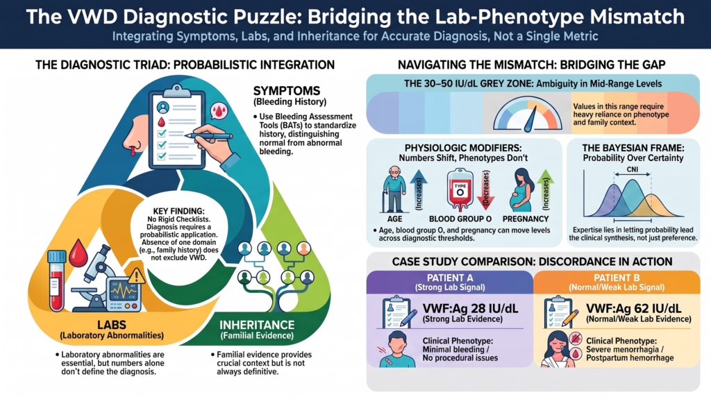
Why this spoke matters
Von Willebrand disease resists reduction.
Not to a number.
Not to a symptom.
Not to a single test.
Diagnosis requires integration of:
- bleeding symptoms
- laboratory abnormalities
- inheritance
This diagnostic triad remains central to VWD identification: a personal history of excessive mucocutaneous bleeding, at least one laboratory abnormality of VWF function, and, when present, evidence of familial inheritance. But the triad is applied probabilistically, not as a rigid checklist.1
Family history strengthens diagnostic confidence when present.
Its absence does not exclude VWD.
Yet in real practice, these domains frequently disagree.
Recognizing mismatch, and resisting premature resolution, is central to expert care.
Two patients, opposite signals
Patient A
VWF:Ag 28 IU/dL
Minimal bleeding history
No procedural bleeding
Patient B
VWF:Ag 62 IU/dL
Severe menorrhagia since adolescence
Postpartum hemorrhage
Which patient has stronger evidence for VWD?
Which patient has stronger evidence for clinically important bleeding?
The reflex answer depends on which signal you privilege.
VWD forces you to choose neither.
Patient A has stronger laboratory evidence of low VWF or VWD-spectrum biology. Patient B has stronger phenotypic evidence of a clinically important bleeding disorder, but the available VWF results do not, by themselves, establish VWD.
Neither case is fully resolved.
That incompleteness is the point.
Why the mismatch exists
Several well-described features of VWF biology create discordance.
1. Continuous distribution of VWF levels
There is no natural biologic divide between normal and abnormal.
VWF levels vary widely in the population, and the distinction between “normal” and “low” is partly conventional.2
Thresholds help clinicians act.
They do not create biological borders.
2. Imperfect correlation between VWF level and bleeding severity
A given VWF level does not determine the bleeding phenotype.
Bleeding risk generally increases as VWF falls, but the signal strengthens most clearly at very low levels. VWF levels below 30 IU/dL provide stronger evidence for VWF-linked disease biology; values in the 30–50 IU/dL range are more ambiguous and require greater reliance on bleeding phenotype, family context, repeat testing, and hemostatic-challenge history.3
3. Physiologic modifiers
Stress, exercise, inflammation, pregnancy, estrogen exposure, aging, thyroid status, and blood group influence measured VWF levels.
ABO blood group is especially important: group O is associated with lower VWF levels than non-O groups.
These modifiers can move a patient across a diagnostic threshold without changing the underlying bleeding story.4
4. Assay limitations
Laboratory assays measure surrogates of function, not in vivo bleeding risk.
VWF antigen (VWF:Ag) measures quantity. Platelet-dependent VWF activity assays, VWF collagen-binding activity (VWF:CB), factor VIII activity (FVIII:C), VWF multimer analysis, VWF activity/VWF:Ag ratios, and VWF–FVIII binding assays (VWF:FVIIIB) each interrogate different parts of VWF biology. None alone captures the full hemostatic phenotype.5
This is why abnormal results should usually be confirmed, especially when the diagnosis is mild, borderline, or clinically discordant. Diagnosis should not rest on a single vulnerable measurement.
5. Phenotypic heterogeneity
Type 1 VWD remains mechanistically heterogeneous and incompletely explained at the molecular level.
In many patients with type 1 VWD, especially milder cases, the phenotype may reflect more than one genetic or environmental determinant rather than a single VWF mutation.6
Mismatch is therefore expected, not exceptional.
The danger of privileging the lab
When clinicians privilege laboratory numbers, patients with important bleeding may be incompletely evaluated.
Heavy menstrual bleeding may be normalized or managed in isolation from a bleeding-disorder workup. Bleeding may be attributed too readily to gynecologic causes alone. Women may experience delayed diagnosis, especially when the laboratory signal is borderline or temporarily normal.7
Guidelines emphasize bleeding-assessment tools because phenotype cannot be inferred from VWF levels alone.8
A normal or near-normal VWF level does not exclude clinically important bleeding or another bleeding disorder.
But it does weaken a VWD diagnosis unless repeat testing, functional assays, family history, or subtype-specific findings support VWF pathology.
That distinction matters.
Patient B may need serious evaluation and treatment planning. But the question is not simply, “Does she bleed?” It is, “Is VWF the explanation?”
The danger of privileging the phenotype
The opposite error also occurs.
Overattribution is the mirror image error: naming disease where biology remains uncertain.
A patient with easy bruising, a mildly elevated bleeding score, VWF activity of 44 IU/dL, blood group O, no family history, and no hemostatic challenges may be labeled with lifelong VWD on the basis of a borderline result and common symptoms.
That label can carry consequences:
- procedural anxiety
- insurance or documentation burden
- family screening
- unnecessary treatment
- premature closure around alternative diagnoses
Mild bleeding symptoms are common in the general population, and low-to-borderline VWF levels may coexist with those symptoms by chance.9
The low VWF debate reflects this tension. Removing diagnostic barriers may improve access to care, especially for symptomatic women. But overdiagnosis may also create unnecessary burden and misclassification.10
Phenotype without laboratory support may still justify action.
It does not automatically justify a VWD diagnosis.
A Bayesian frame
VWD diagnosis is best understood as evidence integration.
Each domain shifts probability:
- bleeding history
- laboratory abnormality
- family pattern
None independently establishes certainty.
Tosetto and Eikenboom explicitly frame VWD diagnosis as Bayesian: the same finding carries different meaning depending on the pre-test setting. Mild bleeding in a sibling of a patient with VWD does not carry the same diagnostic weight as mild bleeding in an unselected older adult.11
Return to the opening cases.
In Patient A, a VWF antigen of 28 IU/dL substantially raises the probability of VWF-linked disease biology, even though the bleeding history is quiet. But the absence of procedural bleeding and family history should temper confidence about clinical severity.
In Patient B, severe reproducible bleeding and postpartum hemorrhage raise the probability of a clinically important bleeding disorder. But VWF levels above 50 IU/dL lower the probability that classic type 1 VWD is the explanation, unless repeat testing, VWF activity/VWF:Ag ratio, FVIII, family history, or subtype-specific findings change the picture.
Mismatch does not invalidate diagnosis.
It recalibrates probability.
The “normalized over time” problem
An especially challenging scenario:
a patient previously diagnosed with type 1 VWD
later testing shows VWF >50 IU/dL
Does the disease disappear?
Guidelines explicitly address how to approach patients whose levels normalize over time.12
Physiologic normalization during pregnancy, inflammation, or aging complicates interpretation. VWF levels below 50 IU/dL may normalize in a substantial proportion of individuals over time, particularly with aging.13
But normalization of the number does not necessarily normalize the bleeding phenotype.
Recent discussions of “undiagnosing” VWD emphasize that the question should not be answered by the current VWF level alone. Bleeding history, prior diagnostic context, repeated testing, age, hemostatic challenges, and patient-specific consequences all matter.14
Here, discordance is not just cross-sectional.
It is longitudinal.
How experts reason in discordant cases
Phenotypic severity and reproducibility
How severe is the bleeding? Has it occurred repeatedly? Does it involve high-signal events such as surgical bleeding, dental extraction bleeding, postpartum hemorrhage, or recurrent iron deficiency?
Objective corroboration
Is there documentation: transfusion, reoperation, emergency care, procedural delay, iron deficiency reflecting chronic blood loss, or treatment requirement?
Laboratory stability
Are VWF antigen, platelet-dependent activity, FVIII:C activity, and activity-to-antigen relationships consistent across time and testing conditions?
Family pattern
Is there a first-degree relative with similar bleeding, low VWF, or confirmed VWD?
Treatment response may clarify mechanism or management, but response does not by itself establish the diagnosis.
Bleeding assessment tools can structure this evaluation. They standardize the history, improve communication, and help distinguish normal from abnormal bleeding. But they do not diagnose VWD by themselves, and they may not distinguish VWD from other mild bleeding disorders.15
Mechanism is inferred through convergence, not single signals.
When mismatch should increase caution
Mismatch warrants diagnostic expansion when:
laboratory abnormalities are profound but bleeding is absent
bleeding is severe but VWF testing is repeatedly normal
inheritance contradicts the laboratory or bleeding pattern
The first two are phenotype-lab mismatches.
The third introduces the inheritance axis, which matters because VWD is not only a laboratory phenotype or a bleeding phenotype. It is also, in most cases, an inherited biology.
In discordant cases, alternative or additional explanations should be considered:
- platelet function disorders
- acquired VWF abnormalities
- gynecologic causes of heavy menstrual bleeding
- connective tissue disorders
- bleeding disorder of unknown cause
VWD is common.
It is not universal.
What we do not know
We still lack:
- precise individual-level bleeding risk prediction from VWF levels
- robust tools to distinguish mild disease from physiologic variation
- definitive thresholds for procedural bleeding risk
Recent “beyond the guidelines” discussions emphasize that many difficult VWD scenarios remain outside strong evidence, including the implications of physiologic normalization and individualized management when clinical and laboratory signals diverge.16
Discordance is therefore a structural feature of the disease, not merely a temporary gap in knowledge.
Clinical synthesis
The lab–phenotype mismatch is not a flaw in the diagnostic process.
It is the process.
Diagnostic confidence emerges not from any single domain, but from convergence over time.
When bleeding history, laboratory pattern, and inheritance move in the same direction, diagnosis stabilizes.
When they diverge, expertise lies in resisting premature closure.
The task is not to choose the number or the story.
It is to let probability, not preference, lead.
Evidence anchor: why lab–phenotype mismatch is expected in VWD
Summary derived from diagnostic guidelines, cohort studies, low VWF literature, and expert reviews. The evidence consistently shows that VWD diagnosis cannot be reduced to a single VWF value, bleeding score, or family-history pattern. Laboratory values, bleeding phenotype, and inheritance each contribute diagnostic information, but each has important limitations.
| Evidence stream | What it shows | Why it matters | Main limitation |
|---|---|---|---|
| Population distribution of VWF | VWF levels vary continuously across the population. There is no natural biologic boundary between “normal” and “low.”17 | Diagnostic thresholds are useful clinical tools, but they do not define a sharp disease border. | A low VWF value may represent disease biology, physiologic variation, or a risk factor rather than a complete diagnosis. |
| Type 1 VWD and low VWF studies | Very low VWF levels are more strongly associated with VWF-linked disease biology, while the 30–50 IU/dL range is heterogeneous.18 | The same VWF value may carry different diagnostic weight depending on bleeding history, family context, and prior hemostatic challenges. | Borderline reductions may coexist with common bleeding symptoms by chance. |
| Bleeding assessment tool studies | BATs structure bleeding history and improve reproducibility.19 | They help clinicians avoid relying on vague impressions of “bleeds” or “does not bleed.” | BATs do not diagnose VWD by themselves and may not distinguish VWD from other mild bleeding disorders. |
| Laboratory assay studies | VWF antigen, platelet-dependent activity, FVIII, ratios, collagen binding, multimer analysis, and VWF–FVIII binding assay (VWF:FVIIIB) interrogate different parts of VWF biology.20 | No single assay captures in vivo bleeding risk or all VWD mechanisms. | Preanalytical variables, physiologic modifiers, and assay variability can mislead interpretation. |
| Aging and normalization studies | VWF levels may rise with age and sometimes normalize in patients previously diagnosed with type 1 VWD or low VWF.21 | Discordance can be longitudinal, not just cross-sectional. | It remains uncertain whether normalization of VWF levels always normalizes bleeding risk. |
Interpretive note: These evidence streams point in the same direction: VWD diagnosis is probabilistic. Low VWF levels, bleeding symptoms, and family history each shift diagnostic confidence, but none is sufficient alone. The strongest diagnosis emerges when laboratory pattern, bleeding phenotype, inheritance, and repeat testing converge. When they diverge, the task is not to force a label, but to recalibrate probability and widen the differential when needed.
Guideline perspective: How diagnostic guidance fits into lab–phenotype mismatch
Guideline organizations and expert sources referenced
ASH/ISTH/NHF/WFH 2021 VWD diagnosis guideline
British Society for Haematology / UKHCDO 2024 laboratory diagnosis guideline
Tosetto and Eikenboom clinical and laboratory diagnosis review
Low VWF and “undiagnosing VWD” expert reviews
Shared guidance themes
Guidelines and expert reviews consistently emphasize that VWD diagnosis requires integration of:
- bleeding phenotype
- VWF laboratory pattern
- family history or inherited context
- repeat testing when results and history disagree
A single VWF value should not be interpreted in isolation.
Where guidance is clearest
VWF levels below 30 IU/dL provide stronger evidence for VWF-linked disease biology.
VWF levels in the 30–50 IU/dL range require more clinical judgment, especially when bleeding history, family history, and repeat testing are not concordant.
VWF levels above 50 IU/dL generally weaken a classic type 1 VWD diagnosis, but do not exclude clinically important bleeding or another bleeding disorder.
Where guidance urges caution
Diagnostic confidence should be reduced when:
- bleeding is severe but VWF testing is repeatedly normal
- VWF is low but bleeding history is minimal or absent
- results are obtained during pregnancy, inflammation, physiologic stress, or other states that may alter VWF levels
- the diagnosis rests on a single borderline result
What guidelines support
- Use of bleeding assessment tools to structure bleeding history
- Initial testing with VWF antigen, platelet-dependent VWF activity, and FVIII:C activity
- Repeat testing when results are borderline, unexpected, or clinically discordant
- Subtype-directed testing when the first-line pattern suggests qualitative VWF dysfunction
- Reconsideration, rather than automatic removal, of a prior VWD diagnosis when VWF levels normalize over time
What guidelines do not support
- Diagnosing VWD from bleeding symptoms alone
- Dismissing serious bleeding solely because VWF is near normal
- Treating a single borderline VWF value as a lifelong disease label without context
- Assuming that normalized VWF levels always mean normalized bleeding risk
Practical takeaway
Guidelines do not eliminate uncertainty in VWD diagnosis.
They teach clinicians how to manage it.
When bleeding phenotype, laboratory pattern, and inheritance converge, diagnostic confidence rises. When they diverge, the task is to repeat, contextualize, and widen the differential rather than forcing a premature label.
Reflect & Apply
Case 1
- VWF antigen 24 IU/dL
- Platelet-dependent VWF activity 22 IU/dL
- No surgical bleeding history
Case 2
- VWF antigen 58 IU/dL
- VWF activity 55 IU/dL
- Heavy menstrual bleeding since menarche
- Postpartum hemorrhage
What does the evidence in each case support?
Where does it conflict?
What additional data would most meaningfully change your confidence:
- repeat testing
- activity-to-antigen ratio
- FVIII activity level
- bleeding assessment score
- family study
- surgical history
- postpartum records
- ferritin
- evaluation for another bleeding disorder?
Test your thinking
A short quiz on lab–phenotype mismatch in VWD.