
How a spectrum became types, and why the lines keep moving
Note: The video and audio linked above were generated with the assistance of AI. Clinical accuracy has been reviewed, but no AI-generated content can be guaranteed to be fully error-free.
Why this spoke matters
Von Willebrand disease is often presented as a tidy classification system.
Type 1.
Type 2A, 2B, 2M, 2N.
Type 3.
Laboratory thresholds can appear equally tidy.
30 IU/dL.
50 IU/dL.
Normal.
Abnormal.
But VWF biology is not tidy.
VWF levels are continuously distributed across the population. They vary with blood group, age, pregnancy, inflammation, stress, hormones, endothelial biology, clearance, and assay method.
Bleeding is also not binary.
Bleeding symptoms are common in the general population. Some are trivial. Some are disabling. Some are never tested by surgery, childbirth, dental extraction, or trauma until later in life.
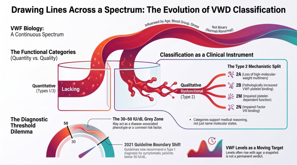
This is the central classification problem in VWD: medicine must draw lines across a spectrum. Every classification system therefore represents a compromise between biological accuracy and clinical usefulness.
The boundaries it creates are necessary. They are also provisional.
The same VWF antigen or platelet-dependent activity result may carry different diagnostic meanings depending on bleeding history, family history, hemostatic challenge, repeat testing, and the guideline era in which the patient is seen.
That is why the history of classification and thresholds matters.
It teaches that VWD categories are clinical decisions as much as diagnostic labels.
Classification as a clinical instrument
Clinicians need classification to communicate mechanism, interpret tests, anticipate risk, select treatment, counsel families, and define research cohorts. No system can serve all of these purposes perfectly. Biological precision and clinical usability rarely increase together. The modern classification is therefore not simply a taxonomy of mutations. It is a clinical instrument designed to support better reasoning.
The first durable divide: quantity versus quality
As immunologic assays, platelet-dependent activity assays, and multimer analysis developed, a fundamental distinction emerged.
Some patients had less VWF.
Others had VWF that was present but functionally abnormal.
This created the durable conceptual divide:
type 1
partial quantitative deficiency
type 2
qualitative VWF dysfunction
type 3
virtually complete VWF deficiency
This distinction was powerful because it mapped clinical categories onto mechanism. Less protein was different from abnormal protein. Virtually absent protein was different again.1
The categories helped clinicians communicate, interpret laboratory patterns, and anticipate treatment.
They also marked a conceptual advance.
VWD was no longer merely a bleeding tendency.
It was a family of VWF failure modes.
Type 2 and the pressure to split
Type 2 VWD became the proving ground for classification.
At first, qualitative VWD was a broad bucket.
But as multimer analysis, platelet-binding studies, FVIII-binding assays, collagen-binding assays, and molecular genetics improved, the bucket began to split.
Different mechanisms became visible:
- loss of high-molecular-weight multimers
- increased platelet binding
- impaired platelet-dependent function not explained by loss of high-molecular-weight multimers
- impaired FVIII binding
These mechanisms became the modern type 2 subtypes:
2A
loss of high-molecular-weight multimers
2B
increased VWF-platelet binding, often with loss of large multimers and sometimes thrombocytopenia
2M
impaired platelet-dependent function despite a relatively normal multimer distribution
2N
impaired FVIII binding, which can mimic mild hemophilia A
The subtypes were not created for elegance.
They were created because different mechanisms have different implications.
A patient with loss of high-molecular-weight multimers is not the same as a patient whose VWF binds platelets too avidly.
A patient whose VWF cannot bind FVIII may resemble a patient with mild hemophilia A, but the mechanism is different and may require VWF or genetic testing to distinguish.2
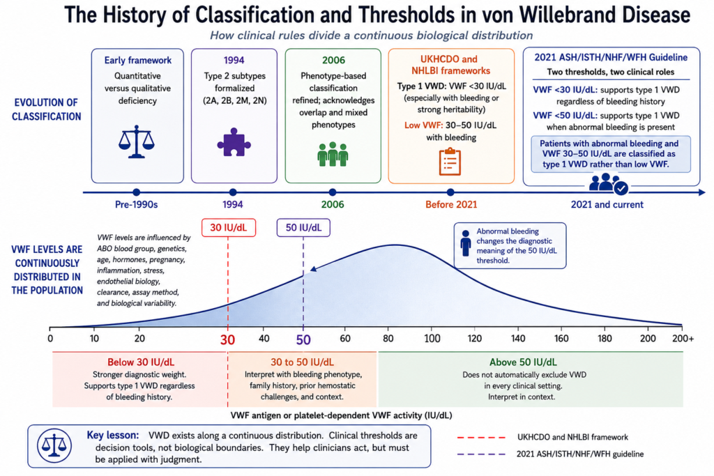
In 1994, J. Evan Sadler and the ISTH subcommittee formalized the type 2A, 2B, 2M, and 2N framework, shifting classification toward identifiable functional mechanisms. The 2006 ISTH SSC classification update was designed to keep classification clinically useful and largely phenotype-based, while acknowledging mixed phenotypes and unresolved heterogeneity.3
That caveat matters.
The modern system looks orderly.
Its history is less orderly.
Thresholds and the illusion of precision
Once assays existed, cutoffs followed.
Medicine needed numbers.
A laboratory value had to be interpreted.
A patient had to be labeled or not labeled.
A procedure had to be planned.
A treatment had to be approved.
A research cohort had to be defined.
Thresholds made those decisions possible.
But VWF levels do not naturally divide into normal and abnormal populations.
They are continuously distributed.
A threshold therefore converts a continuous biological trait into a clinical category.
That conversion is necessary.
It is also imperfect.
A patient at 29 IU/dL and a patient at 31 IU/dL are not biologically transformed by crossing a line.
A patient at 48 IU/dL and a patient at 52 IU/dL may be separated by a label more than by disease biology.
Thresholds help clinicians act.
They do not define nature.
The instability of type 1
Type 1 VWD seems, at first glance, the simplest category.
There is less VWF.
But history made type 1 increasingly complicated.
Patients labeled with type 1 VWD did not all behave the same way.
Some had clearer inheritance.
Some did not.
Some had identifiable VWF variants.
Many did not.
Some showed stable reductions over time.
Others had levels that fluctuated or normalized.
Some bled substantially.
Others barely bled.
The category remained useful, but it was not uniform.
Type 1 VWD sits at the border between inherited disease, quantitative biology, modifier effects, and normal population variation.
That is why the threshold problem eventually became inseparable from classification.
The 30 to 50 IU/dL problem
The low-VWF debate exposed the core threshold problem.
VWF is a continuous trait. Bleeding symptoms are common. Mildly low VWF levels are also common.
The 30 to 50 IU/dL range can behave like a disease-associated phenotype in some patients and like a risk factor or incidental finding in others.
J. Evan Sadler framed the problem with unusual clarity: low VWF can sometimes function like a disease and sometimes like a risk factor. Very low levels are more likely to reflect a heritable VWF disorder with clinically meaningful bleeding. Modestly low levels, especially in the 30 to 50 IU/dL range, are common enough in the population that they may coincide with common bleeding symptoms by chance.4
This distinction reshaped the diagnostic debate.
The rationale was biological as well as clinical.
Patients with lower VWF levels are more likely to have VWF variants and clearer autosomal dominant inheritance. Patients in the 30 to 50 IU/dL range often have less clear inheritance, fewer identifiable VWF variants, and bleeding phenotypes that correlate poorly with residual VWF level.5
That made “low VWF” attractive.
It preserved biological caution.
It acknowledged risk without necessarily declaring disease.
But it created a different problem.
For patients, especially those with heavy menstrual bleeding, postpartum hemorrhage, or repeated procedure-related bleeding, the label “low VWF” could sound less real than VWD.
It could also affect access to care.
When guidelines changed the boundary
The 2021 ASH/ISTH/NHF/WFH diagnostic guidelines made a consequential decision.
Earlier frameworks often reserved type 1 VWD for VWF levels below 30 IU/dL and used the label “low VWF” for patients in the 30 to 50 IU/dL range with bleeding. The 2021 ASH/ISTH/NHF/WFH guideline recommended confirming type 1 VWD at VWF levels below 30 IU/dL regardless of bleeding history, and at levels below 50 IU/dL in patients with abnormal bleeding. This was issued as a strong recommendation despite low certainty in the evidence, reflecting the panel’s emphasis on avoiding missed diagnoses and improving access to care.6
This removed “low VWF” as a separate diagnostic category within the guideline and brought bleeding patients with levels between 30 and 50 IU/dL under the diagnosis of type 1 VWD. The term nevertheless remains common in clinical usage and in parts of the literature.
The decision was controversial.
Not because it was careless.
Because it was value-laden.
The guideline prioritized avoiding missed diagnoses and improving access to care.
That is a legitimate clinical priority.
But critics worried about overdiagnosis, especially because mild bleeding symptoms and borderline VWF levels are both common.
The disagreement was not only about biology.
It was also about access, labeling, diagnostic delay, medicalization, and the consequences of naming disease.
A diagnostic label affects procedure planning, obstetric care, treatment access, anxiety, self-understanding, insurance, and research eligibility.
The boundary is scientific.
It is also practical.
Low VWF as a moving target
The low-VWF debate became more complex when longitudinal data showed that VWF levels can rise with age.
A patient tested in childhood may have VWF levels below 30 IU/dL and be diagnosed with type 1 VWD.
The same patient first tested as an adult may fall into the 30 to 50 IU/dL range.
The same patient first tested later in life may have levels above 50 IU/dL.
The biology has not necessarily disappeared.
The snapshot has changed.
Recent analyses have argued that low VWF may not represent a discrete clinicopathological entity, but rather part of the heterogeneous type 1 VWD spectrum, modified by age, biology, and timing of testing.7 Other investigators continue to view low VWF as a potentially distinct state, citing its less consistent association with pathogenic VWF variants, heterogeneous clearance biology, and variable relationship between residual VWF level and bleeding.
That insight is important.
It does not eliminate diagnostic uncertainty.
It relocates it.
Instead of asking only:
Is this low VWF or type 1 VWD?
the clinician must ask:
- When was the patient tested?
- What bleeding challenges have occurred?
- Has the phenotype evolved?
- Are levels persistently low?
- Does the bleeding history remain clinically meaningful despite normalization?
The 2021 guideline recommends reconsidering rather than automatically removing a previously confirmed type 1 VWD diagnosis when VWF levels normalize with age. This was a conditional recommendation based on very low certainty in the evidence and is best approached through shared decision-making.
A single VWF level is not a verdict.
It is a moment in time.
Genetics: clarification and complication
Genetics has clarified many type 2 and type 3 mechanisms, including defects in multimer assembly, platelet binding, factor VIII binding, secretion, and clearance. Its contribution is less decisive in mild type 1 VWD and the 30 to 50 IU/dL range, where pathogenic variants are identified less consistently and multiple biological modifiers influence VWF levels. Genetics can localize mechanism, but it cannot determine by itself whether a laboratory finding explains the patient’s bleeding.8
Classification and prevalence
Thresholds shape epidemiology.
If a broad laboratory cutoff is used, VWD appears relatively common.
If diagnosis requires bleeding, reproducible abnormal values, family history, and specialist recognition, the apparent prevalence falls dramatically.
The disease has not changed.
The denominator has.
Population screening may identify many people with low VWF levels.
Primary care and referral studies identify fewer people with symptomatic disease.
Tertiary center registries identify fewer still.
Recent classification reviews emphasize that VWD prevalence depends on clinical criteria, laboratory criteria, referral patterns, population selection, and how bleeding history is assessed.9
This affects how the disease is perceived.
Common can sound trivial.
Rare can sound severe.
VWD disrupts both assumptions.
Mild laboratory abnormalities may be common.
Clinically consequential bleeding may be much less common.
Severe type 3 VWD is rare but serious.
Type 1 VWD is common but heterogeneous.
Type 2 VWD is less common but mechanistically diverse.
Bleeding history enters the classification
Because VWF levels alone cannot carry the diagnosis, bleeding history became increasingly central.
Bleeding assessment tools helped standardize the clinical phenotype.
They gave clinicians a way to move beyond vague phrases such as “easy bruising” or “heavy periods.”
But bleeding scores also have limits.
They depend on age, sex, recall, prior hemostatic challenges, cultural expectations, access to care, and whether prophylactic treatment has already altered the natural history.
A young child may have VWD but few bleeding challenges.
A woman with heavy menstrual bleeding may normalize symptoms because they have always been present.
A patient from a bleeding family may underreport symptoms because bleeding feels normal.
A patient with borderline VWF and common symptoms may be overclassified if the laboratory result is treated as proof.
Classification therefore requires a relational approach:
- bleeding history
- VWF antigen
- platelet-dependent VWF activity
- FVIII level
- activity-to-antigen ratio
- multimer pattern
- VWF collagen-binding activity when indicated
- Low-dose RIPA and targeted genetic testing when indicated
- family history
- repeat testing
- clinical context
No single element is sovereign.
The modern classification: useful, imperfect, necessary
The modern classification of VWD remains clinically useful.
Type 1 alerts the clinician to partial quantitative deficiency.
Type 2 alerts the clinician to qualitative dysfunction.
Type 3 alerts the clinician to virtually complete VWF deficiency and often very low FVIII.
Type 2A, 2B, 2M, and 2N help localize the failure:
- multimer loss
- excess platelet binding
- impaired platelet-dependent function
- impaired FVIII binding
This structure helps guide testing and treatment.
But the categories are not borders in nature.
They are interpretive tools.
They are strongest when the phenotype, laboratory pattern, inheritance, and mechanism point in the same direction.
They are weakest at the edges:
- mild type 1
- low VWF
- normalized levels with age
- limited bleeding challenges
- complex genetic backgrounds
- assay variability
- overlap with bleeding disorders of unknown cause
This is why the best use of classification is not mechanical.
It is thoughtful.
Clinical synthesis
The history of VWD classification is the history of converting continuous biological reality into clinically useful categories.
Those categories are indispensable.
They allow clinicians to communicate, plan, treat, counsel, study, and anticipate.
But they are not nature itself.
They simplify.
They organize.
They decide.
And because they decide, they carry consequences.
The central questions remain:
- How low is low enough?
- How much bleeding is enough?
- When does risk become disease?
- When does a label help?
- When does it harm?
These are not failures of classification.
They are the inevitable result of classifying a continuous, context-dependent hemostatic system.
VWD categories are real enough to guide care.
They are not rigid enough to replace judgment.
Guideline perspective: Thresholds are decision tools
Modern guidelines do not erase the historical tension described above. Instead, they represent different ways of managing it.
Shared themes
- Very low VWF levels carry stronger diagnostic weight.
- The 30 to 50 IU/dL range requires integration with bleeding phenotype, family history, repeat testing, and prior hemostatic challenges.
- Physiologic modifiers and assay variability influence interpretation.
- Bleeding assessment tools structure history but do not diagnose VWD.
- Treatment decisions remain individualized even when diagnostic thresholds are met.
Where guidance differs
- Earlier UKHCDO and NHLBI frameworks commonly distinguished type 1 VWD below 30 IU/dL from “low VWF” between 30 and 50 IU/dL, emphasizing biological caution.
- The 2021 ASH/ISTH/NHF/WFH guideline recommends diagnosing type 1 VWD in patients with abnormal bleeding and VWF below 50 IU/dL, prioritizing access to care and avoiding missed diagnosis.
Practical takeaway
Guidelines provide structure, not certainty. Thresholds are best understood as decision tools interpreted through phenotype, context, and time.
Reflect & Apply
A 22-year-old woman is referred for heavy menstrual bleeding since menarche, easy bruising, and prolonged bleeding after wisdom tooth extraction.
Her mother has heavy menstrual bleeding but has never been tested.
Laboratory results, repeated three months apart while well, show:
- VWF antigen: 42 and 46 IU/dL
- platelet-dependent VWF activity: 40 and 43 IU/dL
- FVIII activity: 68 IU/dL
- Platelet-dependent VWF activity-to-antigen ratio: normal
- multimer analysis: normal
Questions for reflection:
- Why is this not simply a “normal versus abnormal” decision?
- How would older low-VWF frameworks have classified her?
- How would the 2021 ASH/ISTH/NHF/WFH diagnostic guideline likely classify her if her bleeding phenotype is abnormal?
- Why might clinicians disagree about the best label?
- What are the risks of underdiagnosis?
- What are the risks of overdiagnosis?
- Why does repeat testing matter?
- Why does family history help but not settle the question?
The central lesson:
A threshold can support a decision.
It cannot carry the diagnosis alone.
Classification does not settle the problem.
It defines the terms on which the problem must still be solved.
Test your thinking
A short quiz on history of classification in VWD.