Note: The video and audio linked above were generated with the assistance of AI. Clinical accuracy has been reviewed, but no AI-generated content can be guaranteed to be fully error-free.
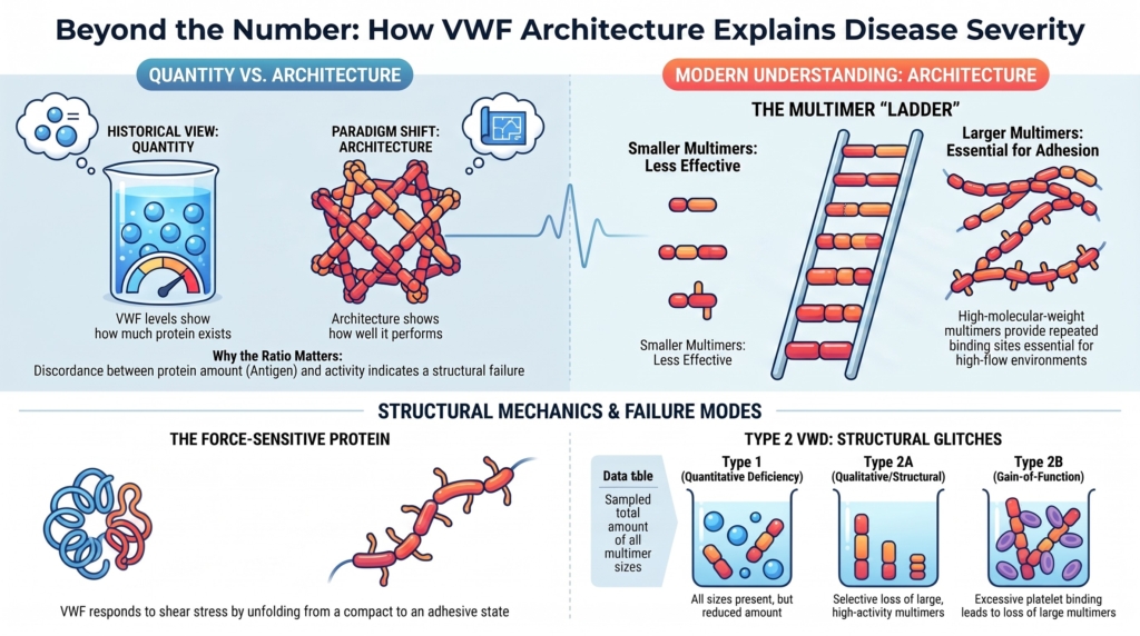
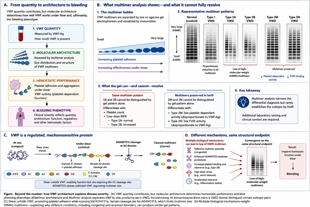
Figure. Beyond the number: how VWF architecture explains disease severity. Figure 1 contrasts VWF quantity, multimer architecture, and hemostatic function, showing why similar antigen levels may produce different bleeding phenotypes. Figure 2 illustrates progressive multimer assembly and representative patterns in type 1, type 2A, type 2B, type 2M, and type 2N VWD. High-molecular-weight multimers support platelet adhesion under high shear, but multimer patterns alone do not distinguish every subtype; platelet-binding studies, FVIII-binding assays, and clinical context remain necessary.
Why this spoke matters
By the 1970s, clinicians could measure more than they once could.
Factor VIII activity (FVIII:C). VWF antigen (VWF:Ag). Platelet-dependent VWF activity (VWF:GPIbM or VWF:GPIbR).
These assays transformed von Willebrand disease. They separated VWD more clearly from hemophilia A, showed that VWF was both a plasma protein and a mediator of platelet adhesion, and made the disease measurable. For the first time, clinicians could compare quantity with function and begin to localize biological defects.
Yet an important mystery remained.
Why did patients with apparently similar amounts of VWF bleed differently?
Some patients had modestly reduced VWF antigen but much lower VWF activity. Some had antigen and activity reduced in parallel. Some had low factor VIII because VWF could not stabilize it. Some had excessive platelet binding rather than impaired platelet binding.
Quantity mattered.
But quantity was not enough.
The decisive shift came when investigators began asking not only:
How much VWF is present?
but:
What kind of VWF is present?
That question changed the disease.
VWD became not only a disorder of amount.
It became a disorder of architecture.
Before multimers: the limits of quantity
The early laboratory history of VWD was dominated by attempts to measure what was missing.
Factor VIII activity connected VWD to plasma coagulation. VWF antigen assays made the protein visible. Ristocetin-based platelet-dependent activity assays (the predecessors of today’s GPIb-based assays) made VWF function measurable.1
These were essential advances.
They showed that VWD had both antigenic and functional dimensions. They helped clinicians distinguish quantitative deficiency from qualitative dysfunction.
But they also exposed a new interpretive problem.
The numbers did not always move together. This discordance was unexpected. If VWF function depended only on the amount of protein present, antigen and activity should have fallen together.
A patient might have VWF antigen near 50 IU/dL but platelet-dependent VWF activity far lower. Another might have proportionally reduced antigen and activity. Another might have a low factor VIII activity level out of proportion to the VWF antigen.
The platelet-dependent activity/VWF:Ag ratiobecame one of the first clues that quantity alone could not explain function. A ratio below approximately 0.7 raises concern for a qualitative VWF defect, although interpretation depends on the assay, laboratory, and clinical context.
When VWF activity fell out of proportion to antigen, the problem was often not simply how much VWF was present. It was how the protein was built, processed, or exposed to platelets.
The laboratory could detect discrepancy.
It could not yet fully explain architecture.
The multimer breakthrough
The recognition that VWF circulates as a series of multimers was one of the decisive conceptual events in the history of VWD.
In 1980, Ruggeri and Zimmerman used multimer analysis to demonstrate abnormal VWF structure across clinically distinct variants of VWD, helping establish that the disorder reflected multiple molecular mechanisms rather than a single quantitative deficiency.
VWF was not a single uniform plasma protein.
It was a population of molecules of different sizes.
Dimers assembled into progressively larger multimers. The largest multimers were especially effective for platelet adhesion and aggregation under shear because they provided repeated binding sites and could span the physical demands of flowing blood.2
That discovery changed the central question.
Not simply:
How much VWF is present?
But:
How is VWF assembled?
Multimer analysis turned a plasma factor into an architecture.
A gel pattern became a biological map.
In multimer analysis, VWF is separated by sodium dodecyl sulfate agarose gel electrophoresis into a ladder of bands according to molecular size. Lower-resolution gels emphasize the overall distribution of multimers of low, intermediate, and high molecular weight, whereas higher-resolution gels can reveal satellite triplet bands produced by proteolytic processing.
Figure. Reading VWF multimer patterns. VWF multimer analysis separates VWF molecules according to molecular size, producing a characteristic ladder of bands. Type 1 VWD shows a proportionately reduced but otherwise preserved ladder. Types 2A and 2B both show loss of high-molecular-weight multimers, whereas types 2M and 2N generally preserve them. Multimer analysis therefore narrows the differential diagnosis but does not distinguish every subtype. Additional laboratory studies—including platelet count, low-dose RIPA, platelet-dependent VWF activity, FVIII activity, and VWF:FVIII-binding assays—are often required. Different biological mechanisms, including defective assembly, enhanced proteolysis, increased platelet binding, high shear, and accelerated clearance, may converge on the same structural endpoint.
The ladder of VWF multimers showed that size distribution was not incidental. It was central to function. High-molecular-weight multimers were especially important under shear, where smaller or poorly assembled forms of VWF could not perform the same adhesive work.3
This was the moment when bleeding severity could be interpreted structurally.
When heterogeneity became mechanism
Before multimer analysis, variation among patients could look like noise.
After multimer analysis, variation became interpretable.
Some patients had all multimer sizes present but reduced in amount. Some lacked high-molecular-weight multimers. Some had abnormal intermediate forms. Some showed patterns suggesting abnormal processing or excessive proteolysis.
The multimer pattern helped explain why patients with similar antigen levels could have different activity and different bleeding phenotypes.
This insight was especially important for what became type 2 VWD.
Type 2 disease is not simply “less VWF.”
It is VWF that fails in a particular way.
In type 2A VWD, high-molecular-weight multimers are reduced because of defective multimer assembly or secretion, increased susceptibility to ADAMTS13-mediated proteolysis, or both. The resulting loss of the most adhesive multimers impairs platelet-dependent function.
In type 2B VWD, gain-of-function VWF-platelet binding leads to loss of large multimers and may cause thrombocytopenia. A similar pattern can occur in platelet-type VWD, where the gain-of-function defect resides in platelet GPIb rather than VWF itself, so RIPA mixing studies and/or genetic testing may be needed.
In type 2M VWD, platelet-dependent function is impaired despite relative preservation of high-molecular-weight multimers, although some variants may affect other ligand interactions.
In type 2N VWD, the defect lies in factor VIII binding rather than platelet adhesion.4
These categories did not appear fully formed.
They emerged as assays made different failure modes visible.
Multimer analysis was therefore not merely a laboratory refinement.
It was a classificatory engine.
It helped transform VWD from one disorder with variable expression into a family of mechanisms.
Why large multimers matter
Large VWF multimers matter because hemostasis is not a static binding problem.
It is a flow problem.Hemostasis occurs in moving blood, not in a test tube.
At sites of vascular injury, platelets must be captured from moving blood. This is especially difficult in the microcirculation and arterial circulation, where shear forces are high. VWF is built for this environment. It binds collagen, tethers platelets through GPIbα, supports platelet accumulation, and stabilizes factor VIII through distinct structural domains.5
The largest multimers are particularly effective because they are multivalent.
They provide repeated binding sites. They span distances. They engage platelets under conditions where smaller proteins would fail. They convert flowing blood into stable platelet adhesion.
This is why loss of high-molecular-weight multimers can produce a bleeding phenotype even when some VWF remains measurable.
Quantity contributes.
Architecture determines performance.
Structure as a clinical explanation
The multimer era made several clinical observations more coherent.
First, it explained why VWF antigen and VWF activity may diverge.
A patient may have measurable antigen but impaired function because the most active multimer forms are missing or because the molecule cannot bind appropriately.
Second, it explained why qualitative variants can be more clinically significant than their antigen level suggests.
The problem is not merely concentration.
It is performance.
Third, it clarified why type 2 disease requires more than a single VWF level.
A low antigen result can suggest deficiency, but it cannot define the mechanism. The relationship between antigen, platelet-dependent activity, collagen binding, FVIII level, RIPA, and multimers localizes the defect.
In practice, unexplained discordance between antigen and activity is a structural clue.
In severe aortic stenosis, high shear can produce an acquired type 2A von Willebrand syndrome with loss of the largest multimers. The biology resembles congenital type 2A VWD not because the gene is abnormal, but because the final structural consequence is similar: loss of the largest, most hemostatically effective multimers.6
This was an important lesson.
The same structural failure can arise from different causes.
Congenital variants, abnormal assembly, enhanced proteolysis, high shear, and accelerated clearance can all converge on loss or dysfunction of the most hemostatically effective multimers.
Different paths can converge on the same hemostatic problem.
The molecule becomes dynamic
The early multimer story could have been interpreted too simply:
large multimers good, small multimers less effective.
That was true, but incomplete.
Later work showed that VWF architecture is not static.
VWF is synthesized, dimerized, multimerized, stored in Weibel-Palade bodies, secreted as ultralarge multimers, unfolded by force, cleaved by ADAMTS13, and cleared from circulation.7
The molecule is not merely structural. It is dynamic.
It has a life cycle.
Severity may reflect reduced synthesis, impaired secretion, defective multimer assembly, loss of high-molecular-weight multimers, increased platelet binding, impaired platelet binding, impaired collagen binding, impaired factor VIII binding, accelerated clearance, or enhanced ADAMTS13-mediated proteolysis.
Structure explains severity, but structure includes more than shape.
It includes assembly, force response, proteolysis, survival, and context.
The force-sensitive protein
One of the most elegant later insights was that VWF is mechanosensitive.
Work by Thomas Springer and others helped establish how force can unfold VWF, expose adhesive regions, and reveal the A2 domain cleavage site for ADAMTS13.
VWF is not only large.
It responds to force.
Work by Thomas Springer and others helped establish that, under shear stress, VWF can elongate from a compact form into an extended adhesive structure. This conformational change exposes functional regions that support platelet binding while also revealing the A2 domain cleavage site for ADAMTS13.8
Activation and regulation occur through the same structural transition.
The same forces that activate VWF for platelet adhesion also make it susceptible to size regulation.
This creates a biological balance.
Too little multimeric VWF, and bleeding occurs. Too much ultralarge VWF, and thrombosis becomes possible.
ADAMTS13 is central to that boundary, trimming ultralarge multimers to limit excessive platelet-rich thrombosis while preserving enough VWF activity for hemostasis. The broad principle is clear, although the spatial and temporal regulation of ADAMTS13 activity in vivo remains incompletely understood.9
This later mechanobiology does not replace the multimer story.
It completes it.
Multimers explained why structure matters.
Shear and ADAMTS13 explained why structure must be regulated.
Why the multimer era still matters clinically
Multimer analysis is not always the first test a clinician orders.
It is specialized. It is technically demanding. It is not needed for every patient with suspected VWD.
But the reasoning it introduced remains central.
Every modern VWD interpretation asks structural questions, even when multimer analysis is not immediately performed.
Is this a proportional quantitative deficiency? Is activity reduced out of proportion to antigen? Are high-molecular-weight multimers missing? Is platelet binding impaired or enhanced? Is factor VIII low because VWF cannot bind it? Is clearance accelerated? Is the pattern congenital or acquired?
These are multimer-era questions.
They reflect the historical shift from measuring VWF to interpreting VWF.
The clinician is no longer simply asking:
Is the level low?
The clinician is asking:
What kind of failure is this?
Clinical synthesis
The history of VWD changed when structure became visible.
Before multimer analysis, VWD could be recognized, measured, and partially separated from hemophilia.
After multimer analysis, VWD could be mechanistically organized.
The disease came to be understood as a set of structural and functional failure modes.
Structure helps explain severity.
But not perfectly. Not alone. Not without bleeding history, clinical context, hemostatic challenge, and judgment.
Multimer analysis explains mechanism better than it predicts bleeding in isolation.
The lesson is not that structure replaces phenotype.
The lesson is that phenotype often becomes intelligible only when structure is considered.
VWF is not merely a concentration.
It is an architecture in motion.
And that architecture helps determine how the patient bleeds.
Reflect & Apply
A 28-year-old woman has lifelong heavy menstrual bleeding, recurrent epistaxis, and postpartum hemorrhage.
Her results are:
VWF antigen: 54 IU/dL
Platelet-dependent VWF activity: 24 IU/dL
FVIII: 62 IU/dL
Activity-to-antigen ratio: low
Multimer analysis: loss of high-molecular-weight multimers
A trainee says:
“Her antigen is only mildly reduced. Why is she bleeding so much?”
Questions for reflection:
Why is VWF antigen alone insufficient here?
What does the low activity-to-antigen ratio suggest?
Why does loss of high-molecular-weight multimers matter?
How does this pattern differ from simple type 1 VWD?
What additional testing might help distinguish type 2A VWD from type 2B VWD or platelet-type VWD?
How does this case illustrate the historical shift from quantity to architecture?
The central lesson:
A VWF level tells you how much protein is present.
A structural interpretation tells you what kind of protein the patient has.
In VWD, that difference can explain severity.
Test your thinking
Apply the concepts from this essay in the companion interactive quiz.