
Splenic infarction occurs when splenic perfusion becomes inadequate enough to produce parenchymal ischemia and tissue necrosis.1
It is uncommon. In one 10-year series, only 32 cases were identified among 196,625 medical admissions, a frequency of approximately 0.016%.2
But rarity is only part of the challenge. Splenic infarction does not have a reliable clinical signature. Left upper quadrant pain is classic, but symptoms are often nonspecific, and some infarcts are painless or discovered incidentally on CT.
More importantly, the infarct may be the first clue to an unsuspected systemic disease. In one cohort of 89 patients with 90 episodes of splenic infarction, evaluation uncovered a previously unknown underlying condition in 34/90 episodes (37.8%).3
That makes splenic infarction less an endpoint than a diagnostic starting point.
Anatomy
The spleen receives its arterial supply from the splenic artery, a branch of the celiac trunk. Near the hilum, the artery divides into terminal branches that subdivide into segmental arteries supplying relatively discrete territories of splenic parenchyma. Blood ultimately traverses the splenic microcirculation and venous sinusoids before draining into the splenic vein and then the portal venous system.
The splenic artery courses along the pancreas. This intimate anatomic relationship helps explain why pancreatic inflammation, infection, or malignancy can affect the splenic vascular bed.4
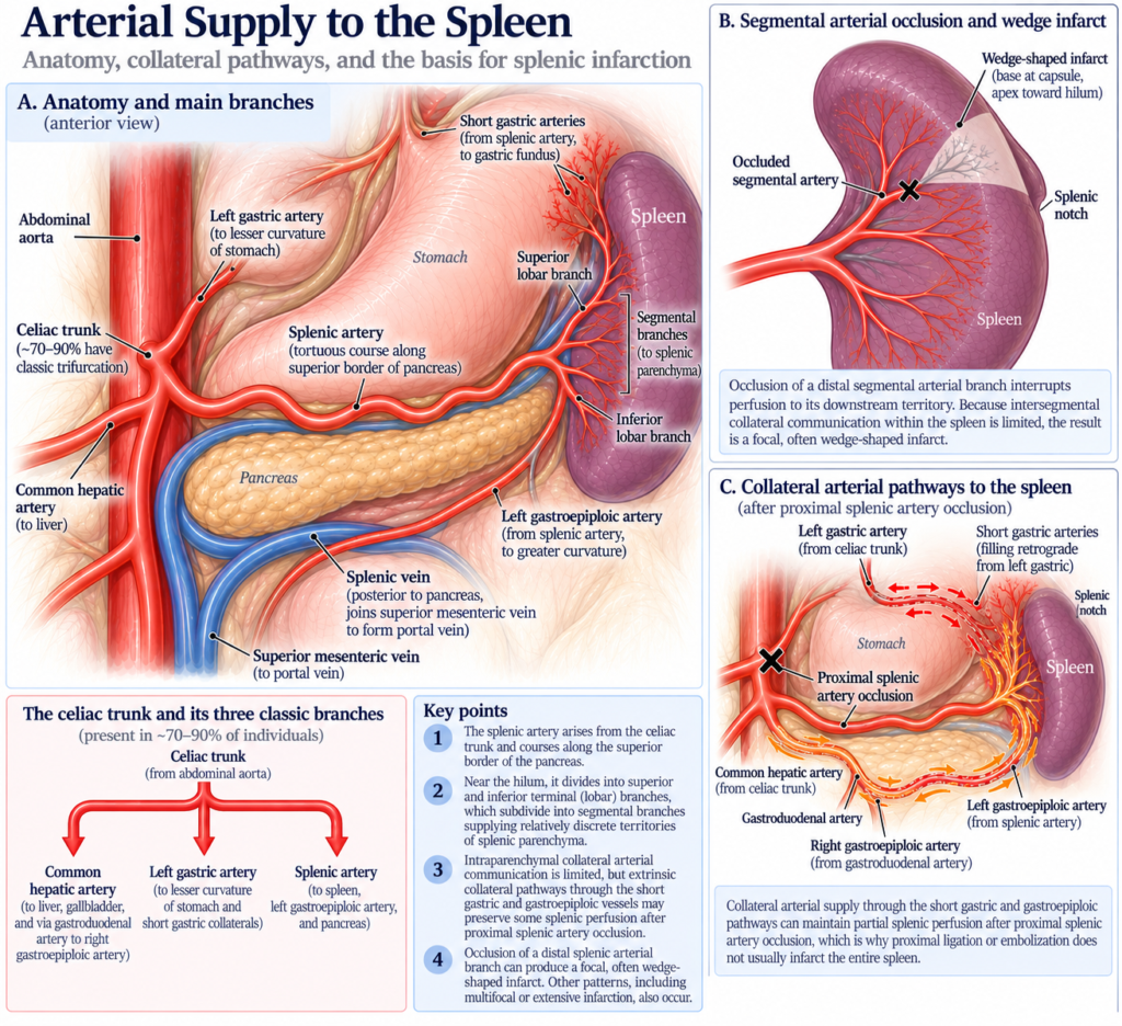
This branching pattern helps explain the classic morphology of splenic infarction. Occlusion of a distal segmental artery deprives its downstream territory of arterial inflow. Because collateral communication between adjacent intrasplenic territories is limited, neighboring territories may not rescue it.
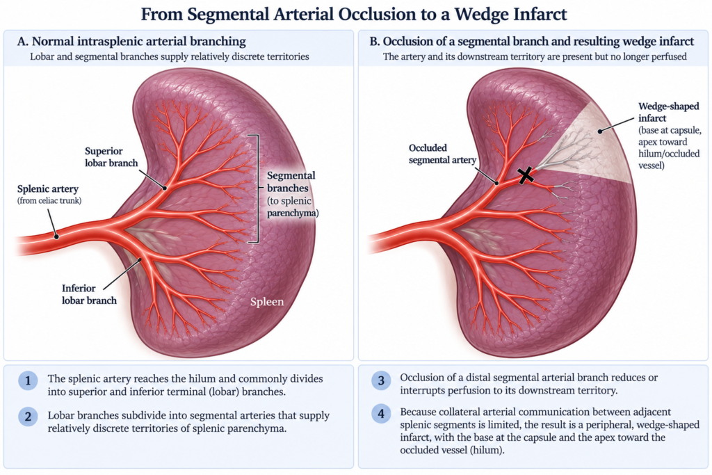
But the classic wedge is not obligatory. Infarcts may also be multiple, irregular, patchy, confluent, extensive, or global.5
The splenic vein matters too. It runs alongside the pancreas and drains into the portal venous system. Splenic vein thrombosis can impair venous outflow, raising sinusoidal and venous pressure and reducing the pressure gradient that normally drives blood through the splenic microcirculation. Severe congestion and impaired microvascular flow may thereby produce ischemia and infarction, particularly in pancreatic and other local abdominal disease. This represents a different vascular pathway from the classic segmental arterial infarct.6
venous obstruction → ↑ venous/sinusoidal pressure → congestion → ↓ arteriovenous perfusion gradient → impaired microvascular flow → ischemia/infarction
For the classic wedge-shaped infarct, however, the arterial side is the key anatomy.
What actually causes the infarct?
The first challenge is to avoid mixing different levels of causation.
An embolus tells us where the occluding material came from.
In-situ thrombosis tells us where the thrombus formed.
Hypercoagulability tells us why thrombosis may have occurred.
And some splenic infarcts are not caused by a conventional platelet-fibrin thrombus at all.
Table 1. Keep the causal levels separate
| Question | What it tells us | Examples |
|---|---|---|
| What physically interrupted perfusion? | Proximate event | Thrombus/embolus, sickled RBCs, parasitized RBCs, mechanical occlusion, venous outflow obstruction |
| Where did a thrombus form? | Location of thrombus formation | Upstream → embolism; locally → in-situ thrombosis |
| Why did thrombosis occur? | Mechanism or predisposition | Abnormal flow, vessel-wall disease/injury, hypercoagulability |
| What other processes can impair splenic perfusion? | Alternative or contributing pathways | Sickling, infection involving RBCs, splenomegaly, pancreatic disease, severe systemic illness |
Where did the arterial thrombus come from?
Upstream: embolism
The thrombus or other embolic material forms elsewhere and travels into the splenic arterial circulation.
Potential sources include atrial fibrillation, left ventricular thrombus, structural or valvular heart disease, infective endocarditis, aortic thrombus or atheroembolic material, cardiac or vascular procedures, and paradoxical embolism from the venous circulation through a right-to-left shunt.7
Cardioembolism was the leading identified cause in several series. In the Wand cohort, a cardioembolic source was identified in 49/90 episodes (54.4%).8 In the Schattner cohort, cardiogenic emboli accounted for 20/32 cases (62.5%).9
A single normal ECG does not exclude paroxysmal atrial fibrillation. Schattner and colleagues specifically raised occult episodic AF as a possible source in patients in whom AF was not documented during the initial evaluation.10
Rarely, the embolus may originate in the venous circulation:
DVT → right heart → PFO/right-to-left shunt → systemic arterial circulation → splenic embolus
This is paradoxical embolism.
Locally: in-situ arterial thrombosis
Alternatively, thrombus forms within the arterial circulation supplying the spleen.
Abnormalities of the vessel wall or lumen may provide the substrate, including celiac or splenic artery atherosclerosis, vasculitis, arterial dissection, and procedural or traumatic arterial injury.
In the Brett cohort, celiac or splenic artery atherosclerosis was present in 34/163 patients (21%) and was the only identified associated condition in 6/163 (3.7%).11
Routine contrast-enhanced CT may reveal major vascular abnormalities, but dedicated vascular imaging may be needed when the specific question is celiac or splenic arterial stenosis, occlusion, dissection, aneurysm, or another vascular lesion.
Why did the thrombus form?
Now we move one level upstream.
The causes of thrombosis can be understood through the familiar components of Virchow’s triad:
Abnormal blood flow: stasis or turbulence
→ atrial fibrillation and intracardiac stasis
Vessel-wall or endothelial abnormality
→ atherosclerosis, vasculitis, dissection, procedural injury
Hypercoagulability
→ APS, MPN, malignancy, and other acquired prothrombotic states
These are mechanisms through which underlying diseases promote thrombosis, not diagnoses themselves.
Multiple contributors are common. In the Brett cohort, 65/163 patients (40%) had more than one plausible associated condition.12
Table 2. Important causes of splenic infarction
| Condition | Principal route to infarction | Useful clue |
|---|---|---|
| Atrial fibrillation | Cardiac thrombus → embolism | Known or occult AF |
| LV thrombus | Cardiac thrombus → embolism | LV dysfunction, recent MI |
| Infective endocarditis | Septic embolism | Fever, bacteremia, valve disease, other emboli |
| Aortic disease | Thromboembolism or atheroembolism | Aortic plaque or mural thrombus |
| Local arterial disease | In-situ thrombosis or arterial compromise | Splenic/celiac atherosclerosis, dissection, vasculitis |
| MPN | Acquired prothrombotic state | Thrombocytosis, erythrocytosis, leukocytosis; may be occult |
| APS | Acquired arterial or venous thrombophilia | Persistent aPL, other thrombosis |
| Malignancy | Hypercoagulability ± local disease | Cancer history or CT findings |
| SCD/SCT | Sickled RBC microvascular obstruction | HbS; hypoxia/altitude particularly relevant in SCT |
| Babesiosis | Parasitized RBCs, splenic sequestration, inflammation ± microthrombosis | Hemolysis, tick exposure, parasitemia |
| Malaria | Parasitized RBC sequestration and microvascular dysfunction | Travel/exposure, parasitemia |
| EBV/CMV | Uncertain; splenic enlargement/inflammation ± transient prothrombotic effects | Mononucleosis syndrome |
| Pancreatic disease | Local venous and/or arterial involvement | Pancreatitis, tumor, peripancreatic inflammation |
| Splenic vein thrombosis | Venous outflow obstruction | Pancreatic disease, splanchnic thrombosis |
| Iatrogenic/mechanical | Direct interruption of flow | Embolization, surgery, vascular procedure, torsion |
The table is deliberately heterogeneous: some entries are diseases, some are vascular lesions, and some represent distinct mechanisms. Its purpose is to summarize the clinical differential, not to imply that all causes occupy the same level in the causal chain.
The relative frequency of these conditions varies substantially among series, reflecting differences in patient populations and how associated conditions were classified. Selected cohort data are shown below.
Table 3. Conditions associated with splenic infarction in selected cohorts
| Study | N | Selected associated conditions |
|---|---|---|
| Schattner 2015 | 32 | Cardioembolic 20/32 (63%); APS 3/32 (9%); other causes… |
| Wand 2018 | 90 episodes | Cardioembolic 49/90 (54%); MPN 10/90 (11%); APS 5/90 (6%) |
| Brett 2020 | 163 | Celiac/splenic atherosclerosis 34/163 (21%); sepsis/septic shock 27/163 (17%); pancreatitis 10/163 (6%); sickling disorder 10/163 (6%); pancreatic cancer 8/163 (5%) |
Infective endocarditis deserves special attention
The spleen is a classic target of systemic embolization from infective endocarditis.
The diagnosis can be difficult because fever and leukocytosis may accompany tissue infarction itself, while endocarditis may initially lack dramatic infectious findings.
In the Wand cohort, CT, ECG, echocardiography, and blood cultures were among the investigations the authors considered particularly useful in identifying an underlying cause.13
In the multicenter Yen cohort of 130 patients, infective endocarditis identified a higher-risk subgroup, including a high rate of ICU admission.14
So:
splenic infarction + fever/bacteremia/valvular disease/multiorgan emboli → think endocarditis
Myeloproliferative neoplasms
MPN is particularly important because thrombosis may precede recognition of the underlying disorder.
In the Wand cohort, an MPN was identified in 10/90 episodes (11.1%), including 7/90 (7.8%) with a documented JAK2-mutated MPN. Five JAK2-positive MPNs were newly diagnosed during the evaluation.15
Typical clues were not always present. Erythrocytosis and splenomegaly did not distinguish these patients, whereas thrombocytosis was strongly enriched among patients with JAK2-positive MPN.16
Thus, JAK2 V617F testing is reasonable in otherwise unexplained splenic infarction, particularly when persistent thrombocytosis or another MPN phenotype is present.
If JAK2 V617F is negative and suspicion for a Philadelphia-negative MPN persists, additional molecular testing should be phenotype-directed, including CALR and MPL. BCR::ABL1 belongs on a separate branch and should be considered when the CBC and smear suggest CML.
Antiphospholipid syndrome
APS is an acquired prothrombotic disorder capable of producing both arterial and venous thrombosis.
In the Wand cohort, APS was identified in 5/90 episodes (5.6%), including 3/90 (3.3%) newly diagnosed cases.17
In the Schattner cohort, APS was ultimately identified in 3/32 patients. The reported antibody profiles were not uniformly triple-positive: two patients were described as lupus anticoagulant positive, while another was diagnosed later after persistently positive anticardiolipin and anti-β2-glycoprotein I antibodies were identified.18
An objectively confirmed splenic arterial thrombosis counts as an arterial thrombotic event. Persistent positivity for a qualifying antiphospholipid antibody, such as lupus anticoagulant on two occasions at least 12 weeks apart, together with such an event satisfies the traditional Sydney classification framework for thrombotic APS.19
The 2023 ACR/EULAR classification criteria use a weighted system that considers alternative explanations for arterial thrombosis, including conventional cardiovascular risk factors, rather than treating every arterial event as equally supportive of APS.20 These are classification criteria, not diagnostic rules. Clinically, however, an otherwise unexplained splenic arterial thrombosis together with persistent lupus anticoagulant positivity should raise strong concern for thrombotic APS.
The distinction matters therapeutically. For definite APS with a first arterial thrombosis, EULAR recommends treatment with a vitamin K antagonist rather than aspirin alone; the intensity of anticoagulation is individualized according to recurrent-thrombosis and bleeding risk. DOACs are generally not recommended in APS with arterial events.21
What about inherited thrombophilia?
Factor V Leiden, prothrombin G20210A, and deficiencies of protein C, protein S, and antithrombin predominantly predispose to venous, not arterial, thrombosis.
They therefore have a clearer relationship to splenic, portal, mesenteric, or other venous thrombosis than to an isolated splenic arterial infarct.
In the Wand cohort, conventional inherited thrombophilia testing was unrevealing in all 24 patients tested, whereas selectively ordered APS and JAK2 testing identified previously unrecognized disease.22
Inherited thrombophilia can still matter indirectly:
venous thrombophilia → DVT → PFO → paradoxical embolism → splenic arterial infarction
But that is very different from saying that factor V Leiden ordinarily causes in-situ splenic arterial thrombosis.
A thrombophilia panel is not the same thing as a thoughtful thrombosis evaluation.
When the red cell is part of the problem
Not every splenic infarct is a platelet-fibrin story.
Sickle cell disease and sickle cell trait
Sickling provides a fundamentally different route to splenic ischemia:
HbS polymerization → RBC sickling → impaired deformability and microvascular obstruction → ischemia
In the Brett cohort, sickle cell disease or a sickling variant was present in 10/163 patients (6%).23
Splenic infarction is particularly well described in sickle cell trait (SCT) during exposure to high altitude or hypoxemia. Jefferson and colleagues systematically reviewed published SCT-associated splenic infarctions from 1970 through 2020.24
The association is also supported by observational experience. In one single-center series of 27 patients who developed splenic infarction in a high-altitude setting, 25/27 (92.5%) had sickle cell trait and 85% developed their infarct after exposure to very high altitude.25
Interestingly, thrombus within the splenoportal axis was demonstrated in 22.2% of patients in that series, reminding us that microvascular sickling and conventional thrombosis need not be mutually exclusive.26
Babesiosis
Babesiosis provides another RBC-centered route to splenic infarction.
The exact mechanism remains uncertain. Proposed contributors include altered deformability and splenic sequestration of parasitized erythrocytes, destruction of infected RBCs, inflammatory and endothelial activation, and coagulation activation with microthrombus formation.27
Malaria
Malaria is another infection in which abnormal erythrocytes and the splenic microcirculation meet.
Splenic infarction has been reported with several malaria species. Proposed mechanisms include sequestration and reduced deformability of parasitized erythrocytes, splenic enlargement, inflammation, and altered microvascular flow.28
The broader lesson is:
Sometimes the circulating red cell, rather than fibrin alone, is central to the obstruction.
Viral infection and acute splenic enlargement
EBV and CMV are additional infectious associations with splenic infarction, but unlike babesiosis and malaria, they do not act through infected erythrocytes.
In the Antopolsky cohort, acute EBV or CMV infection was identified in 4/49 episodes of splenic infarction.29
The mechanism is uncertain. Rapid splenic enlargement, inflammation, altered local perfusion, and transient prothrombotic phenomena, including antiphospholipid antibodies, have all been proposed.
This illustrates a broader point: infection can cause splenic infarction through very different mechanisms.
Endocarditis → embolization
Babesia/malaria → RBC and microvascular effects
EBV/CMV → splenic enlargement/inflammation ± transient prothrombotic effects
What about splenomegaly itself?
Marked splenomegaly frequently accompanies hematologic disorders associated with splenic infarction, particularly myelofibrosis and other MPNs.
But cause and association are difficult to separate.
The enlarged spleen may itself become more susceptible to ischemia through increased metabolic demand or altered microcirculation. Alternatively, splenomegaly may simply mark an underlying hematologic disorder that independently predisposes to thrombosis. Older pathology-based series cannot reliably disentangle those possibilities.30
Local pancreatic and intra-abdominal disease
The pancreas lies in intimate contact with both the splenic artery and splenic vein.
In the Brett cohort of 163 patients with splenic infarction, pancreatitis was present in 10/163 patients (6%), and pancreatic cancer in 8/163 (5%).31
Splenic venous involvement is particularly well recognized in pancreatic disease. Local inflammation, infection, tumor, or mass effect may produce splenic vein thrombosis. Splenic arterial involvement may occur as well.
The available splenic-infarction series do not establish how often infarction in pancreatic disease results from venous thrombosis, arterial thrombosis, simultaneous involvement of both vascular beds, or another local process.
What about hypoperfusion?
Sepsis, shock, and hypotension are associated with splenic infarction, but their precise mechanistic role is less certain.
In the Brett cohort, sepsis or septic shock was present in 27/163 patients (17%), and hypotension without sepsis in 11/163 (7%).32
A classic wedge-shaped infarct maps naturally to the territory of an arterial branch. Systemic hypotension does not selectively interrupt one segmental artery.
The available series therefore do not establish that hypoperfusion alone produces the classic wedge-shaped splenic infarct.
In critically ill patients, hypoperfusion may instead lower the threshold for infarction in a spleen already vulnerable because of arterial disease, hypercoagulability, inflammation, or another process.
And not every arterial obstruction is thrombotic
Splenic arterial inflow can occasionally be interrupted by mechanical or iatrogenic means rather than spontaneous thrombosis.
Reported settings include intentional splenic artery embolization, vascular or abdominal procedures, surgery, traumatic arterial injury, and torsion of the splenic pedicle.33
Thus:
arterial occlusion → infarction
does not invariably mean:
spontaneous platelet-fibrin thrombus → infarction
How does splenic infarction present?
The classic presentation is left upper quadrant abdominal pain.
But real patients are considerably less tidy.
Table 4. Clinical and laboratory findings in selected series
| Finding | Study | Frequency |
|---|---|---|
| Abdominal/left flank pain | Antopolsky | 39/49 episodes (80%) |
| Isolated LUQ pain | Antopolsky | 16/49 episodes (33%) |
| No pain | Antopolsky | 10/49 episodes (20%) |
| Fever or chills | Antopolsky | 14/49 episodes (27%) |
| Nausea or vomiting | Antopolsky | 11/49 episodes (22%) |
| LUQ tenderness | Antopolsky | 18/49 episodes (35%) |
| Abdominal pain | Wand | 74/90 episodes (82%) |
| Elevated LDH | Wand | 72% of those tested |
| Leukocytosis | Wand | 67% of those tested |
| LUQ pain as primary presenting symptom | Brett | 32/163 patients (20%) |
| Abdominal pain elsewhere | Brett | 77/163 patients (47%) |
| No abdominal pain | Brett | 54/163 patients (33%) |
As Table 4 shows, abdominal pain is common in some series, yet isolated LUQ pain is much less consistent, and a substantial minority of patients have no abdominal pain at all. The variation across cohorts reinforces that splenic infarction has no reliable clinical signature.343536
Left upper quadrant pain should make you think about splenic infarction. Its absence should not make you stop thinking about it.
What do the labs tell us?
FINDINGS THAT MAY REFLECT THE INFARCT
↑ LDH
Ischemic tissue injury and necrosis can release intracellular LDH.
Leukocytosis
May accompany the inflammatory response to tissue injury.
↑ inflammatory markers
May accompany acute tissue injury.
FINDINGS THAT MAY POINT TOWARD THE CAUSE
Thrombocytosis, erythrocytosis, or leukocytosis → consider MPN
Anemia/hemolysis → consider a sickling disorder, babesiosis, malaria, or another RBC process
Thrombocytopenia → may point toward infection, systemic illness, malignancy, or hematologic disease
Bacteremia → consider endocarditis
aPL positivity → consider APS
The distinction is not absolute. Laboratory abnormalities may reflect the infarct, the disease causing the infarct, or both. Laboratory testing is generally more useful for understanding etiology and context than for confirming the infarct itself.
Why does LDH rise?
The most straightforward explanation is ischemic cellular injury and necrosis with release of intracellular LDH.
That mechanism is physiologically plausible rather than directly established in the splenic-infarction cohorts. LDH also rises with infarction in other organs and can be elevated by hemolysis, liver injury, malignancy, infection, and other forms of tissue damage.
There is no need to invoke the spleen’s large RBC content specifically.
How is splenic infarction diagnosed?
Contrast-enhanced abdominal CT is the imaging workhorse.
The classic lesion is a peripheral wedge-shaped region of decreased enhancement, but infarcts may also be irregular, patchy, heterogeneous, multiple, subtotal, or global.37
CT may also reveal clues to the extent or cause of disease. In the Wand cohort, additional potentially relevant CT abnormalities were present in 45/90 episodes (50%), including renal infarction in 20/90 (22%) and intestinal ischemia in 6/90 (7%).38
Thus:
spleen + kidney + bowel infarction → think systemic arterial embolization
Routine CT may suggest a vascular abnormality, but CTA or another dedicated vascular study may be needed when the specific question is celiac or splenic arterial occlusion, dissection, aneurysm, or another vascular lesion.
What about ultrasound?
Ultrasound is considerably less reliable during the acute phase.
In the Antopolsky series, ultrasound was diagnostic in only 3/17 patients (18%) who underwent the examination despite CT-confirmed infarction.39
Once you see the infarct, don’t stop
The CT establishes what happened to the spleen.
It does not establish why.
In the Wand cohort, evaluation uncovered a previously unknown underlying condition in 34/90 episodes (37.8%).40
There is no prospectively validated diagnostic algorithm. A reasonable framework, derived largely from retrospective data and etiologic reasoning, is:
Table 4. Practical evaluation after splenic infarction
| Question | Evaluation | What are you looking for? |
|---|---|---|
| What does the CT already tell us? | Review infarct distribution and associated findings | Multiple territories, other visceral infarcts, pancreatic disease, malignancy, hemorrhage |
| Is there a vascular lesion? | CTA/vascular imaging when indicated | Celiac/splenic occlusion, dissection, aneurysm, aortic disease |
| Could this be embolic? | ECG ± rhythm monitoring | AF, including paroxysmal AF |
| Could the heart be the source? | TTE; selective TEE | Intracardiac thrombus, valve disease, endocarditis |
| Could this be infection? | Blood cultures when appropriate | Bacteremia/endocarditis |
| Is there an MPN clue? | CBC, smear, JAK2 ± CALR/MPL | Persistent thrombocytosis, erythrocytosis, leukocytosis |
| Could this be CML? | BCR::ABL1 when phenotype suggests it | Leukocytosis, left shift, basophilia |
| Could this be APS? | LA, aCL, anti-β2GPI | Acquired arterial thrombophilia |
| Could RBC biology be responsible? | Targeted testing based on context | SCD/SCT, babesiosis, malaria |
TTE is a reasonable initial cardiac study when a cardiac source is suspected. TEE should be used selectively, particularly when infective endocarditis, valvular pathology, or another embolic source remains a concern after the initial assessment.
How is splenic infarction treated?
There is no single treatment because splenic infarction describes an ischemic event, not its cause.
HOW DO I TREAT IT?
Support the uncomplicated infarct
Analgesia
Hydration as appropriate
Antiemetics
Observation
Anticoagulate when the mechanism warrants it
AF or intracardiac thrombus
APS-associated arterial thrombosis
Venous thrombosis
Paradoxical embolism
Other established thromboembolic indications
Treat the underlying cause
Endocarditis → antimicrobial/source management
MPN → disease-specific and thrombosis-directed therapy
SCD/SCT → address precipitating hypoxia and provide supportive care
Babesiosis/malaria → appropriate antimicrobial therapy
Pancreatic/local disease → treat the local process
Escalate when complicated
Hemorrhage or rupture
Abscess or persistent sepsis
Hemodynamic instability
Selected proximal arterial occlusion → vascular/interventional evaluation
Key point: Anticoagulation is an etiologic decision, not a radiologic reflex.
Anticoagulate or not?
The retrospective splenic-infarction literature does not support a blanket rule that every patient should receive anticoagulation.
In one multicenter retrospective cohort of 86 patients, 45/86 (52.3%) received anticoagulation. Anticoagulant therapy was associated with lower long-term mortality without a statistically significant increase in bleeding, but the study was observational and cannot establish that anticoagulation caused the survival difference.41
Conversely, in a cohort of 206 patients with cancer and splenic infarction, recurrence was uncommon and anticoagulation was not associated with a clear reduction in subsequent thromboembolic events.42
So the question is not simply:
Does the patient have a splenic infarct?
It is:
What caused it, is there an independent indication for anticoagulation, and what is the bleeding risk?
For APS-associated arterial thrombosis, long-term anticoagulation is generally part of treatment. For atrial fibrillation or intracardiac thrombus, treatment follows the underlying cardioembolic indication. By contrast, infarction associated with hemorrhage, trauma, severe thrombocytopenia, infective endocarditis, arterial injury, or a mechanical/local process requires a different risk-benefit assessment.
Treat the cause, not the CT image.
Is there a role for thrombolysis or thrombectomy?
There is no established routine role for catheter-directed thrombolysis or thrombectomy in the usual completed peripheral splenic infarct.
Endovascular reperfusion has been reported in highly selected cases involving acute proximal celiac or splenic arterial occlusion, but the evidence is case-based. A large proximal occlusion threatening multiple visceral territories is a different problem from a completed distal wedge-shaped infarct.
What complications matter?
Most uncomplicated infarcts do not require splenic intervention.
Reported complications include:
- splenic abscess
- hemorrhage
- pseudocyst formation
- splenic rupture
In the Antopolsky series of 49 episodes, there were no serious complications and no in-hospital deaths.43
Worsening pain, hemodynamic instability, falling hemoglobin, peritoneal findings, persistent fever or bacteremia, or concern for rupture or abscess should prompt reassessment and consideration of surgical or interventional management.
What determines prognosis?
The prognosis of splenic infarction varies markedly among studies because the underlying diseases and clinical settings vary markedly.
In the Brett cohort, 32/163 patients (20%) died during hospitalization, but mortality differed substantially according to the associated condition.44
In the multicenter Yen cohort, 9/130 patients (6.9%) died during hospitalization.45
The difference is informative rather than contradictory.
The prognosis of splenic infarction is largely the prognosis of the disease that caused it.
Putting it together
Splenic infarction is best understood by keeping different causal levels separate.
What happened?
Splenic perfusion failed.
For the classic wedge-shaped lesion, the most straightforward explanation is segmental arterial occlusion.
If the occlusion is thrombotic, where did the thrombus form?
Upstream → embolism
or
Locally → in-situ thrombosis
Why did thrombosis occur?
Think about the underlying diseases acting through:
abnormal blood flow
vessel-wall or endothelial abnormality
hypercoagulability
And is thrombosis even the right model?
Sometimes it is not.
Sickled erythrocytes can obstruct the microcirculation.
Babesia- and Plasmodium-infected erythrocytes can alter splenic microvascular flow.
Splenic venous thrombosis can impair venous outflow.
Pancreatic disease can affect either side of the splenic vascular bed.
Mechanical and iatrogenic processes can interrupt arterial flow directly.
And severe systemic hypoperfusion may contribute without necessarily explaining a classic territorial wedge by itself.
The splenic infarct therefore tells us what happened to the spleen.
It does not tell us why it happened.
When the CT report says “splenic infarction,” the diagnostic work is not finished.
The next question is:
What interrupted splenic blood flow, and what does that reveal about the patient?