For larger image, click here.
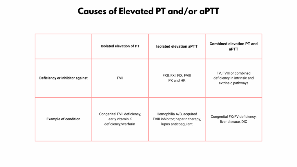
Overview of PT and PTT
| Test | Measures | Pathway | Factors Tested |
|---|---|---|---|
| PT | Time to clot formation after adding tissue factor | Extrinsic + common | VII, X, V, II (prothrombin), I (fibrinogen) |
| aPTT | Time to clot after contact activation | Intrinsic + common | XII, XI, IX, VIII, X, V, II, I |
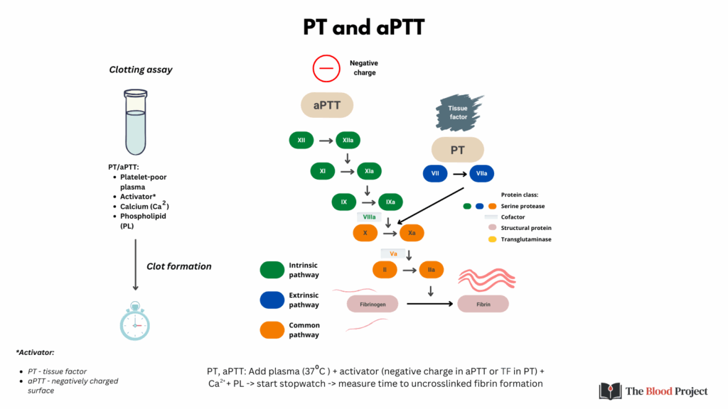
How the Tests Are Performed
- Sample Collection
- Citrate tube (blue top) is used: contains sodium citrate, which chelates calcium to prevent clotting.
- Blood is centrifuged to obtain platelet-poor plasma.
- Prothrombin Time (PT) – Step-by-Step:
- Recalcification: Calcium is added back to the plasma.
- Tissue Factor + phospholipids (Thromboplastin) is added—this mimics injury by activating the extrinsic pathway.
- Add calcium
- Timer starts: The time it takes for a visible clot to form is measured.
- Results are reported in seconds and as the International Normalized Ratio (INR) for warfarin monitoring.
- Activated Partial Thromboplastin Time (aPTT or PTT) – Step-by-Step
- Recalcification: Calcium is again added to citrated plasma.
- Activator (e.g., kaolin, silica, or ellagic acid) is added to mimic contact activation (starts intrinsic pathway).
- Partial thromboplastin (phospholipids without tissue factor) is added to substitute for platelet surfaces.
- Add calcium – re-enables coagulation, as calcium is essential for multiple steps; triggers clotting after the appropriate activating agents are added (tissue factor for PT, contact activator for aPTT).
- Timer starts, and time to fibrin clot formation is recorded.
- Called “partial” because it lacks tissue factor.