
A patient has a hemoglobin of 4 g/dL.
He is sitting comfortably. His blood pressure is preserved. His oxygen saturation is normal. His heart rate is not particularly fast.
How is that possible?
At a hemoglobin of 4 g/dL, the oxygen-carrying capacity of blood is profoundly reduced. Yet patients whose anemia develops gradually can sometimes remain remarkably comfortable at rest even at very low hemoglobin concentrations.1
Classic studies of chronic severe anemia included patients with hemoglobin concentrations as low as 1.5 to 3 g/dL.2
The surprising thing is not simply that some of these patients are not tachycardic.
It is that oxygen consumption can remain relatively well defended despite an enormous reduction in the oxygen-carrying capacity of the blood.
How?
Rather than memorize a list of adaptations, we can work through the physiology one step at a time.
At each step, we will ask two questions:
What changes?
Why?
START WITH OXYGEN DELIVERY
Systemic oxygen delivery is:
DO₂ = cardiac output × arterial oxygen content
or:
DO₂ = CO × CaO₂
Severe anemia primarily affects one side of that equation: arterial oxygen content.
Step 1: Arterial oxygen content falls
Arterial oxygen content is approximately:
CaO₂ = (1.34 × Hb × SaO₂) + (0.003 × PaO₂)
Most oxygen in arterial blood is carried by hemoglobin. Only a small amount is physically dissolved in plasma.
Consider someone with:
Hb = 4 g/dL
SaO₂ = 98%
PaO₂ = 100 mmHg
Hemoglobin-bound oxygen is approximately:
1.34 × 4 × 0.98 = 5.25 mL O₂/dL
Dissolved oxygen contributes:
0.003 × 100 = 0.3 mL O₂/dL
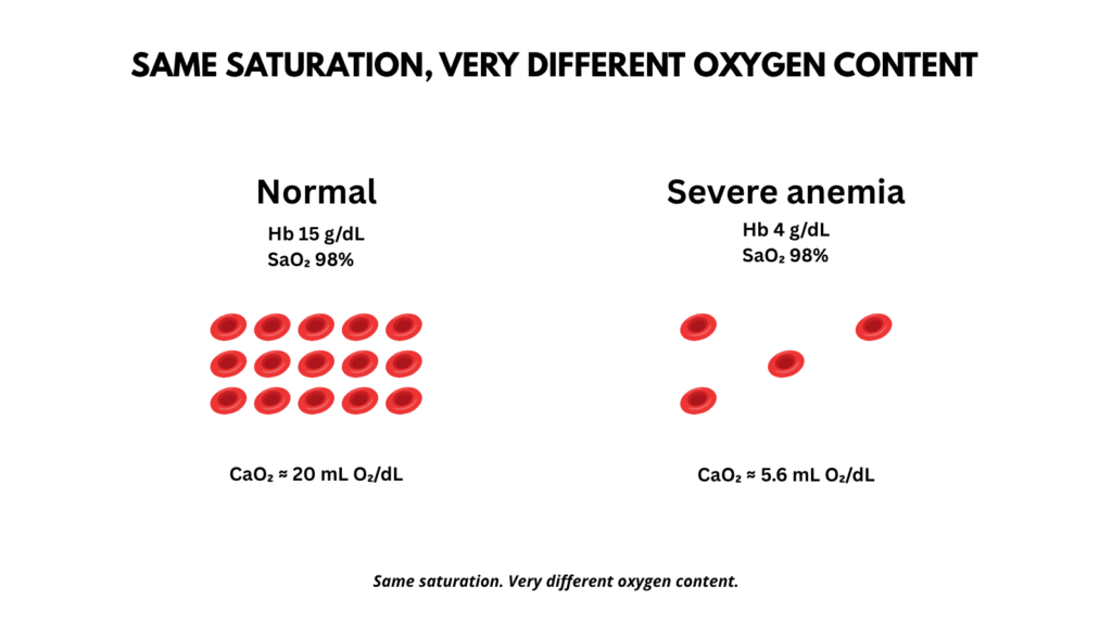
So arterial oxygen content is only about 5.6 mL O₂/dL, compared with roughly 20 mL/dL at a hemoglobin of 15 g/dL.
Notice what has not necessarily changed.
PaO₂ and SaO₂ may remain entirely normal. A pulse oximeter can read 98% while arterial oxygen content is profoundly reduced.
The problem is not necessarily getting oxygen into the blood.
There simply is not enough hemoglobin carrying it.
And there is relatively little room for the body to compensate by increasing arterial oxygen content itself.
SaO₂ is already close to its ceiling. Raising PaO₂ increases dissolved oxygen, but under ordinary conditions the absolute contribution remains small because oxygen has limited solubility in plasma.3
At very low hemoglobin concentrations, dissolved oxygen becomes proportionally more important, but under normal atmospheric conditions it still cannot replace the large amount of oxygen normally carried by hemoglobin.
Short of substantially increasing dissolved oxygen under unusual conditions, such as hyperbaric oxygen, the major way to restore arterial oxygen content is to increase the hemoglobin concentration.
Without transfusion, therefore, the body has to compensate somewhere else.
Return to:
DO₂ = CO × CaO₂
If CaO₂ falls, one major way to defend oxygen delivery is to increase cardiac output.
Step 2: Cardiac output increases
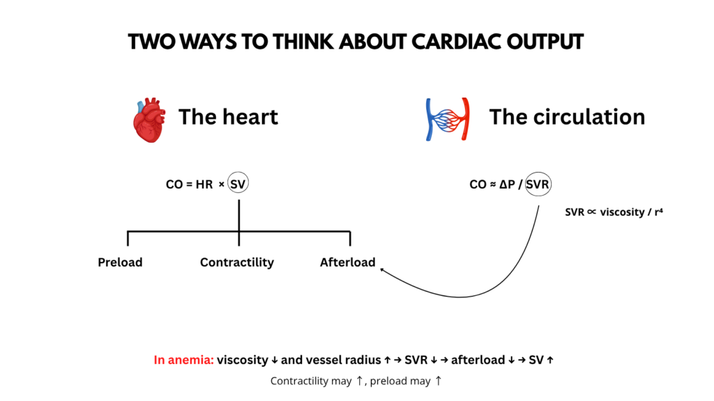
There are two complementary ways to interrogate cardiac output, and it is useful to keep them separate.
The first asks:
How does the heart generate more flow?
For that, we use:
CO = HR × SV
The second asks:
How does the systemic circulation permit more flow?
For that, we use:
CO ≈ ΔP / SVR
These are two different views of the same circulation.
They can be mapped onto one another, but they should not be conflated.
Let us take them one at a time.
VIEW 1: HOW DOES THE HEART GENERATE MORE FLOW?
Cardiac output is:
CO = heart rate × stroke volume
or:
CO = HR × SV
Both variables can change.
In chronic severe anemia, however, the increase in cardiac output can be driven importantly by a larger stroke volume, rather than requiring marked resting tachycardia.
Roy and colleagues studied 51 patients with chronic severe anemia. Heart rate correlated poorly with cardiac output, whereas stroke volume correlated much more closely. One patient with a hemoglobin of 2 g/dL had a heart rate of only 65 beats/min.4
A later review similarly concluded that the increased cardiac output of chronic anemia principally reflects a larger stroke volume because tachycardia is frequently absent.5
This differs from the pattern observed in acute isovolemic anemia.
In healthy resting subjects whose hemoglobin was rapidly lowered to approximately 5 g/dL, both heart rate and stroke volume increased. The investigators estimated that about 75% of the increase in cardiac output came from heart rate and about 25% from stroke volume.6
Thus, the hemodynamic pattern of acute isovolemic anemia differs importantly from that described in chronic severe anemia.
Why does stroke volume rise?
A useful way to think about stroke volume is in terms of:
preload
contractility
afterload
The evidence is not equally strong for all three.
A) DOES PRELOAD INCREASE?
This is less certain than is often implied.
It would be too simple to write:
chronic anemia → plasma volume expansion → preload ↑
In chronic anemia without overt congestion, plasma volume may be only modestly increased, while total blood volume and central blood volume may be unchanged or reduced. Right atrial and pulmonary wedge pressures are often normal.7
Acute isovolemic anemia provides an especially clean example. Stroke volume increased while central venous and pulmonary wedge pressures remained unchanged.8
So increased filling pressure or expansion of total blood volume is not required for stroke volume to rise.
One proposed mechanism is that lower hematocrit, reduced viscosity, and systemic vasodilation reduce resistance to venous return.
That may allow more blood to return to the heart for a given driving pressure. Sympathetic venoconstriction may also mobilize blood from the venous reservoir when arterial pressure is threatened.9
A proposed sequence is:
Hct ↓ + viscosity ↓ + systemic vasodilation
↓
resistance to venous return ↓
↓
venous return may increase
↓
ventricular filling may increase
↓
stroke volume ↑
The important word is may.
This is physiologically plausible, but it is not as directly demonstrated in humans as some of the other adaptations.
Marked plasma-volume expansion can occur in patients with very severe anemia, particularly those who develop edema and neurohormonal activation.
Anand and colleagues studied four untreated patients with severe chronic anemia and edema. Their mean hematocrit was only 13%.
They found:
- plasma volume about 70% above normal
- extracellular volume about 32% above normal
- total body water about 14% above normal
- exchangeable sodium about 30% above normal
These patients also had neurohormonal activation, including increased norepinephrine, renin activity, aldosterone, vasopressin, growth hormone, and atrial natriuretic peptide.11
Marked volume expansion should therefore be viewed as an additional feature of some patients with advanced severe anemia, especially those with edema, rather than as the universal explanation for increased stroke volume.
B) CONTRACTILITY MAY INCREASE
Experimental studies suggest that myocardial contractility can increase during anemia.
But the mechanism and its quantitative importance in chronic human anemia remain uncertain.
In experimental models, increased stroke volume persisted despite beta-adrenergic blockade and even cardiac denervation, suggesting that sympathetic activation is not required for the response, although it may contribute.12
Other work has suggested roles for catecholamine and non-catecholamine inotropic factors.13
So:
contractility ↑ may contribute
but its quantitative importance is less securely established.
C) AFTERLOAD FALLS
The left ventricle also ejects into a lower-resistance circulation.
That means afterload falls.
This favors more complete ventricular emptying and a larger stroke volume.
But why does vascular resistance fall?
For that, it is better to switch explicitly to our second view of cardiac output, rather than trying to derive vascular resistance from the preload, contractility, afterload framework.
VIEW 2: HOW DOES THE SYSTEMIC CIRCULATION PERMIT MORE FLOW?
A second way to think about cardiac output is:
CO ≈ ΔP / SVR
where:
ΔP = the pressure gradient driving systemic flow
SVR = systemic vascular resistance
For a given pressure gradient, flow increases as resistance falls.
One of the most consistent findings in severe anemia is a marked reduction in systemic vascular resistance.
Why?
Two major factors are especially important:
blood viscosity falls
and:
resistance vessels dilate
A) BLOOD VISCOSITY FALLS
Resistance to flow depends in part on viscosity.
A simplified form of Poiseuille’s relationship is:
Resistance ∝ viscosity / radius⁴
As hematocrit falls:
Hct ↓ → viscosity ↓ → resistance ↓
A classic human series found that a 44% decrease in hematocrit was accompanied by about a 39% reduction in measured blood viscosity.14
Experiments that separated oxygen-carrying capacity from viscosity are especially informative.
When oxygen-carrying capacity was reduced with methemoglobinemic blood without a comparable reduction in viscosity, cardiac output did not rise substantially. In contrast, dextran exchange, which lowered hematocrit and viscosity, produced a marked rise in cardiac output.15
That tells us something fundamental:
Anemia changes the circulation mechanically, not merely through tissue hypoxia.
B) RESISTANCE VESSELS DILATE
Vessel radius also increases.
Several mechanisms have been proposed, including:
- local hypoxia-related metabolic vasodilation
- flow-mediated endothelial vasodilation
- nitric oxide and other endothelial relaxing mechanisms
The exact contribution of individual mediators is less securely established than the overall vasodilated phenotype.16
Higher flow itself may promote further vasodilation through increased endothelial shear stress and release of relaxing factors.17
There is also an intriguing nitric oxide hypothesis.
Hemoglobin avidly binds nitric oxide. Anand and colleagues proposed that very low hemoglobin concentrations reduce inhibition of endothelium-derived relaxing activity, thereby contributing to generalized vasodilation. Increased flow could then further stimulate endothelial relaxing-factor release.18
This remains a plausible mechanism rather than a fully established explanation.
So:
Hct ↓ → viscosity ↓
and:
reduced oxygen delivery → vasodilation
and:
increased flow → flow-mediated vasodilation
Together:
SVR ↓
For a given driving pressure:
SVR ↓ → systemic flow ↑
The importance of vascular resistance is supported by intervention studies. In patients with chronic severe anemia, increasing peripheral vascular resistance with methoxamine reduced cardiac output. When atropine was added to prevent reflex bradycardia, cardiac output still did not return fully to its previous level.19
These experiments support an important role for reduced vascular resistance, although they do not prove whether vasodilation initiates the high-output state or also develops as a consequence of increased flow.
VIEW 2: HOW DOES THE SYSTEMIC CIRCULATION PERMIT MORE FLOW?
We can now connect the two approaches without confusing them.
From the systemic-circulation perspective:
SVR ↓ → flow ↑
From the ventricular perspective:
SVR ↓ → afterload ↓ → stroke volume ↑
So the same reduction in systemic vascular resistance can be described in two different ways.
It permits more flow through the systemic circulation, and it reduces the load against which the left ventricle ejects.
These are connected consequences of the same physiologic change, but they belong to two different conceptual frameworks.
That distinction is useful because it prevents us from treating:
CO = HR × SV
and:
CO ≈ ΔP / SVR
as though one were simply an expansion of the other.
FROM OXYGEN DELIVERY TO OXYGEN UNLOADING
So far, we have asked how the body responds to the fall in arterial oxygen content by changing blood flow.
But getting oxygen to the tissues is not enough. Oxygen must also be released from hemoglobin.
Chronic anemia therefore produces another adaptation: it changes the affinity of hemoglobin for oxygen, making oxygen easier to unload where it is needed.
Step 3: Hemoglobin releases oxygen more easily
Increasing cardiac output partly compensates for the smaller amount of oxygen carried in each unit of blood.
But the red cell itself also adapts.
In chronic anemia, erythrocyte 2,3-bisphosphoglycerate, or 2,3-BPG, typically increases.
2,3-BPG binds preferentially to deoxygenated hemoglobin and stabilizes its lower-affinity state.
Thus:
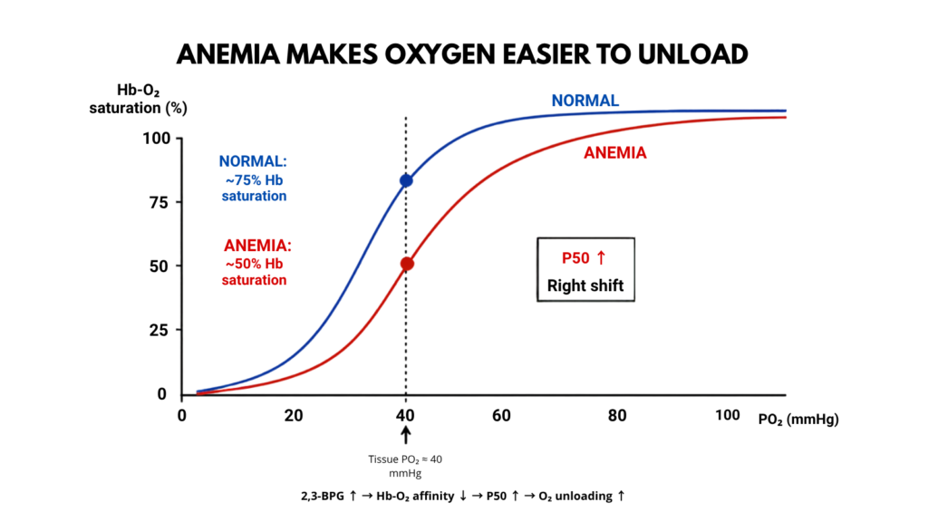
2,3-BPG ↑ → hemoglobin-O₂ affinity ↓ → P50 ↑
The oxygen-hemoglobin dissociation curve shifts to the right.
At a given PO₂, hemoglobin releases more oxygen.
The classic anemia literature links this rightward shift to increased intraerythrocytic 2,3-DPG.20Later reviews similarly identify increased 2,3-DPG as the principal adaptive determinant of reduced hemoglobin-oxygen affinity in chronic anemia.21
This should be distinguished from the Bohr effect.
Local increases in CO₂ and H⁺ in metabolically active tissues also shift the oxygen dissociation curve to the right and facilitate unloading.
But chronic anemia does not need to cause systemic acidosis for its adaptive right shift to occur.
FROM OXYGEN UNLOADING TO OXYGEN CONSUMPTION
Releasing oxygen from hemoglobin makes it available to the tissues. But availability is not the same as use.
The oxygen dissociation curve tells us how readily oxygen can be released from hemoglobin at a given PO₂. The next question is what the tissues do with that oxygen: how much of the oxygen delivered do they actually extract and consume?
This brings us to the Fick equation:
VO₂ = CO × (CaO₂ − CvO₂)
CaO₂ tells us what enters the tissue circulation. CvO₂ tells us what leaves. The difference tells us how much oxygen was extracted along the way.
Step 4: Tissues extract a larger fraction of the oxygen delivered
We now move from oxygen unloading to oxygen consumption.
The Fick equation is:
VO₂ = CO × (CaO₂ − CvO₂)
where VO₂ is oxygen consumption and CaO₂ − CvO₂ is the amount of oxygen removed from each unit of blood as it passes through the tissues.
Another useful way to express this is the oxygen extraction ratio (OER):
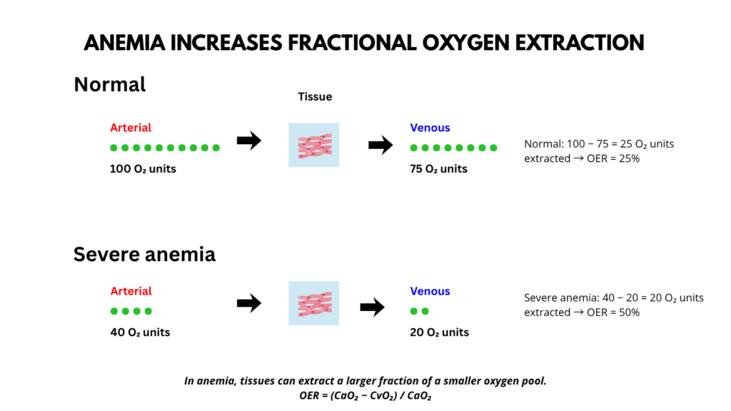
OER = (CaO₂ − CvO₂) / CaO₂
OER is the fraction of the oxygen delivered in arterial blood that is removed by the tissues. For example, an OER of 0.25 means that the tissues extract 25% of the oxygen entering the systemic circulation.
Why does OER increase in anemia?
Part of the answer follows directly from the equations. Since:
OER = VO₂ / DO₂
if oxygen delivery falls while resting oxygen consumption is maintained, the tissues must extract a greater fraction of the oxygen that reaches them.
But how is that accomplished?
Oxygen is continuously consumed by mitochondria, keeping tissue PO₂ below capillary PO₂. This creates the gradient that drives oxygen from blood into tissue.
As dissolved oxygen leaves the plasma, capillary PO₂ falls.
That, in turn, causes hemoglobin to release more oxygen.
In chronic anemia, the rise in 2,3-BPG shifts the oxygen dissociation curve to the right, so hemoglobin releases more oxygen at a given PO₂. Venous blood therefore leaves the tissue with less of its available oxygen, increasing the fraction extracted.
So the sequence is:
mitochondrial O₂ consumption → tissue PO₂ remains low
↓
O₂ diffuses from capillary blood into tissue
↓
capillary PO₂ falls
↓
hemoglobin unloads more O₂
↓
right-shifted ODC facilitates unloading
↓
fractional oxygen extraction ↑
Thus, the increase in OER reflects both the need to preserve VO₂ as DO₂ falls and the ability to unload a greater fraction of the oxygen carried by hemoglobin.
Roy and colleagues showed that as oxygen transport fell in chronic severe anemia, the percentage of delivered oxygen extracted by tissues increased.22
But there is an important distinction.
Increased fractional extraction does not necessarily mean an increased absolute arterial-venous oxygen-content difference.
Why?
Because arterial oxygen content starts so low.
In Roy’s patients, the average absolute A-V oxygen-content difference was actually lower than in healthy controls, even though the fraction of available oxygen extracted was greater.23
So:
oxygen extraction ratio ↑
but:
absolute CaO₂ − CvO₂ does not necessarily ↑
A useful way to say it is:
In severe anemia, tissues extract a larger fraction of a much smaller oxygen pool.
Step 5: Oxygen consumption can be defended
Return to:
VO₂ = CO × (CaO₂ − CvO₂)
As hemoglobin falls:
CaO₂ ↓
But:
cardiac output ↑
and:
fractional oxygen extraction ↑
Together, these adaptations can defend resting oxygen consumption.
In Roy’s chronic severe anemia cohort, oxygen consumption remained within the normal range even among patients whose average hemoglobin was approximately 3 g/dL.24
Something similar was seen in healthy people with acute isovolemic anemia to approximately 5 g/dL.
Their oxygen consumption did not fall.
It actually increased modestly, from about 3.01 to 3.42 mL O₂/kg/min, while plasma lactate did not rise.25
The investigators attributed at least part of this increase to the metabolic cost of tachycardia and sympathetic activation.
That makes an interesting point:
Compensation itself consumes oxygen.
Does oxygen demand also fall?
Potentially, but this does not appear to be the dominant whole-body adaptation in chronic severe anemia.
Classic studies found resting whole-body oxygen consumption to be largely preserved even in very severe anemia.26
At the organ level, however, oxygen consumption may fall when delivery cannot be completely maintained. Cerebral oxygen consumption, for example, has been reported to decrease modestly in chronic anemia.27
A symptomatic patient may also reduce total oxygen demand simply by reducing activity.
That is different from saying that chronic anemia causes a generalized adaptive reduction in basal metabolic rate.
The better-supported sequence is:
defend oxygen delivery
plus:
increase fractional extraction
with some reduction in oxygen demand occurring regionally or behaviorally when reserve becomes limited.
Step 6: Regional blood flow adapts
So far, we have treated cardiac output as a whole-body variable. But cardiac output is not distributed evenly across organs.
In anemia, preserving oxygen delivery depends not only on how much blood the heart pumps, but also on where that blood goes.
Different vascular beds therefore adapt differently according to local oxygen demand.
Cerebral blood flow increases in chronic anemia. Coronary blood flow also increases substantially.28
The coronary response is particularly important because the myocardium already extracts a large fraction of delivered oxygen under normal conditions. It therefore has relatively little extraction reserve, making increased coronary flow an important compensatory mechanism as arterial oxygen content falls.
But increased flow does not necessarily restore oxygen delivery completely.
In the brain, for example, increased cerebral blood flow does not fully compensate for the reduced oxygen-carrying capacity of the blood. Cerebral oxygen delivery may remain slightly reduced, and cerebral oxygen consumption may fall.29
The kidney can behave quite differently.
In Anand’s patients with severe chronic anemia and edema, systemic vascular resistance was markedly reduced, yet renal blood flow fell by approximately 46%, while renal vascular resistance increased.30
Thus:
systemic vasodilation does not mean uniform vasodilation of every organ.
Regional blood flow is governed by local metabolic requirements and organ-specific regulation.
Step 7: Chronic adaptation can change structure
If anemia persists, adaptation is no longer limited to moment-to-moment changes in flow.
Chronic increases in blood flow and shear stress can produce vascular remodeling. Microvascular recruitment and angiogenesis have also been described, although some of this evidence comes from experimental models and from patients with chronic kidney disease rather than isolated anemia.31
roduce eccentric left ventricular hypertrophy.
This introduces an important distinction:
Acute compensation changes flow.
Chronic compensation can also change structure.
The same changes that initially help preserve oxygen delivery may eventually contribute to cardiovascular pathology if the anemia persists.
Step 8: Compensation has a limit
Cardiac output cannot rise indefinitely.
Oxygen extraction cannot increase indefinitely.
Vasodilation cannot increase indefinitely.
And eventually the heart itself becomes limited by oxygen supply.
Experimental studies demonstrate a characteristic relationship between oxygen delivery and oxygen consumption.
At ordinary levels of oxygen delivery:
DO₂ ↓ → VO₂ remains relatively stable
But below a critical level:
DO₂ ↓ → VO₂ ↓
Oxygen consumption becomes supply dependent, and lactate begins to rise.
Cilley and colleagues demonstrated this pattern in anesthetized dogs when oxygen delivery was reduced by anemia, hypoxemia, or low cardiac output.32
The absolute threshold from those experiments should not be transferred directly to patients.
The animals were anesthetized, and in the anemia experiments the investigators deliberately restrained the normal increase in cardiac output with cardiac tamponade. Splenectomy was also performed to prevent splenic autotransfusion.
In other words, this was partly a model in which normal compensation was deliberately blocked.
Healthy conscious humans can tolerate lower measured oxygen-delivery values than the animal threshold would suggest. In the Weiskopf study, some subjects had oxygen delivery below 10 mL O₂/kg/min, with a lowest value of approximately 6.5 mL O₂/kg/min, without developing lactic acidosis.33
So the number is less important than the principle:
Eventually oxygen delivery becomes insufficient to sustain oxygen consumption.
Older chronic-anemia data suggest that cardiac output tends to plateau at extremely low hemoglobin concentrations, particularly below approximately 3 g/dL.34
At that point, an important compensatory reserve is approaching its limit.
Step 9: Resting compensation is not the same as reserve
Now return to the patient with a hemoglobin of 4 g/dL.
He is sitting comfortably.
Rest is the key word.
At rest, oxygen consumption is relatively low.
The patient may already have recruited multiple mechanisms to maintain it:
cardiac output ↑
stroke volume ↑
systemic vascular resistance ↓
oxygen unloading ↑
fractional oxygen extraction ↑
regional blood flow altered
These mechanisms may be sufficient while the patient is sitting quietly.
Exercise changes the equation.
VO₂ rises.
The circulation must now increase cardiac output and regional blood flow even further.
In chronic anemia, the increase in cardiac output required for a given increase in oxygen consumption during exercise is substantially greater than normal. Classic studies therefore found exercise to be a more sensitive probe of abnormal circulatory physiology than resting cardiac output.35
Weiskopf and colleagues made the same point from the opposite direction. Their findings applied to healthy subjects at rest and were explicitly not intended to be extrapolated to exercise, cardiovascular disease, impaired cardiac reserve, or impaired regional blood flow.36
So a patient can appear remarkably well at rest not because oxygen delivery is normal, but because compensatory mechanisms are sufficient for the current oxygen demand.
The clinically important question is how much reserve remains.
PUTTING IT ALL TOGETHER
Start with the primary disturbance:
Hb ↓
↓
CaO₂ ↓
The body has very little ability to restore arterial oxygen content without restoring hemoglobin.
So it compensates elsewhere.
Increase cardiac output: ventricular view
CO = HR × SV
In chronic severe anemia, stroke volume may contribute importantly to the rise in cardiac output.
Stroke volume is favored by:
afterload ↓
with possible contributions from:
venous return/filling ↑
and:
contractility ↑
Increase cardiac output: systemic-circulation view
CO ≈ ΔP / SVR
Anemia causes:
Hct ↓ → viscosity ↓
and:
vasodilation → vessel radius ↑
↓
SVR ↓
↓
systemic flow ↑
The two views intersect because:
SVR ↓ → afterload ↓
but they remain conceptually distinct.
Improve oxygen unloading
2,3-BPG ↑
↓
P50 ↑
↓
hemoglobin-O₂ affinity ↓
↓
oxygen unloading ↑
Extract more of what arrives
oxygen extraction ratio ↑
Redistribute flow
organ-specific blood flow changes
Together:
cardiac output ↑ + fractional oxygen extraction ↑
↓
VO₂ defended despite CaO₂ ↓
At least for a time.
BACK TO THE PATIENT
A patient with a hemoglobin of 4 g/dL can have:
normal oxygen saturation
preserved blood pressure
a surprisingly normal heart rate
and relatively few symptoms while sitting quietly
because the hemoglobin concentration is only one variable in a much larger oxygen-transport system.
His blood is less viscous.
His systemic vascular resistance is lower.
His circulation can move more blood per minute.
His red cells release oxygen more readily.
His tissues extract a larger fraction of the oxygen delivered.
Regional blood flow adapts.
And if the anemia has been present long enough, structural adaptation may have occurred as well.
That does not make a hemoglobin of 4 g/dL safe.
It explains why the hemoglobin concentration alone does not tell us how close the patient is to the limit of compensation.
THE TAKE HOME
The fundamental problem in severe anemia is easy to state:
arterial oxygen content falls.
The remarkable part is everything that happens next.
DO₂ = CO × CaO₂
Cardiac output can then be examined through two complementary lenses:
CO = HR × SV
and:
CO ≈ ΔP / SVR
They describe different aspects of the same high-output circulation and should not be treated as interchangeable equations.
Finally:
VO₂ = CO × (CaO₂ − CvO₂)
As hemoglobin falls, the heart, systemic circulation, red cells, microcirculation, and tissues all adjust to defend oxygen use.
Those adaptations can be remarkably successful at rest.
But they are finite.
Severe chronic anemia is therefore not just a low hemoglobin. It is a test of how much physiologic reserve remains.