
How observation, assays, multimers, and molecular biology changed what von Willebrand disease meant
Note: The video and audio linked above were generated with the assistance of AI. Clinical accuracy has been reviewed, but no AI-generated content can be guaranteed to be fully error-free.
Why this spoke matters
Von Willebrand disease did not enter medicine as a molecule.
It entered as a pattern.
A family bled. Women bled. Children bled. The bleeding looked inherited, but it did not behave like classic hemophilia.
That is the historical lesson.
Before there was a VWF gene, before multimer analysis, before ristocetin assays, before type 2 subtypes, there was bedside observation.
The history of VWD is therefore not just a chronology. It is a lesson in how disease categories are shaped by the tools available to see them.
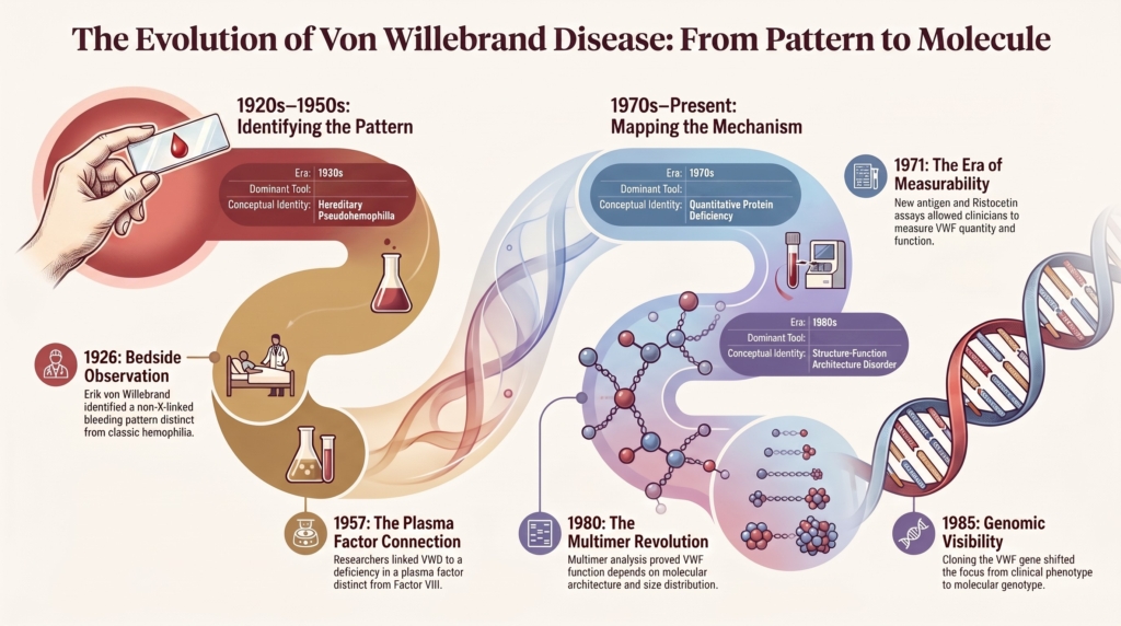
Each era changed what clinicians thought VWD was: first a familial bleeding tendency, then hereditary pseudohemophilia, then a platelet and vessel-wall disorder, then a plasma factor abnormality, then a platelet-adhesion disorder, then a multimer disorder, then a molecularly heterogeneous condition.
The disease did not simply become better understood.
It became differently understood.
Pattern before mechanism
Erik Adolf von Willebrand’s original description was grounded in clinical observation. He first evaluated Hjördis, a young girl from Föglö in the Åland Islands, in 1924 and published his account in 1926 under the title Hereditär pseudohemofili in Finska Läkaresällskapets Handlingar.
Von Willebrand traced the disorder across 66 family members and identified 23 with abnormal bleeding. Three of Hjördis’s siblings had already died from hemorrhage. Hjördis herself experienced recurrent epistaxis, gingival bleeding, bruising, and prolonged bleeding after minor trauma. She died at age 14 from uncontrolled bleeding during her fourth menstrual period.1
The key features were not molecular. They were relational: bleeding across generations, mucocutaneous rather than deep muscular bleeding, prominent bleeding in females, variable severity within a family, and a pattern distinct from classic hemophilia.
That is why the origin story still matters. The first act of recognition was phenotypic, not biochemical. A clinician saw that this was not simply hemophilia under another name. The disorder looked hereditary but not X-linked, caused bleeding but not the same pattern of bleeding, and affected families without following the expected script.
VWD began as a diagnostic discomfort because the available categories were not enough.
The first hypothesis: platelets and the vessel wall
Von Willebrand did not know about VWF.
No one did.
But using the tools available in the 1920s, he made a perceptive inference. The most important laboratory abnormality was a prolonged bleeding time. The platelet count was generally not low enough to explain the bleeding. The clotting time was normal. Clot retraction was normal.
This pattern pointed away from classic hemophilia and toward primary hemostasis.
Von Willebrand proposed that the bleeding could be explained by disturbed platelet function together with a more general abnormality of the capillary wall.2
The hypothesis was incomplete but directionally correct. Modern VWF biology would eventually show that the missing factor sits at the interface of platelets, endothelium, subendothelial matrix, blood flow, and coagulation. Von Willebrand did not identify the molecule, but he identified the biological territory in which the defect must lie.
The shadow of hemophilia
For much of early hematology, hemophilia provided the dominant template for inherited bleeding disorders.
That template was powerful. It linked severe bleeding, male inheritance, and deficiency of a plasma coagulation factor.
VWD did not fit cleanly. Its bleeding was often mucosal. Its inheritance could affect women. Its severity varied. Its laboratory profile was unsettled. Its relationship to factor VIII was confusing.
This is why terms such as “hereditary pseudohemophilia” were historically useful, but also limiting. They named the resemblance without explaining the difference.
The disease passed through several identities before becoming VWD. Depending on the dominant hypothesis of the era, it was described as pseudohemophilia, vascular hemophilia, angiohemophilia, hereditary capillary purpura, or Willebrand-Jürgens thrombopathy.3
Each name carried an assumption. Was the problem in the platelet, the vessel wall, the plasma, the coagulation cascade, or some combination of these?
The word “pseudo” placed VWD in hemophilia’s shadow. It suggested imitation rather than independent biology.
But VWD was not false hemophilia.
It was a different window into hemostasis.
When plasma factor biology changed the question
The next major shift came when VWD became linked to plasma factor biology.
By the 1950s, methods for measuring antihemophilic factor, later factor VIII, became available. Investigators found that some patients with the clinical pattern of VWD also had reduced factor VIII activity.
This was puzzling.
The bleeding phenotype looked like a primary hemostatic disorder.
But the laboratory pointed toward a coagulation factor.
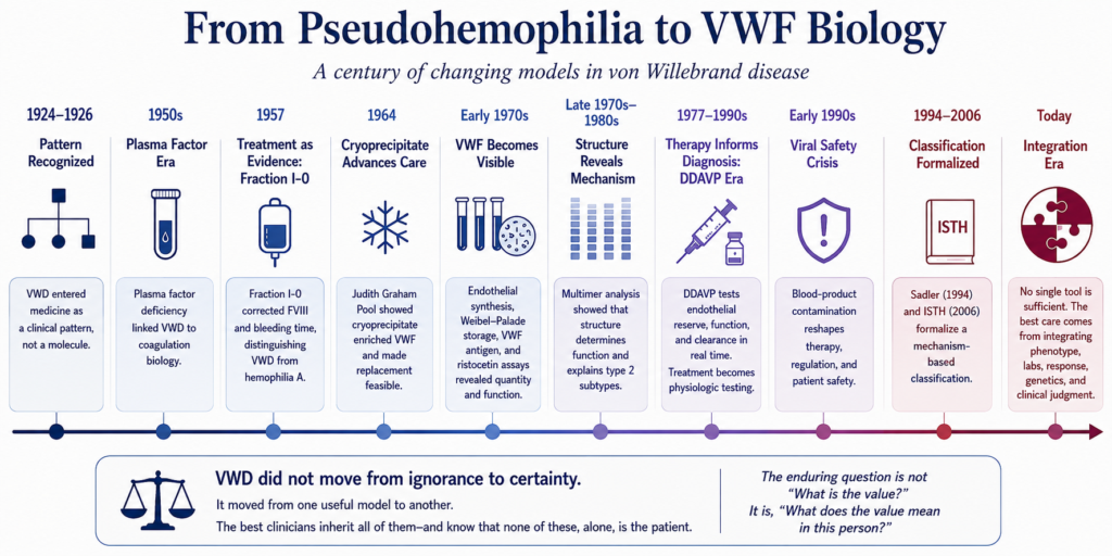
In 1957, Inga Marie Nilsson, Margareta Blombäck, Birger Blombäck, and colleagues revisited the Åland family and confirmed that affected relatives shared the same clinical and laboratory pattern seen in other Swedish patients. Their therapeutic experiment also became biological evidence: plasma fraction I-0 corrected both factor VIII activity and the prolonged bleeding time in patients with VWD, while preparations that corrected hemophilia A did not fully reproduce this effect. Treatment therefore helped show that VWD involved a plasma factor related to, but distinct from, the factor missing in classic hemophilia A.4
The question changed.
It was no longer simply:
Why do these families bleed?
It became:
What is the relationship between this disorder, factor VIII, platelets, and the vessel wall?
That question was transformative.
VWD began to occupy an interface position. It was not only a platelet-like bleeding disorder, not only a coagulation-factor disorder, and not simply vascular fragility. It connected all three.
That interface position remains the key to modern reasoning about VWD.
VWF supports platelet adhesion under shear and stabilizes factor VIII in circulation. Once that dual role became visible, the clinical paradox began to make sense. Mucocutaneous bleeding reflected failure of platelet-dependent primary hemostasis. Low factor VIII in some patients reflected failure of VWF carrier function.
One molecule connected two hemostatic systems.
The vessel-wall hypothesis also acquired a remarkable molecular vindication. In the early 1970s, investigators demonstrated that VWF is synthesized by endothelial cells, and subsequent work localized VWF to Weibel-Palade bodies, the endothelial storage organelles from which it can be released. The vessel wall was therefore not merely a passive surface affected by the disorder. It was one of the principal sites where the missing protein was made and stored.
This discovery completed an arc that had begun with von Willebrand’s original bedside inference. He had proposed a disorder involving platelets and the capillary wall before the molecule existed as a measurable concept. Endothelial biology later showed why that intuition had been so productive.
1971: the disease becomes measurable
The early 1970s transformed VWD diagnosis.
In 1971, Zimmerman and colleagues developed an antiserum against highly purified factor VIII preparations and identified a plasma protein then called factor VIII-related antigen. This protein was present in healthy individuals and in patients with hemophilia A, but reduced or absent in many patients with VWD. It later became clear that this antigen was VWF antigen.5
Around the same time, Howard and Firkin observed that ristocetin induced platelet agglutination in normal platelet-rich plasma but not in platelet-rich plasma from patients with VWD. Soon afterward, quantitative ristocetin cofactor assays were developed, allowing investigators to measure the platelet-dependent activity of the missing plasma factor.67
VWD could now be measured in two complementary ways: how much VWF-like protein was present and how well that protein supported platelet interaction.
The disease could now be separated more clearly from hemophilia A.
It could also be divided into different forms.
The laboratory could now ask not only how much VWF was present, but whether it worked.
Ristocetin and the accidental history of testing
Ristocetin was not developed to diagnose VWD. It was an antibiotic whose clinical use was curtailed by platelet-related toxicity, but that unwanted effect became diagnostically valuable because ristocetin could induce interaction between VWF and platelet GPIbα in vitro.
That historical turn matters because it illustrates how measurement reshapes disease.
Once clinicians could test platelet-dependent VWF activity, VWD became easier to distinguish from disorders of VWF quantity alone. The assay helped move VWD from a quantitative disorder toward a structure-function disorder.
It also introduced new complexity. A low activity result could reflect reduced protein, defective platelet binding, loss of high-molecular-weight multimers, assay behavior, or another mechanism.
Ristocetin also revealed that VWF-platelet interaction could be abnormal in more than one direction. In some patients, later classified as type 2B VWD, VWF binds platelets too avidly. In platelet-type VWD, the abnormality lies not in VWF but in the platelet GPIb receptor. These disorders can look similar unless the laboratory asks the right mechanistic question.8
The test clarified the disease while creating new interpretive problems.
That is the recurring historical pattern.
Every new assay makes something visible.
Every new assay also creates a new gray zone.
When structure entered the story
The recognition of VWF multimers changed VWD classification profoundly.
VWF was no longer just a plasma factor with a measurable level. It was a multimeric adhesive protein whose size distribution mattered.
Large multimers were especially important for platelet adhesion under shear. Their absence or dysfunction helped explain why some patients had disproportionately impaired VWF activity despite measurable antigen.
VWD could now be understood in structural terms. Some patients had too little VWF. Some had abnormal assembly or loss of high-molecular-weight multimers. Some had excessive platelet binding. Others had impaired platelet binding, impaired collagen binding, impaired factor VIII binding, or accelerated clearance. These were not merely different test patterns. They were different ways for a large adhesive protein to fail.
In 1980, Ruggeri and Zimmerman demonstrated abnormal multimeric structure in different VWD variants, including patterns that helped distinguish what would become type 2A and type 2B VWD.9
The multimer era transformed VWD from a disorder defined by quantity into one defined by architecture.
How much protein was present still mattered.
But how that protein was assembled mattered too.
Classification began to reflect mechanism.
Type 1, type 2, and type 3 were not merely labels. They were attempts to map clinical and laboratory patterns onto biological failure modes.
Later, type 2 disease was subdivided into 2A, 2B, 2M, and 2N, reflecting distinct mechanisms: loss of high-molecular-weight multimers, increased platelet binding, impaired platelet-dependent function without loss of multimers, and impaired factor VIII binding.10 This mechanistic framework was formalized by Sadler and the ISTH subcommittee in 1994 and revised in 2006 as molecular and laboratory understanding evolved. Classification had become more than descriptive pattern recognition. It was now an explicit attempt to map phenotype onto mechanism. It allowed clinicians to reason more effectively about diagnosis, prognosis, and therapy.
But it also carried a risk.
Once categories became more precise, they could seem more complete than they were.
The multimer era taught that structure matters.
It did not prove that structure explains everything.
Patients with similar laboratory patterns could bleed differently. Patients within the same subtype could have different clinical trajectories. Some patients sat uneasily between categories. Others changed over time as VWF levels rose with age, pregnancy, inflammation, or other physiologic modifiers.
Structure explained severity better than earlier models had.
But it did not eliminate judgment.
Therapy changed diagnosis
The history of VWD is also a history of treatments that functioned as biological experiments. The 1957 fraction I-0 studies had already shown that therapeutic response could clarify disease biology.
In 1964, Judith Graham Pool showed that slowly thawed plasma yielded a cryoprecipitate enriched in antihemophilic factor and VWF. Cryoprecipitate made effective replacement possible with far less volume than plasma and became an important practical advance for patients with VWD and hemophilia.11
Cryoprecipitate and plasma-derived concentrates transformed care, but products manufactured before effective viral inactivation also transmitted HIV and hepatitis viruses to many people with inherited bleeding disorders. This history permanently altered perceptions of replacement therapy, product safety, institutional trust, and informed consent. The broader history of blood-product contamination deserves separate treatment and is beyond the scope of this essay.
Desmopressin introduced a different therapeutic idea.
In 1977, Mannucci and colleagues brought DDAVP into clinical use for patients with mild hemophilia A and VWD. Instead of replacing VWF, DDAVP released endogenous VWF from endothelial stores.12
Like the earlier fraction I-0 experiments, DDAVP changed more than treatment. It turned therapeutic response into a physiological test of endogenous VWF biology.
A response could reveal stored, releasable, functional VWF. A short-lived response could suggest accelerated clearance. A poor response could shape procedural planning. A thrombocytopenic response could raise concern for type 2B disease.
DDAVP turned endothelial biology into a bedside experiment.
Plasma-derived VWF/FVIII concentrates, and later recombinant VWF, made treatment more specific and safer, especially for patients with severe VWD or those unresponsive to DDAVP.13
Therapy also made diagnosis more consequential. A label could open access to preventive treatment, procedural planning, and family counseling. But it could also bring lifelong caution, anxiety before procedures, treatment exposure, insurance consequences, and altered self-understanding.
Naming disease became an intervention.
The genomic era and the limits of molecular certainty
Cloning of the VWF gene in 1985 added another layer of visibility.
Investigators could now move from phenotype to genotype, linking variants, domains, inheritance patterns, and laboratory behavior.14
This clarified many mechanisms, especially in qualitative type 2 variants and severe type 3 disease.
It also revealed the complexity of mild quantitative disease.
Genetic testing clarified many type 2 and type 3 mechanisms, but it did not eliminate diagnostic uncertainty in mild quantitative VWF deficiency, where VWF variants are less consistently found and non-VWF modifiers contribute. Genotype-phenotype correlation is often weak in mild type 1 VWD and low VWF.15
ABO blood group, clearance biology, endothelial regulation, platelet biology, age, inflammation, pregnancy, anemia, and environmental context all shape the phenotype.
The genomic era did not replace clinical reasoning.
It refined it.
Molecular testing is powerful when it answers a specific question: whether a low FVIII phenotype reflects type 2N VWD rather than mild hemophilia A, whether enhanced platelet interaction reflects type 2B VWD rather than platelet-type VWD, whether severe deficiency has implications for family counseling, or whether a variant explains the observed laboratory pattern.
But genetics does not abolish the boundary problem.
In some patients, especially those with mild quantitative reductions, the central question remains clinical:
Does this person have a bleeding disorder, a risk state, or physiologic variation with coincidental symptoms?
The gene can help.
It cannot always decide.
Clinical synthesis
The 30–50 IU/dL range is not merely a laboratory interval. It is the historical residue of a continuous biological trait being translated into categorical disease language.
Modern VWD diagnosis inherited a century of increasingly powerful tools. Clinical observation identified the bleeding pattern. Bleeding time placed the disorder in the world of platelets and vessels. Factor VIII assays revealed its connection to coagulation. VWF antigen made the protein measurable. Ristocetin made platelet-dependent function visible. Multimer analysis revealed molecular architecture. DDAVP response made endothelial reserve and clearance testable. Genetics connected phenotype with variants and domains.
Each tool clarified a different dimension of the disorder, but no tool made the boundary between variation and disease disappear. VWF is continuously distributed across the population and is modified by blood group, age, inflammation, pregnancy, stress, hormones, clearance, and other genetic and environmental influences. Bleeding symptoms are also common, particularly heavy menstrual bleeding, epistaxis, and easy bruising.
For some patients, a VWD or low-VWF label explains a lifelong bleeding pattern, guides procedural management, supports family counseling, and prevents dismissal of symptoms. For others, the label may imply more biological certainty than the findings justify. A patient whose VWF rises with age reminds us that biology changes while diagnoses persist. A patient with low factor VIII reminds us that VWF is also a carrier protein. A patient with a qualitative defect reminds us that quantity and function are not equivalent. A patient with no identifiable VWF variant reminds us that molecular testing can clarify mechanism without defining disease in every case.
The history of VWD is therefore not a progression from ignorance to certainty. It is a succession of useful models, each revealing something the previous model could not see. The best clinicians inherit all of them without treating any one as complete. They know that phenotype matters, assays matter, structure matters, treatment response matters, and genes matter. They also know that none of these, alone, is the patient.
The enduring question is not simply, “What is the value?” It is, “What does the value mean in this person?”
Reflect & Apply
A 36-year-old woman has lifelong heavy menstrual bleeding, postpartum hemorrhage, and recurrent iron deficiency.
Her VWF antigen is 46 IU/dL.
Her platelet-dependent VWF activity is 43 IU/dL.
FVIII is 58 IU/dL.
Multimers are normal.
No clearly pathogenic VWF variant is identified.
A trainee asks:
“Does she really have VWD?”
Questions for reflection:
- What would you call this: type 1 VWD, low VWF, bleeding disorder of uncertain cause, or something else?
- What does the label help you do for her?
- What could the label cost her?
- How much weight should you give her bleeding history compared with the laboratory values?
- What does normal multimer analysis clarify, and what does it not clarify?
- What does the absence of a pathogenic VWF variant mean?
- How would your answer change before major surgery, pregnancy, or dental extraction?
This case illustrates the historical lesson of VWD:
each tool changes the question.
None ends the conversation.
The question is not only whether she fits the category.
The question is whether the category helps manage her bleeding.
Test your thinking
Apply the concepts from this essay in the companion interactive quiz.