
What the initial panel can and cannot tell us
Note: The video and audio linked above were generated with the assistance of AI. Clinical accuracy has been reviewed, but no AI-generated content can be guaranteed to be fully error-free.
Why this spoke matters
The first VWD laboratory panel is often treated as the diagnostic moment.
It is not.
It is the beginning of interpretation.
First-line testing does not answer a single question.
It answers several smaller ones:
- How much VWF is present?
- Does the VWF present function normally?
- Is FVIII preserved?
- Is there thrombocytopenia?
- Is another hemostatic pathway abnormal?
- Was the sample obtained under conditions that make interpretation difficult?
These answers matter only when read together.
A VWF antigen value by itself is not VWD.
A low activity value by itself is not a subtype.
A normal FVIII level does not exclude VWD.
A normal PT, aPTT, or platelet count does not exclude VWD.
First-line testing creates a map.
Clinical reasoning still has to read it.
Where first-line testing belongs
Testing should follow phenotype.
The question is not:
Which panel diagnoses VWD?
The better question is:
Does this patient’s bleeding pattern justify evaluating the VWF system?
First-line VWF testing is most useful when the clinical story suggests a disorder of primary hemostasis:
- epistaxis
- easy bruising
- oral mucosal bleeding
- heavy menstrual bleeding
- postpartum hemorrhage
- excessive bleeding after dental work
- excessive bleeding after procedures
- family history of similar bleeding
Modern guidelines therefore emphasize bleeding history and, in some settings, formal bleeding assessment tools as part of the diagnostic process.1
But the history does not replace the laboratory.
And the laboratory does not replace the history.
VWD sits at the intersection of both.
The core first-line panel
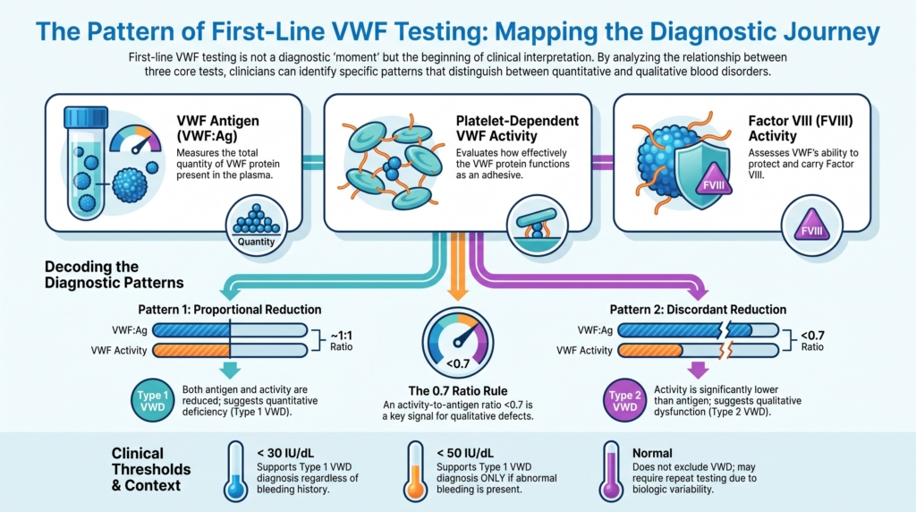
A practical first-line evaluation usually centers on three measurements:
- VWF antigen (VWF:Ag)
- platelet-dependent VWF activity
- FVIII activity (FVIII:C)
These should be interpreted alongside:
- bleeding phenotype
- family history
- CBC and platelet count
- PT and aPTT
- physiologic state at the time of sampling
- blood group, if known
Modern diagnostic guidelines consistently place VWF:Ag, platelet-dependent VWF activity, and FVIII:C at the center of first-line testing.23
Three patterns to recognize
Most first-line VWF panels fall into one of three broad patterns.
Pattern 1: Proportional reduction
- VWF:Ag reduced
- VWF activity reduced
- VWF activity/VWF:Ag ratio relatively preserved
Think quantitative deficiency.
Type 1 VWD or low VWF become leading considerations.
Pattern 2: Discordant reduction
- platelet-dependent VWF activity reduced more than VWF:Ag
- VWF activity/VWF:Ag ratio reduced
Think qualitative dysfunction.
Type 2 VWD moves higher in the differential diagnosis and second-line testing becomes necessary.
Pattern 3: Normal panel despite convincing bleeding
- VWF studies normal
- bleeding phenotype remains compelling
Think repeat testing, physiologic modifiers, platelet disorders, connective tissue disorders, acquired causes, or alternative explanations.
Most interpretation ultimately comes back to recognizing which of these three patterns is present.
VWF antigen: how much protein is present?
VWF antigen (VWF:Ag) estimates the amount of VWF protein in plasma.
It answers a quantity question:
How much VWF is there?
A low antigen level may support quantitative deficiency.
But it does not explain mechanism.
A low value may reflect:
- inherited quantitative deficiency
- low VWF
- blood group effects
- physiologic variation
- increased clearance
- preanalytic artifact
VWF antigen therefore provides an estimate of quantity, not function.
It does not tell you whether the protein works normally.
It does not tell you whether the patient bleeds.
It does not tell you whether a borderline value is stable over time.
Platelet-dependent VWF activity: what the protein can do
Platelet-dependent VWF activity assays estimate how effectively VWF supports platelet adhesion
They ask a function question:
Does the VWF present behave as an adhesive protein?
Historically this was assessed using VWF ristocetin cofactor activity (VWF:RCo).
Although VWF:RCo transformed VWD diagnosis, it has important limitations:4
- substantial variability
- reduced performance at low VWF levels
- dependence on ristocetin rather than physiologic shear stress
- susceptibility to certain VWF sequence variants that lower assay results without producing clinically important dysfunction
For these reasons, modern guidelines prefer newer platelet-dependent activity assays such as VWF:GPIbM or VWF:GPIbR over VWF:RCo when available.5
Different laboratories may report platelet-dependent activity under different names:
- VWF:RCo (ristocetin cofactor activity)
- VWF:GPIbM: VWF glycoprotein IbM activity, using gain-of-function recombinant GPIb without ristocetin
- VWF:GPIbR: VWF glycoprotein IbR activity, using recombinant GPIb with ristocetin
Clinicians should understand which assay their laboratory uses because assay performance characteristics differ.
No static laboratory assay perfectly reproduces what VWF does under flowing blood.
Nevertheless, activity testing remains indispensable because VWD can be a disease of quality as well as quantity.
FVIII activity: the carrier function of VWF
FVIII activity (FVIII:C) is included because VWF serves as FVIII’s carrier protein.
When VWF is markedly reduced, FVIII may fall because it is no longer adequately protected from clearance.
This is why:
- severe type 3 VWD can resemble hemophilia A
- type 2N VWD can mimic mild hemophilia A
FVIII:C therefore asks:
Is VWF adequately performing its carrier role?
A low FVIII:C with low VWF:Ag may be expected.
A disproportionately low FVIII:C with relatively preserved VWF:Ag raises concern for type 2N VWD or hemophilia A.
A normal FVIII:C does not exclude VWD.
CBC, platelet count, PT, and aPTT: context, not exclusion
CBC, platelet count, PT, and aPTT remain useful.
But they are not reliable rule-out tests for VWD.
A CBC may reveal iron deficiency from chronic blood loss.
The platelet count may be low in type 2B VWD.
The aPTT may be prolonged if FVIII is sufficiently reduced.
However, many patients with VWD have:
- normal CBC
- normal platelet count
- normal PT
- normal aPTT
A normal aPTT does not exclude VWD.
A normal platelet count does not exclude VWD.
These tests provide context.
They do not replace VWF-specific testing.6
The VWF activity/VWF:Ag ratio
The VWF activity/VWF:Ag ratio is one of the most important interpretive tools in first-line testing.
It asks:
Is the VWF that is present working as expected?
In type 1 VWD:
- VWF:Ag and platelet-dependent VWF activity typically decrease proportionately
- the ratio remains relatively preserved
In type 2 VWD:
- platelet-dependent VWF activity falls disproportionately relative to VWF:Ag
- the ratio decreases
Current guidance commonly uses a ratio below approximately 0.7 as a trigger to suspect type 2A, 2B, or 2M VWD.7
A low ratio does not complete the diagnosis.
It changes the next question.
It signals the need for second-line testing.
Borderline values require context
Many first-line evaluations produce results near diagnostic boundaries.
This is where interpretation becomes difficult.
The 2021 ASH/ISTH/NHF/WFH guideline recommends:8
- VWF:Ag and/or platelet-dependent VWF activity <30 IU/dL supports type 1 VWD regardless of bleeding history
- VWF:Ag and/or platelet-dependent VWF activity <50 IU/dL supports type 1 VWD in patients with abnormal bleeding
Regardless of the diagnostic framework used, values in the 30–50 IU/dL range require careful interpretation.
The key question is not:
Is the value low?
The key question is:
Does the value explain this patient’s bleeding phenotype and future bleeding risk?
Why repeat testing is often a feature, not a failure
VWF is dynamic.
Levels increase with:
- inflammation
- acute illness
- exercise
- pregnancy
- postoperative stress
- aging
As a result, a single value can be misleading.
A normal result obtained during a VWF-elevating state may mask underlying disease.
Modern guidelines therefore emphasize that VWD diagnosis should generally be based on repeat testing obtained at baseline health.9
Repeat testing is not indecision.
It is biology-aware diagnosis.
Preanalytic pitfalls
Some errors occur before the assay ever begins.
Problems may include:
- improper tube filling
- delayed processing
- inappropriate transport conditions
- specimen mishandling
The BSH guideline specifically warns against transporting whole blood on ice because cold exposure may affect VWF and FVIII measurements and lead to misclassification.10
When the laboratory pattern does not fit the phenotype, sample quality belongs in the differential diagnosis.
When first-line testing is normal
Normal first-line results lower the likelihood of VWD.
But they do not explain every bleeding phenotype.
When suspicion remains high, the next step may include:
- repeat VWF testing
- review of sample quality
- platelet function testing
- evaluation for connective tissue disorders
- evaluation for acquired causes
- medication review
- investigation for gynecologic or structural causes of bleeding
The first-line panel evaluates the VWF system.
It does not evaluate every cause of bleeding.
When first-line testing suggests type 2 VWD
A reduced VWF activity/VWF:Ag ratio often prompts second-line testing.
Depending on the pattern, this may include:
- VWF collagen-binding activity (VWF:CB)
- VWF multimer analysis
- RIPA or targeted genetic testing for type 2B
- VWF–FVIII binding assay (VWF:FVIIIB) or targeted genetic testing for type 2N
The first-line panel rarely establishes a definitive type 2 subtype.
Instead, it identifies which second-line studies are most likely to be informative.
What first-line testing can do
First-line testing can:
- support quantitative VWF deficiency
- identify severe VWF deficiency
- suggest qualitative VWF dysfunction
- identify low FVIII:C related to impaired VWF carrier function
- raise suspicion for type 2N VWD
- identify thrombocytopenia suggestive of type 2B VWD
- guide second-line testing
- inform peri-procedural planning
What first-line testing cannot do
First-line testing cannot:
- diagnose VWD without clinical context
- exclude all bleeding disorders
- reliably subtype all type 2 variants
- distinguish type 2B from platelet-type VWD
- distinguish type 2N from hemophilia A without additional testing
- explain every borderline VWF value
- predict future bleeding risk by itself
It is a first-line map.
Not the whole territory.
Clinical synthesis
First-line VWF testing is not a verdict.
It is a structured conversation with VWF biology.
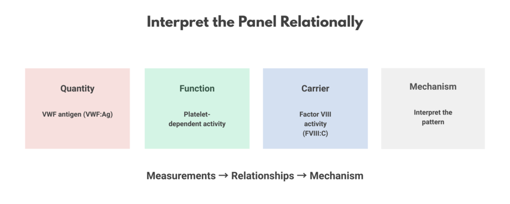
VWF:Ag asks how much protein is present.
Platelet-dependent VWF activity asks whether that protein functions normally.
FVIII:C activity asks whether VWF is performing its carrier role.
CBC, platelet count, PT, aPTT, physiologic state, and specimen quality help determine whether the pattern is coherent.
The most useful question is not:
What is the number?
It is:
What pattern do these numbers create?
Diagnosis begins with that pattern.
Not with a single value.
With a pattern that makes biological and clinical sense.
Evidence anchor: why first-line testing is a pattern-recognition exercise
Summary derived from diagnostic guidelines, laboratory studies, assay evaluations, cohort studies, and expert reviews. The evidence consistently shows that no individual first-line laboratory value adequately captures VWF biology. Diagnostic information emerges from the relationships among antigen, activity, FVIII, bleeding phenotype, and repeat testing.
| Evidence stream | What it shows | Why it matters | Main limitation |
|---|---|---|---|
| Quantitative vs qualitative VWF defects | VWD may result from reduced VWF quantity, impaired VWF function, or both. VWF:Ag and platelet-dependent VWF activity therefore provide complementary information rather than interchangeable measurements.11 | A normal antigen does not exclude dysfunctional VWF, and a low antigen does not define mechanism. | Antigen and activity may both be influenced by physiologic and technical factors. |
| VWF activity/VWF:Ag relationships | Qualitative VWF defects often produce activity levels that are disproportionately reduced relative to antigen levels, whereas quantitative deficiencies usually reduce both measurements proportionately.12 | The ratio often provides more diagnostic information than either value alone. | Ratios are screening tools, not definitive subtype diagnoses. |
| FVIII:C as a carrier-function marker | FVIII:C reflects VWF’s role as a carrier protein and may identify patterns suggestive of severe quantitative deficiency or type 2N VWD.13 | First-line testing assesses both platelet-related and coagulation-related consequences of VWF abnormalities. | FVIII:C may remain normal in many patients with VWD. |
| Biologic variability of VWF | VWF levels rise with inflammation, stress, exercise, pregnancy, acute illness, and aging.14 | A single result may not represent a patient’s baseline VWF biology. | Repeat testing may still leave uncertainty near diagnostic thresholds. |
| Preanalytic and assay limitations | Sample handling, transport conditions, assay methodology, and sequence variants can influence measured VWF activity.15 | Laboratory values must be interpreted within technical as well as biologic context. | Assay performance differs among laboratories and platforms. |
Interpretive note: These evidence streams converge on a common lesson: first-line VWF testing is fundamentally relational. Antigen, activity, FVIII, bleeding phenotype, and clinical context each contribute partial information. Diagnostic confidence increases when these elements point in the same direction. When they diverge, the task is not to force a diagnosis from a single number, but to determine whether the pattern is coherent, reproducible, and clinically meaningful.
Guideline perspective: what first-line testing is designed to do
Based on major diagnostic guidance from NHLBI, UKHCDO/BSH, ASH/ISTH/NHF/WFH, Nordic Haemophilia Council recommendations, and contemporary laboratory guidance.
Shared guidance themes
- VWF antigen (VWF:Ag), platelet-dependent VWF activity, and factor VIII activity (FVIII:C) form the core first-line laboratory panel.
- Laboratory results should be interpreted together with bleeding phenotype and family history.
- Normal PT, aPTT, platelet count, or FVIII do not exclude VWD.
- VWF activity/VWF:Ag relationships help identify patients who may require second-line testing for qualitative VWF defects.
- Testing should ideally occur when patients are at baseline health and not during major VWF-raising states.
- Repeat testing is frequently required when results are borderline or inconsistent with the clinical picture.
- Sample quality and preanalytic handling are important determinants of result reliability.
Where guidance has evolved
Older diagnostic algorithms relied heavily on VWF:RCo activity assays. More recent guidelines increasingly favor newer GPIb-based platelet-dependent VWF activity assays, such as VWF:GPIbM and VWF:GPIbR, when available because of improved reproducibility and reduced susceptibility to certain assay-related artifacts.16
Contemporary guidance also places greater emphasis on interpreting first-line results as patterns rather than isolated abnormalities and on integrating laboratory findings with bleeding phenotype and diagnostic probability.
What guidelines do not eliminate
- biologic variability in VWF levels
- assay-related limitations
- uncertainty near diagnostic thresholds
- the need for repeat testing
- the need for second-line testing when first-line patterns are discordant
- the role of clinical judgment
Practical takeaway: First-line testing is designed to identify diagnostic patterns, not to establish every diagnosis. The purpose of the panel is to determine whether VWF biology appears quantitatively reduced, qualitatively abnormal, or sufficiently normal that alternative explanations should be considered. Clinical interpretation remains essential.
Reflect & Apply Case
A 28-year-old woman with heavy menstrual bleeding has:
- VWF antigen 52 IU/dL
- Platelet-dependent VWF activity: 24 IU/dL
- Factor VIII activity (FVIII:C): 78 IU/dL
What is the most important observation?
Answer: The activity is disproportionately reduced compared with antigen, producing a low activity-to-antigen ratio. This pattern raises concern for a qualitative VWF defect and should prompt evaluation for type 2 VWD rather than simple quantitative deficiency.
The key insight is not the absolute value.
It is the relationship between the values.
Test your thinking
A short quiz on first-line testing in VWD.