Pappenheimer bodies are named after Alwin M. Pappenheimer Jr. (1908–1995), an American pathologist and immunologist. Though best known for his work on diphtheria toxin, his name became associated with these intracellular iron-containing inclusions found in red blood cells.
- Clinical Significance:
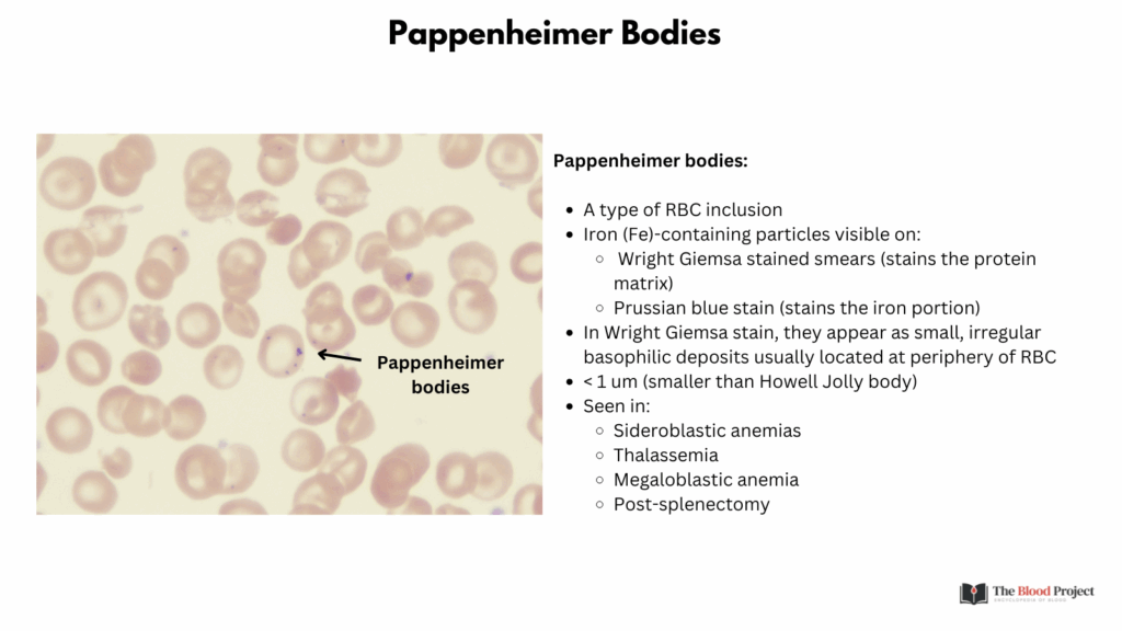
- Pappenheimer bodies are visible with Wright or Giemsa stain but are best confirmed with a Prussian blue iron stain, which distinguishes them from other inclusions like basophilic stippling.
- They are composed of aggregates of ferritin or hemosiderin, and are typically seen in conditions involving disordered iron metabolism such as:
- Sideroblastic anemia
- Hemolytic anemia
- Post-splenectomy
- Myelodysplastic syndromes
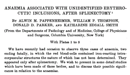
- Origin of the Eponym:1
- In 1945, Pappenheimer et al. described three patients whose red blood cells, after splenectomy, showed inclusions when stained with Giemsa or Wright stain.
- The bodies were described as red purple, usually coccoid, and adjacent to the cell membrane, and they demonstrated that the cells stained for iron with Prussian blue stain.
- They distinguished them from basophilic stippling, Howell-Jolly bodies, and overlying platelets but discussed in detail the possibility that they were Bartonella microorganisms. They ultimately concluded that the inclusions were not microorganisms

References:
Sears DA and Udden MM. Pappenheimer bodies: a brief historical review. Am J Hematol. 2004 Apr;75(4):249-50.